. 2013 Jul 4;8(7):e68775.
doi: 10.1371/journal.pone.0068775. Print 2013.

A huntingtin peptide inhibits polyQ-huntingtin associated defects

Affiliations

A huntingtin peptide inhibits polyQ-huntingtin associated defects

Yoan Arribat et al. PLoS One. .

Abstract

Background: Huntington's disease (HD) is caused by the abnormal expansion of the polyglutamine tract in the human Huntingtin protein (polyQ-hHtt). Although this mutation behaves dominantly, huntingtin loss of function also contributes to HD pathogenesis. Indeed, wild-type Huntingtin plays a protective role with respect to polyQ-hHtt induced defects.

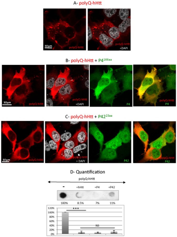
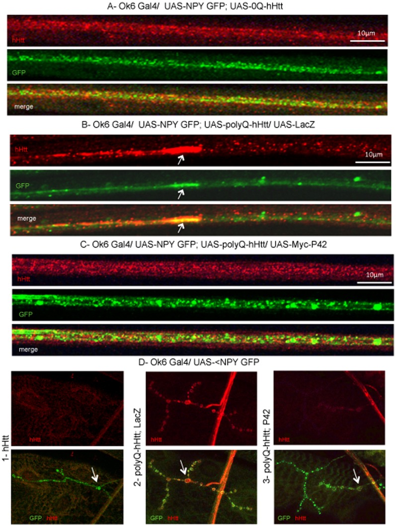
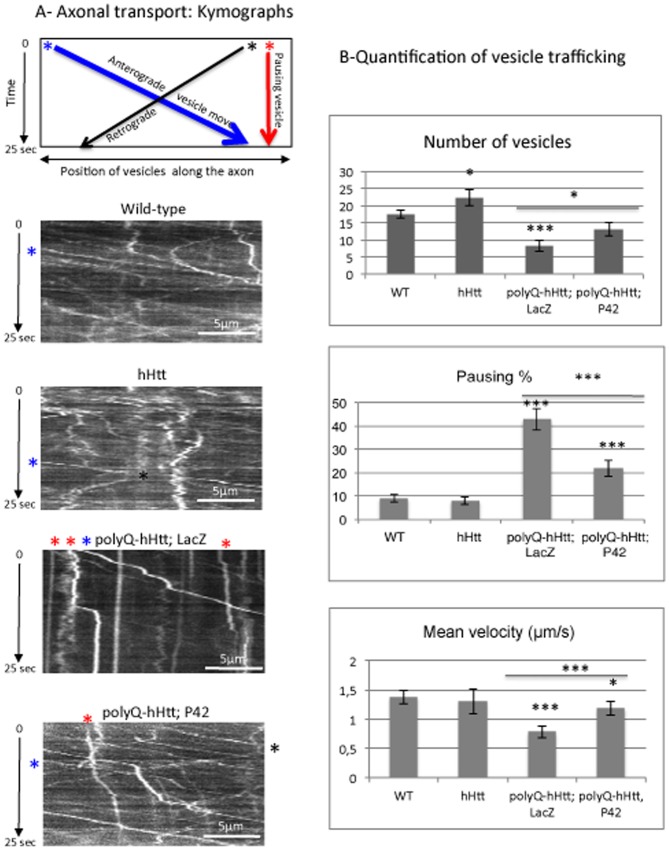
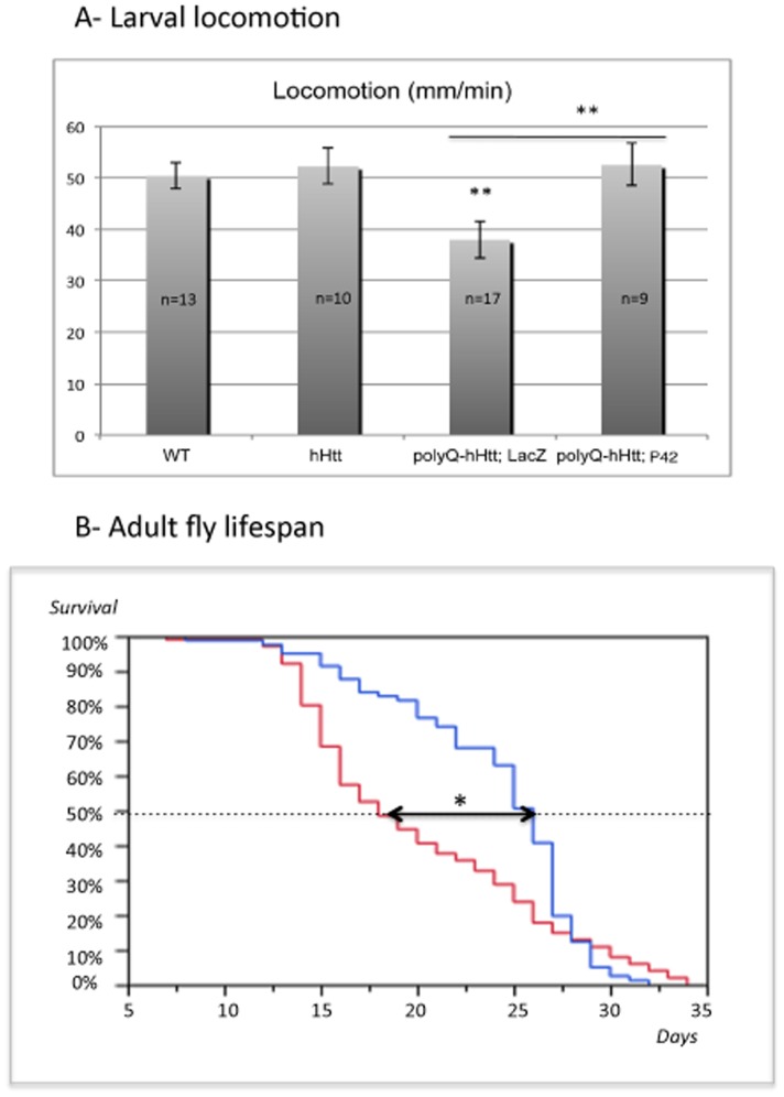
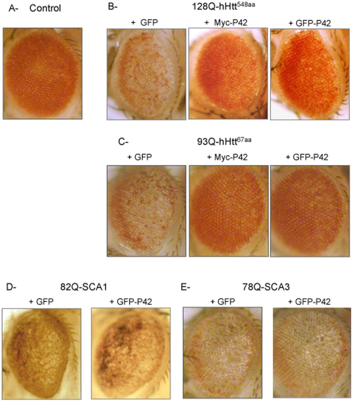
Methodology/principal findings: The question that we addressed here is what part of the wild-type Huntingtin is responsible for these protective properties. We first screened peptides from the Huntingtin protein in HeLa cells and identified a 23 aa peptide (P42) that inhibits polyQ-hHtt aggregation. P42 is part of the endogenous Huntingtin protein and lies within a region rich in proteolytic sites that plays a critical role in the pathogenesis process. Using a Drosophila model of HD, we tested the protective properties of this peptide on aggregation, as well as on different polyQ-hHtt induced neuronal phenotypes: eye degeneration (an indicator of cell death), impairment of vesicular axonal trafficking, and physiological behaviors such as larval locomotion and adult survival. Together, our results demonstrate high protective properties for P42 in vivo, in whole animals. These data also demonstrate a specific role of P42 on Huntington's disease model, since it has no effect on other models of polyQ-induced diseases, such as spinocerebellar ataxias.

Conclusions/significance: Altogether our data show that P42, a 23 aa-long hHtt peptide, plays a protective role with respect to polyQ-hHtt aggregation as well as cellular and behavioral dysfunctions induced by polyQ-hHtt in vivo. Our study also confirms the correlation between polyQ-hHtt aggregation and neuronal defects. Finally, these results strongly suggest a therapeutic potential for P42, specific of Huntington's disease.


Conflict of interest statement

Competing Interests: M.L. Parmentier is an Academic Editor for PLOS ONE. This does not alter the authors' adherence to all the PLOS ONE policies on sharing data and materials.

Figures

Figure 1
Figure 1. Schematic diagram of the 548 aa N-terminal part of human Huntingtin.
A- The different domains of human Huntingtin protein (hHtt) are indicated: Polyglutamine tract (PolyQ), N17 and Proline rich (PRR) domains covering exon 1, as well as the HEAT repeats that share homologies with Drosophila Huntingtin; the sites of cleavage by caspase (in red), calpain (in green) or metallomatrixprotease (MMP); and posttranslational modifications, such as sumoylation (S), palmitoylation (palm), acetylation (Ac) and some of the phosphorylation (P) sites . The positions and/or sizes of P1, P2, P3, P4, P41, P42 and P43 are indicated. Note that P1 is 98aa long, and does not contain any polyQ (0Q). B- The amino acid sequence of P42 is shown (in blue), as well as the caspase (in red) and calpain (in green) sites that surround P42 and that are otherwise indicated in A. Note that the aa position is numbered according to a protein containing 23Q.
Figure 2
Figure 2. P42 reduces aggregate formation of polyQ-hHtt in HeLa cells.
HeLa cells were transfected with GFP-138Q-hHtt171aa (detected in red), in the presence of: A- empty vector; B- Cherry-P4166aa; or C- Cherry-P4223aa expressing vectors detected by the Cherry tag (in green). DAPI staining is shown in grey. D- Quantification of aggregation by dot-blot filtration retardation assay on cells transfected with GFP-138Q-hHtt171aa, in the presence of empty vector (-), 0Q-hHtt548aa, P4166aa or P4223aa as indicated. Aggregates were detected with an anti-GFP antibody. Quantification was performed on 3 independent experiments and is reported on a graph as percentages with respect to the control (-) set up at 100%. Data were analyzed by using the Student’s t-test: ***p<0.001. The presence of hHtt covering either 548 aa, 166 aa or 23 aa leads to equivalent rescues that are not significantly different (NS).
Figure 3
Figure 3. P42 reduces aggregate formation of polyQ-hHtt in salivary glands.
A–C: Anti-HA detection (in red) of HA-138Q-hHtt171aa expressed in salivary glands of MS1096-Gal4, UAS-HA-138Q-hHtt171aa/+ larva, in the presence of either A- one copy of a membrane-associated GFP neutral transgene (UAS-mGFP) (in green) or B- one copy of P42 (UAS-GFP-P42) (in green). Focal plan is at the level of the nuclei. Merged images are shown as indicated. A high magnification zoom from another focal plane is shown in C (HA staining only). D- Quantification of aggregation by dot-blot filtration assay performed on two independent sets of experiments; the percentage of aggregates formed in presence of P42 was determined with respect to the control (+mGFP) set up at 100%.
Figure 4
Figure 4. Distribution of neuropeptide Y-GFP vesicles along the axons and at the neuromuscular junctions (NMJ) of Drosophila larvae.
Vesicular neuropeptide-Y-GFP (NPY-GFP) expression was visualized by anti-GFP immunostaining (in green) in larval motoneurons expressing hHtt548aa detected by anti-hHtt (Hu-4C8) (in red), in different genotypes: A- OK6-Gal4/+; UAS-NPY-GFP/UAS-0Q-hHtt548aa (hHtt). B- OK6-Gal4/UAS-128Q-hHtt548aa; UAS-NPY-GFP;/UAS-LacZ (polyQ-hHtt; LacZ). C- OK6-Gal4/UAS-128Q-hHtt548aa; UAS-NPY-GFP/UAS-P42 (polyQ-hHtt; P42). Merged images are shown. D- Muscle 4 NMJs are shown in the different genetic backgrounds: 1- OK6-Gal4/+; UAS-NPY-GFP/UAS-0Q-hHtt548aa (hHtt). 2- OK6-Gal4/UAS-128Q-hHtt548aa; UAS-NPY-GFP;/UAS-LacZ (polyQ-hHtt; LacZ). 3- OK6-Gal4/UAS-128Q-hHtt548aa; UAS-NPY-GFP/UAS-P42 (polyQ-hHtt; P42). When expressing wild-type hHtt, NPY-GFP and 0Q-hHtt548aa are uniformly distributed along the axons and 0Q-hHtt548aa did not reach the NMJ boutons whereas NPY-GFP did (arrows in D1). In contrast to this control, 128Q-hHtt548aa is found in large aggregates (see arrows in B and Figure S7) and NPY-GFP vesicles form clogs along the axons, and 128Q-hHtt548aa accumulates at the NMJ boutons (arrows in D2). Note that the presence of P42 restores the normal distribution of NPY-GFP and 128Q-hHtt548aa both along the axons and at the NMJ boutons (arrows in D3).
Figure 5
Figure 5. Axonal transport of neuropeptide Y-GFP vesicles.
A- A representative schematic kymograph of vesicular movement along the axons is shown. The data were obtained for vesicles going in anterograde (* in blue) or retrograde (* in black) directions, as well as for vesicles that do not move (* in red). The starting point of vesicles is shown as examples and follow a trajectory according to the color code. Kymographs of vesicular movement are shown for four genotypes: Wild-type, OK6-Gal4/+; UAS-NPY-GFP/UAS-0Q-hHtt548aa (hHtt), OK6-Gal4/UAS-128Q-hHtt548aa; UAS-NPY-GFP/UAS-LacZ (polyQ-hHtt; LacZ), and OK6-Gal4/UAS-128Q-hHtt548aa; UAS-NPY-GFP/UAS-P42 (polyQ-hHtt; P42). Note that only few vesicles are detectable in (polyQ-hHtt; LacZ) larvae and most of them are pausing (* in red), which is not the case in presence of P42. B- Quantification of different parameters (number of vesicles, % of pausing and mean velocity of vesicles) for NPY-GFP vesicular movement was performed in the genetic backgrounds described in A (n = 9-13 larvae). Mean values are compared to wild-type conditions (WT), or to polyQ-hHtt; LacZ. Data were analyzed with the Student’s t-test (*p<0.05, ***p<0.001). As compared to the control (WT), the presence of 128Q-hHtt548aa reduced vesicle number, increased the percentage of pausing, and reduced the mean velocity. Note that the presence of P42 rescued the defects induced by 128Q-hHtt548aa.
Figure 6
Figure 6. P42 rescues physiological behaviors induced by polyQ-hHtt.
A- Larval locomotion was examined during 2 min in four different genetic backgrounds: Wild-type (WT), OK6-Gal4/+; UAS-NPY-GFP/UAS-0Q-hHtt548aa (hHtt), OK6-Gal4/UAS-128Q-hHtt548aa; UAS-NPY-GFP/UAS-LacZ (polyQ-hHtt; LacZ), and OK6-Gal4/UAS-128Q-hHtt548aa; UAS-NPY-GFP/UAS-P42 (polyQ-hHtt; P42). Mean values are calculated on (n = 9 to 17), as indicated. Data were analyzed with the Student’s t-test (** p<0.01). Compared to the WT control, the presence of 128Q-hHtt548aa reduced larval locomotion, whereas P42 rescued the locomotion defect. B- Analysis of adult lifespan for UAS-128Q-hHtt548aa ; elav-Gal4/UAS-LacZ (polyQ-hHtt; LacZ in blue) and UAS-128Q-hHtt548aa ; elav-Gal4/UAS-P42 (polyQ-hHtt; P42 in red) flies are shown. The number of flies that survived from each cohort was evaluated once per day. Mean, median and maximum survival times (in days) were calculated from survival curves by using the Kaplan-Meier analysis; percentage of survival is provided. The arrow indicates the difference in age at which 50% of the flies have died; the median survival is increased in the presence of P42. P value was calculated by using log rank statistics (*p = 0.0148).
Figure 7
Figure 7. Influence of P42 on eye toxicity induced by different polyQ mutant proteins.
A- As a control to examine eye toxicity, we used GMR-Gal4/UAS-GFP flies that exhibit normal eyes. Adult eye phenotypes were analyzed in four different genetic backgrounds: B- GMR-Gal4/UAS-128Q-hHtt548aa, C- GMR-Gal4/+; UAS-93Q-hHtt67aa/+, D- GMR-Gal4/+; UAS-82Q-SCA1/+, and E- GMR-Gal4/+; UAS-78Q-SCA3/+, which was tested either in the presence of the UAS-GFP neutral transgene, or in the presence of P42 tagged with Myc or GFP, as indicated. PolyQ-hHtt flies carrying the UAS-GFP neutral transgene exhibited eye degeneration, with depigmented ommatidia. On the contrary, polyQ-hHtt flies exhibited normal eyes in presence of P42, regardless if this was tagged with Myc or GFP (in B and C). Note that other models of polyQ-induced ataxias exhibited eye degeneration in the presence of either a neutral UAS-GFP transgene or the UAS-P42 transgene (in D and E), indicating that the P42 rescue effect is specific to polyQ-hHtt.

References

    1. Cowin RM, Roscic A, Bui N, Graham D, Paganetti P, et al. (2012) Neuronal aggregates are associated with phenotypic onset in the R6/2 Huntington's disease transgenic mouse. Behav Brain Res 229: 308–319. - PubMed
    1. Yamamoto A, Lucas JJ, Hen R (2000) Reversal of neuropathology and motor dysfunction in a conditional model of Huntington's disease. Cell 101: 57–66. - PubMed
    1. Goldberg YP, Nicholson DW, Rasper DM, Kalchman MA, Koide HB, et al. (1996) Cleavage of huntingtin by apopain, a proapoptotic cysteine protease, is modulated by the polyglutamine tract. Nat Genet 13: 442–449. - PubMed
    1. Martindale D, Hackam A, Wieczorek A, Ellerby L, Wellington C, et al. (1998) Length of huntingtin and its polyglutamine tract influences localization and frequency of intracellular aggregates. Nat Genet 18: 150–154. - PubMed
    1. Wellington CL, Ellerby LM, Gutekunst CA, Rogers D, Warby S, et al. (2002) Caspase cleavage of mutant huntingtin precedes neurodegeneration in Huntington's disease. J Neurosci 22: 7862–7872. - PMC - PubMed

Publication types

MeSH terms