. 2013 Jun 3;2(7):703-10.
doi: 10.1242/bio.20135132. Print 2013 Jul 15.

transparent, a gene affecting stripe formation in Zebrafish, encodes the mitochondrial protein Mpv17 that is required for iridophore survival

Affiliations

transparent, a gene affecting stripe formation in Zebrafish, encodes the mitochondrial protein Mpv17 that is required for iridophore survival

Jana Krauss et al. Biol Open. .

Erratum in

Abstract

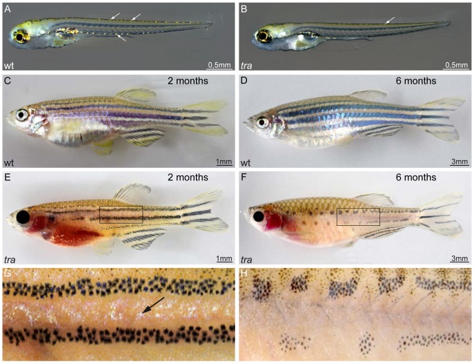
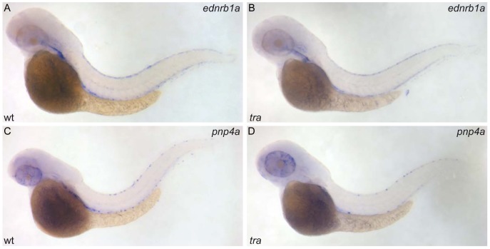
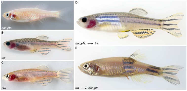
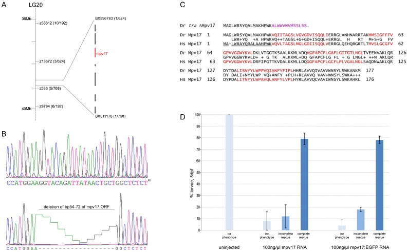
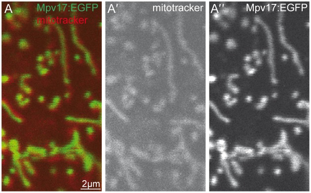
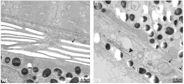
In the skin of adult zebrafish, three pigment cell types arrange into alternating horizontal stripes, melanophores in dark stripes, xanthophores in light interstripes and iridophores in both stripes and interstripes. The analysis of mutants and regeneration studies revealed that this pattern depends on interactions between melanophores and xanthophores; however, the role of iridophores in this process is less understood. We describe the adult viable and fertile mutant transparent (tra), which shows a loss or strong reduction of iridophores throughout larval and adult stages. In addition, in adults only the number of melanophores is strongly reduced, and stripes break up into spots. Stripes in the fins are normal. By cell transplantations we show that tra acts cell-autonomously in iridophores, whereas the reduction in melanophores in the body occurs secondarily as a consequence of iridophore loss. We conclude that differentiated iridophores are required for the accumulation and maintenance of melanophores during pigment pattern formation. The tra mutant phenotype is caused by a small deletion in mpv17, an ubiquituously expressed gene whose protein product, like its mammalian and yeast homologs, localizes to mitochondria. Iridophore death might be the result of mitochondrial dysfunction, consistent with the mitochondrial DNA depletion syndrome observed in mammalian mpv17 mutants. The specificity of the tra phenotype is most likely due to redundancy after gene multiplication, making this mutant a valuable model to understand the molecular function of Mpv17 in mitochondria.

Keywords: Chimeras; Iridophore; Mitochondria; Mpv17.


Conflict of interest statement

Competing interests: The authors have no competing interests to declare.

Figures

Fig. 1.
Fig. 1.. transparent mutants show a reduction in iridophore pigmentation.
(A,C,D) Wild type and (B,E–H) tra mutants. At 5 dpf, mutant larvae show strong reduction in iridophores in dorsal and ventral positions (B) as compared to wild type (A). Arrows in panels A and B highlight the appearance and position of iridophores in larvae. No other defects are apparent. Two months old wild type animals (C) present a fully developed pigment pattern, while tra individuals (E) developed only few iridophores, visible between the dark stripes. (G) Close-up of the region boxed in panel E. At that age, melanophores are reduced in number in the mutants, however, the typical four stripes developed. While the mutant fish grow, the melanophore stripes break up into spots (F, close up in H), compare six months old tra mutant (F) to wild type (D) of the same age. The abdominal cavity, typically covered by a thick sheet of iridophores in wild type, lacks this cell type in tra mutants. Iridophores of the eyes are strongly reduced throughout life in the mutants. Pigment patterning of fins and scales appear normal. Scale bars: 0.5 mm (A,B); 1 mm (C,E), 3 mm (D,F).
Fig. 2.
Fig. 2.. transparent affects pigment synthesis of iridophores, but not their specification and initial differentiation.
ednrb1a RNA hybridization of wild type (A) and tra mutant larva (B), pnp4a RNA hybridization of wild type (C) and tra mutant (D); 48 hpf.
Fig. 3.
Fig. 3.. transparent is required cell-autonomously in iridophores.
(A–C) Mutants used in transplantation experiments: (A) nac;pfe, (B) tra, (C) rse. (D) nac;pfe mutant which received tra mutant xanthophores and melanophores, showing that both cell types contribute normally to stripes. (E) tra mutant which received nac;pfe mutant iridophores, showing that the supply with iridophores is sufficient to normalize the pigment pattern.
Fig. 4.
Fig. 4.. transparent encodes Mpv17.
(A) tra maps to chromosome 20. The genomic interval was mapped by the indicated microsatellites (z-marker) and further refined by SNPs located on contigs BX596783 and BX511178. Brackets give the number of recombinants in relation to the total number of mutant individuals. In this closest interval, 7 genes are annoted. (B) Sequencing revealed a deletion in the mpv17 coding sequence in tra mutants, resulting in a frameshift (D, amino acids in pink) and an early stop codon. (C) Comparison of zebrafish (Dr) Mpv17 protein sequence and its human homolog (Hs). The proteins show 69% amino acid identity. Amino acids forming the transmembrane domains are highlighted in red. The mitochondrial target signal in the human protein sequence is underlined. (D) Injection of mpv17 or mpv17:egfp encoding mRNA into tra mutants rescue larval iridophore pigmentation (n(uninjected) = 167, n(mpv17) = 299, n(mpv17:egfp) = 300).
Fig. 5.
Fig. 5.. Zebrafish Mpv17 colocalizes with mitochondria.
(A) Mpv17 C-terminally tagged with EGFP (green, A″) colocalizes with mitochondria labelled by mitotracker (red, A′). Image was taken from larval epidermal cell at 4 dpf. Scale bar: 2 µm.
Fig. 6.
Fig. 6.. transparent mutant iridophores display characteristics of apoptotic cells.
EM sections of wild type (A) and transparent mutant iridophores (B) in the eye of 5 dpf larvae. (A) Wild type iridophores develop stacks of iridosomes nearly completely filling the cells. Iridosomes are not contrasted and appear empty as the guanine crystals are lost during the sectioning process (arrow). (B) Iridosomes are also visible in tra mutants. In the cell shown, only two iridosomes seem to contain guanine crystals (arrows), whereas others contain contrastable material (arrowhead), indicating that guanine deposition is not completed. Importantly and in contrast to wild type, tra mutant iridophores contain many vesicles, a sign for apoptosis. Scale bars: 0.5 µm.

Similar articles

Cited by

References

    1. AlSaman A., Tomoum H., Invernizzi F., Zeviani M. (2012). Hepatocerebral form of mitochondrial DNA depletion syndrome due to mutation in MPV17 gene. Saudi J. Gastroenterol. 18, 285–289 10.4103/1319-3767.98439 - DOI - PMC - PubMed
    1. Brand M., Granato M., Nüsslein-Volhard C. (2002). Keeping and raising zebrafish. Zebrafish: A Practical Approach Nüsslein-Volhard C, Dahm R, ed7–37Oxford; New York, NY: Oxford Universtiy Press.
    1. Budi E. H., Patterson L. B., Parichy D. M. (2008). Embryonic requirements for ErbB signaling in neural crest development and adult pigment pattern formation. Development 135, 2603–2614 10.1242/dev.019299 - DOI - PMC - PubMed
    1. Budi E. H., Patterson L. B., Parichy D. M. (2011). Post-embryonic nerve-associated precursors to adult pigment cells: genetic requirements and dynamics of morphogenesis and differentiation. PLoS Genet. 7, e1002044 10.1371/journal.pgen.1002044 - DOI - PMC - PubMed
    1. Dooley C. M., Mongera A., Walderich B., Nüsslein-Volhard C. (2013). On the embryonic origin of adult melanophores: the role of ErbB and Kit signalling in establishing melanophore stem cells in zebrafish. Development 140, 1003–1013 10.1242/dev.087007 - DOI - PubMed