. 2013 Jun 11;2(7):728-38.
doi: 10.1242/bio.20135173. Print 2013 Jul 15.

The C-terminus of S. pombe DDK subunit Dfp1 is required for meiosis-specific transcription and cohesin cleavage

Affiliations

The C-terminus of S. pombe DDK subunit Dfp1 is required for meiosis-specific transcription and cohesin cleavage

Anh-Huy Le et al. Biol Open. .

Abstract

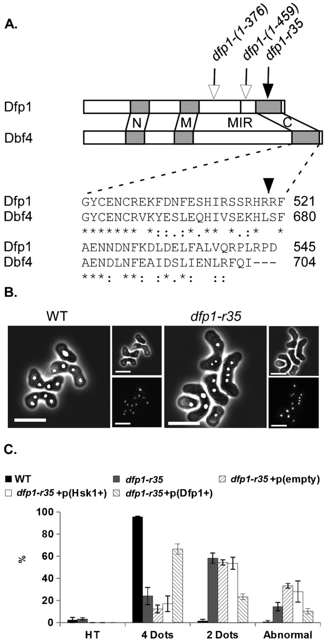
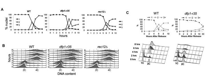
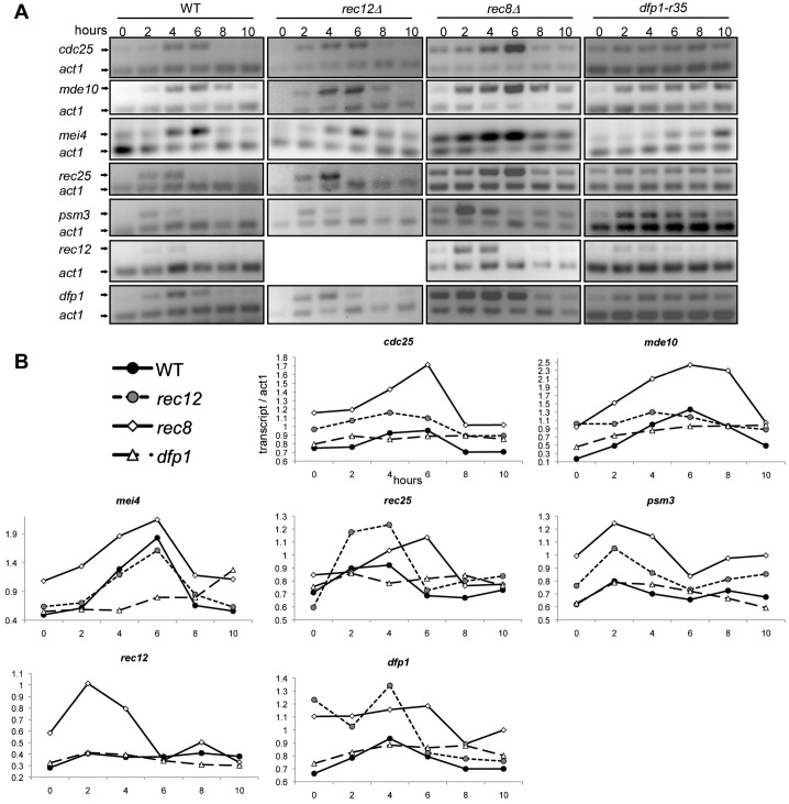
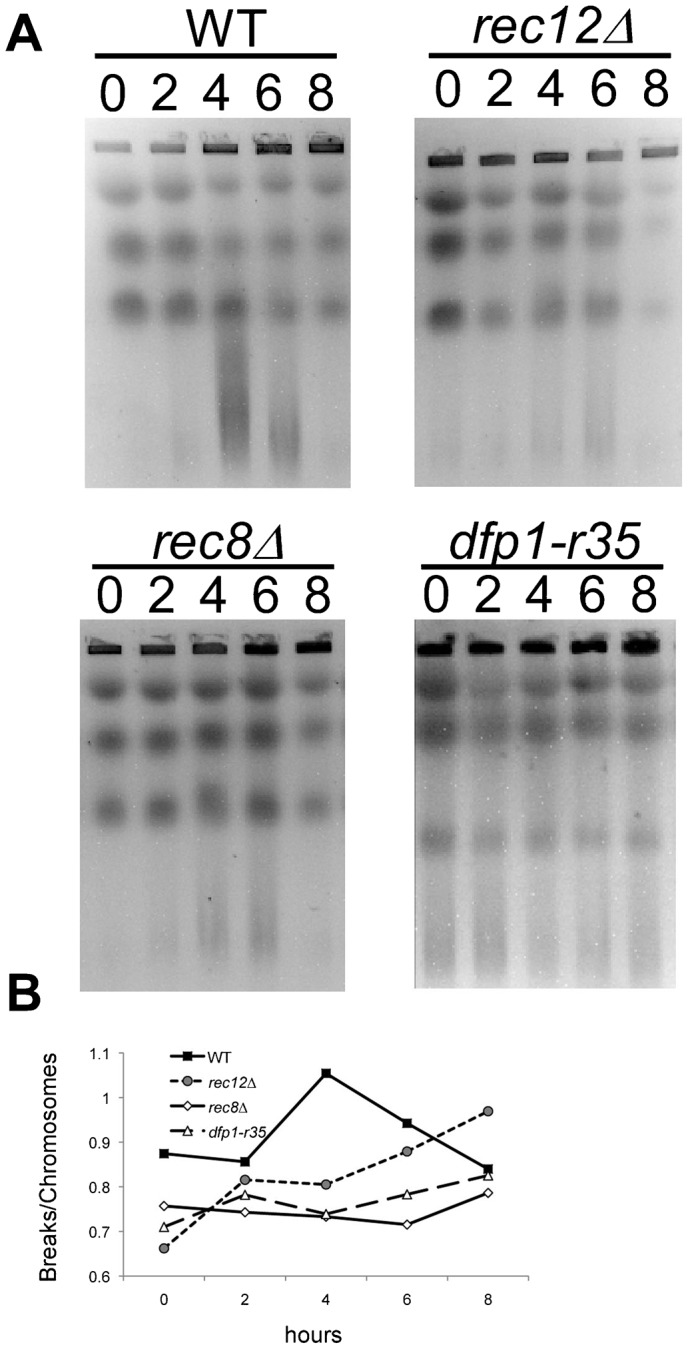
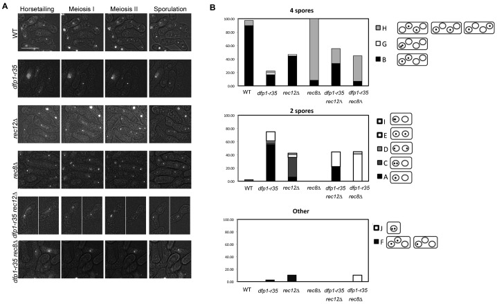
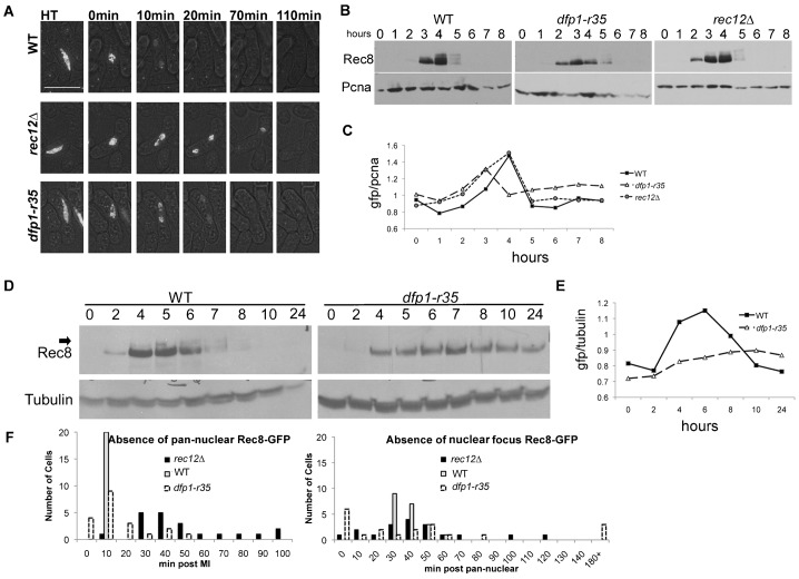
The DDK complex is a conserved kinase complex, consisting of a catalytic subunit, Hsk1 (Cdc7), and its regulatory subunit Dfp1 (Dbf4). This kinase is essential for DNA replication. In this work, we show that dfp1-r35, which truncates the Dfp1 C-terminus zinc finger, causes severe meiotic defects, including reduced spore viability, reduced formation of programmed double strand breaks, altered expression of meiotic genes, and disrupted chromosome segregation. There is a high frequency of dyad formation. Mutants are also defective in the phosphorylation and degradation of the meiotic cohesion, Rec8, resulting in a failure to proceed through the MII division. These defects are more pronounced in a haploid meiosis model than in a normal diploid meiosis. Thus, several critical meiotic functions are linked specifically to the C-terminus of Dfp1, which may target specific substrates for phosphorylation by Hsk1.

Keywords: Cdc7; Dbf4; Fission yeast; Hsk1; Meiosis; Rec8.


Conflict of interest statement

Competing interests: The authors have no competing interests to declare.

Figures

Fig. 1.
Fig. 1.. Terminal meiotic phenotype of dfp1-r35 mutants.
(A) Structure of Dfp1. dfp1-r35 mutation is a truncation within a zinc-finger domain that is conserved between S. pombe and S. cerevisiae. (B) Nuclear morphology of terminal meiotic products of dfp1-r35 (FY1154) compared to WT (FY155) visualized with DAPI staining. Scale bar: 10 mm. (C) Quantification of terminal meiotic phenotypes as horsetailing (HT), 2 DAPI stained bodies, 4 DAPI stained bodies, or abnormal. Defects of dfp1-r35 are suppressed by ectopically expressed dfp1+. 300 asci for each strain were analyzed and error bars represent standard deviation.
Fig. 2.
Fig. 2.. Synchronous meiosis in mat2-102/h pat1-114/pat1-114 diploids and pat1-114 haploids.
(A) Comparison of DAPI stained profiles categorizing 1, 2, or ≥3 DAPI stained bodies for WT, dfp1-r35, and rec12Δ following meiotic induction using pat1-114. (B) FACS profiles demonstrating the progression of meiotic replication through meiotic induction. DNA content moves from 2C to 4C. (C) Comparison of haploid meiotic induction of dfp1-r35 compared to wild type. Both FACs and DAPI staining was done as in panels A and B to monitor meiotic progression. Strains: wild type (FY6332/FY6336), dfp1-r35 (FY6347/FY6378), rec12Δ (FY6530/FY6531), wild type (FY4129), dfp1-r35 (FY4396).
Fig. 3.
Fig. 3.. Transcriptional expression of meiotic genes.
RT-PCR of pat1-114 (FY4129), pat1-114 rec12Δ (FY2008), pat1-114 rec8Δ (FY1955) and pat1-114 dfp1-r35 (FY4396) cells undergoing synchronous haploid meiosis shows expression with altered timing of meiotic markers: mid/late transcripts (cdc25+, mde10+, mei4+), early rep1-independent (rec25+) and -dependent (psm3+, rec12+, dfp1+). (A) Visualization of RT-PCR using SYTOX green on agarose gel. (B) Quantification and graphical representation of panel A.
Fig. 4.
Fig. 4.. prDSBs in dfp1-r35 mutants.
(A) Pulsed field gel electrophoresis (PFGE) of WT (FY4129), rec12Δ (FY2008), rec8Δ (FY1955) or dfp1-r35 (FY4396) pat1-114 cells undergoing synchronous meiosis. dfp1-r35 mutants, similar to rec12Δ mutants, cannot induce prDSBs. (B) Quantification of PFGE in panel A using the ratio of breaks/chromosome signal as previously described by Borde et al. (Borde et al., 2000a).
Fig. 5.
Fig. 5.. Chromosome segregation in dfp1-r35 mutants compared to rec8Δ and rec12Δ.
(A) Representative images from live cell analysis of LacI-GFP chromosome segregation assay in diploid cells (supplementary material Movies 1–6). The indicated strains heterozygous for lys1+::lacO lacI-GFP adjacent to centromere I were grown at (32°C) and plated on agarose pads for image acquisition (see Materials and Methods). Strains: wild type (FY6221×FY6331), rec8Δ (FYFY5916×FY6131), rec12Δ (FY5197×FY6134), dfp1-r35 (FY6236×FY6441), dfp1-r35 rec8Δ (FY6204×FY6416), dfp1-r35 rec12Δ (FY6143×FY6175). Scale bar: 10 µm. Selected panels display predominate segregation phenotype for each mutant observed. (B) Graphical representation of quantification of live cell imaging classes. See supplementary material Table S6 for class descriptions and quantities.
Fig. 6.
Fig. 6.. Rec8-GFP stability.
(A) Representative images from live cell imaging observing diploid meiosis in cells containing Rec8-GFP (supplementary material Movies 7–9). The indicated strains were grown at (32°C temperature) and plated on agarose pads for image acquisition (see Materials and Methods). Strains: wild type (FY6173×FY6174), dfp1-r35 (FY6241×FY6242), rec12Δ (FY6217×6218). Scale bar: 10 µm. (B) Western blot of Rec8-GFP during synchronous meiosis in h/mat2-102 pat1-114/pat1-114 stable diploid. Strains: wild type (FY6332×FY6336), dfp1-r35 (FY6347×FY6378) rec12Δ (FY6530×FY6531). (C) Quantification of panel B using GFP signal/PCNA. (D) Western blot of Rec8-GFP in haploid pat1-114 meiosis. Strains: wild type (FY4129) and dfp1-r35 (FY4396). (E) Quantification of panel D as done in panel C. (F) Graphical representation of quantification of Rec8-GFP timing in live cell imaging in panel A. Pan-nuclear signal disappearance occurred at an average of 10 mins, 15.5 mins, and 50.5 mins post MI for wild type, dfp1-r35, and rec12Δ respectively with P-values using a two-tailed t-test of 0.0000008 and 0.102 for rec12Δ and dfp1-r35 respectively. For the disappearance of the nuclear focus relative to the disappearance of the pan-nuclear signal the timing was an average of 48 mins, 56.6 mins, and 55.5 mins for wild type, dfp1-r35, and rec12Δ with P-values using a two-tailed t-test of 0.258 and 0.599 for rec12Δ and dfp1-r35 respectively.

Similar articles

Cited by

References

    1. Bailis J. M., Forsburg S. L. (2004). S phase assembly of centromeric heterochromatin and cohesion. Cell Cycle 3, 416–418 10.4161/cc.3.4.799 - DOI - PubMed
    1. Bailis J. M., Bernard P., Antonelli R., Allshire R. C., Forsburg S. L. (2003). Hsk1-Dfp1 is required for heterochromatin-mediated cohesion at centromeres. Nat. Cell Biol. 5, 1111–1116 10.1038/ncb1069 - DOI - PubMed
    1. Borde M., Bonansco C., Fernández de Sevilla D., Le Ray D., Buño W. (2000a). Voltage-clamp analysis of the potentiation of the slow Ca2+-activated K+ current in hippocampal pyramidal neurons. Hippocampus 10, 198–206 10.1002/(SICI)1098-1063(2000)10:2<198::AID-HIPO9>3.0.CO;2-F - DOI - PubMed
    1. Borde V., Goldman A. S., Lichten M. (2000b). Direct coupling between meiotic DNA replication and recombination initiation. Science 290, 806–809 10.1126/science.290.5492.806 - DOI - PubMed
    1. Brown G. W., Kelly T. J. (1999). Cell cycle regulation of Dfp1, an activator of the Hsk1 protein kinase. Proc. Natl. Acad. Sci. USA 96, 8443–8448 10.1073/pnas.96.15.8443 - DOI - PMC - PubMed

LinkOut - more resources