. 2013 Jun 21;2(7):750-9.
doi: 10.1242/bio.20135249. Print 2013 Jul 15.

Olfactory ensheathing glia are required for embryonic olfactory axon targeting and the migration of gonadotropin-releasing hormone neurons

Affiliations

Olfactory ensheathing glia are required for embryonic olfactory axon targeting and the migration of gonadotropin-releasing hormone neurons

Perrine Barraud et al. Biol Open. .

Abstract

Kallmann's syndrome is caused by the failure of olfactory axons and gonadotropin-releasing hormone (GnRH) neurons to enter the embryonic forebrain, resulting in anosmia and sterility. Sox10 mutations have been associated with Kallmann's syndrome phenotypes, but their effect on olfactory system development is unknown. We recently showed that Sox10 is expressed by neural crest-derived olfactory ensheathing cells (OECs). Here, we demonstrate that in homozygous Sox10(lacZ/lacZ) mouse embryos, OEC differentiation is disrupted; olfactory axons accumulate in the ventromedial olfactory nerve layer and fewer olfactory receptor neurons express the maturation marker OMP (most likely owing to the failure of axonal targeting). Furthermore, GnRH neurons clump together in the periphery and a smaller proportion enters the forebrain. Our data suggest that human Sox10 mutations cause Kallmann's syndrome by disrupting the differentiation of OECs, which promote embryonic olfactory axon targeting and hence olfactory receptor neuron maturation, and GnRH neuron migration to the forebrain.

Keywords: GnRH neurons; Kallmann's syndrome; Olfactory ensheathing glia; Sox10.


Conflict of interest statement

Competing interests: The authors have no competing interests to declare.

Figures

Fig. 1.
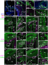
Fig. 1.. During mouse olfactory system development, Sox10 expression is restricted to OECs (and, at late stages, to Bowman's gland/duct cells).
(A–B2) At E10.5, in situ hybridization (ISH) on parasagittal sections followed by immunostaining for neuronal βIII tubulin (nβ3tub) reveals Sox10 expression in non-neuronal cells associated with the “migratory mass” of olfactory axons and migrating neurons emerging from the olfactory epithelium. Sox10 is not expressed by neurons in either the olfactory epithelium or the migratory mass (examples of each are indicated, respectively, by arrowheads and arrows). (C–F) At E11.5, when the developing vomeronasal organ evaginates from the ventromedial olfactory epithelium (Cuschieri and Bannister, 1975), ISH shows (C–E1) Sox10 expression in non-neuronal cells (OECs; arrowheads in E,E1) associated with olfactory and vomeronasal (adjacent to the vomeronasal organ) nerves, but no Sox10 expression in the neurons within either the olfactory or vomeronasal epithelia; and (F) GnRH1-positive cells within the vomeronasal organ epithelium (arrow) and in the region of the vomeronasal nerve (arrowhead; compare with position of neurons in D1–E1). (G–J1) At E12.5, ISH on coronal sections followed by immunostaining for nβ3tub shows Sox10 expression in non-neuronal cells associated with the olfactory and vomeronasal nerves, but no above-background Sox10 expression within either the olfactory or vomeronasal epithelia. (K–L1) At E14.5, immunostaining on coronal sections reveals Sox10-positive, p75NTR-positive OECs surrounding olfactory nerve fascicles in the lamina propria, but no Sox10 expression in neurons in the olfactory epithelium. (M–N2) At E16.5 (M–M2) and E17.5 (N–N2), ISH for Sox10 on coronal sections followed by immunostaining for nβ3tub shows non-neuronal Sox10-positive cells in the olfactory epithelium (arrowheads): these are developing Bowman's gland/duct cells, which begin to protrude from the basal epithelium from E17.5 (Cuschieri and Bannister, 1975). (O,O1) In neonates, immunostaining for Sox10 and nβ3tub on coronal sections shows that Bowman's gland/duct cells (arrowheads) maintain Sox10 expression after birth. Abbreviations: fb, forebrain; lp, lamina propria; nβ3tub, neuronal βIII tubulin; neo, neonatal; ob, olfactory bulb; oe, olfactory epithelium; on, olfactory nerve; vno, vomeronasal organ. Scale bars: 100 µm (C,D,F,G,K), 50 µm (A,H,I,J,M,N,O), 20 µm (B,L), 10 µm (E).
Fig. 2.
Fig. 2.. After Sox10 deletion, neural crest cells colonize the olfactory nerve but are missing from the lamina propria by E16.5.
(A–B1) In homozygous Sox10lacZ/lacZ embryos at E11.5 (A–A3) and E12.5 (B,B1), immunostaining on parasagittal sections reveals β-gal-positive cells associated with the olfactory nerve (arrows, A2,B1). β-gal expression in mesenchymal cells beneath the olfactory epithelium (A1) may reflect protein perdurance in neural crest-derived cells. (C–D2) At E16.5, immunostaining on coronal sections shows β-gal-positive cells (arrows) throughout the olfactory nerve from the lamina propria to the ONL in heterozygous Sox10lacZ/+ embryos (C–C2), but absent from the lamina propria after Sox10 deletion (D–D2). Arrowheads in C1,D1 indicate β-gal-positive prospective Bowman's gland/duct cells in the epithelium, which express Sox10 (Fig. 1N–O1). Fainter β-gal immunoreactivity in cribriform plate and nasal septum cartilage (C,C1,D,D1) may be specific: weak β-gal staining was previously reported in limb cartilage condensations in Sox10lacZ embryos (Britsch et al., 2001). (E–F2) At E16.5, immunostaining on coronal sections reveals S100 expression (arrows) throughout the olfactory nerve from the lamina propria to the ONL in wild-type embryos (E–E2), but absent from the lamina propria after Sox10 deletion (F–F2). (G–H2) At E16.5, immunostaining on coronal sections shows p75NTR expression (arrows) throughout the olfactory nerve from the lamina propria to the ONL in wild-type embryos (G–G2), but absent from olfactory nerve fascicles in the lamina propria after Sox10 deletion (H–H2). Abbreviations: βgal, β-galactosidase; cp, cribriform plate; fb, forebrain; lp, lamina propria; nβ3tub, neuronal βIII tubulin; ns, nasal septum; ob, olfactory bulb; oe, olfactory epithelium; on, olfactory nerve; onl, olfactory nerve layer; vn, vomeronasal nerve; vno, vomeronasal organ. Scale bars: 100 µm (A,B,C,D,E,F,G,H), 50 µm (A1,B1,C1,C2,D1,D2,E1,E2,F1,F2,G1,G2,H1,H2), 25 µm (A2,A3).
Fig. 3.
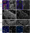
Fig. 3.. After Sox10 deletion, OEC differentiation is defective.
All images show E16.5 coronal sections. (A–B1) Expression of the early glial differentiation marker BLBP is seen in OECs ensheathing olfactory nerve fascicles (labelled by immunostaining for olfactory marker protein) in the wild-type lamina propria (A,A1), but not after Sox10 deletion (B,B1). (C,D) BLBP expression in the ONL is much stronger in wild-type (C) than homozygous Sox10lacZ/lacZ embryos (D). (E–F2) At E16.5, NPY expression is seen in the inner ONL of wild-type embryos (arrows, E1,E2) but undetectable after Sox10 deletion (F1,F2). Abbreviations: epl, external plexiform layer; lp, lamina propria; ob, olfactory bulb; OMP, olfactory marker protein; on, olfactory nerve; onl, olfactory nerve layer; onl-i, inner olfactory nerve layer; onl-o, outer olfactory nerve layer. Scale bars: 100 µm (E,E1,F,F1), 50 µm (C,D), 25 µm (E1 inset), 10 µm (A,B).
Fig. 4.
Fig. 4.. Sox10 deletion disrupts olfactory axon targeting and ORN maturation.
All images show E16.5 coronal sections. (A–E) Olfactory mucosa sections immunostained for the maturation marker OMP plus (A–B1) NCAM or (C–E) neuronal βIII tubulin. Relative to wild-type (A,A1,C) or heterozygote embryos (D), homozygous Sox10lacZ/lacZ embryos (B,B1,E) displayed defasciculated olfactory nerve bundles and inappropriately migrating axons within the lamina propria (asterisk in B,B1), and fewer OMP-positive neurons (compare A1,B1; C–E). (F–H) Bar charts showing the mean/embryo + s.e.m. for wild-type/heterozygote embryos (2/genotype) versus homozygous Sox10lacZ/lacZ embryos, of: (F) OMP-positive cells/100 µm of olfactory epithelium (**P = 0.0014; 2-tailed unpaired t-test); (G) neuronal βIII tubulin-positive cells/100 µm of epithelium; (H) olfactory epithelial thickness. Sox10 deletion only affects the number of mature (OMP-positive) ORNs. (I,J) Olfactory bulb sections immunostained for the axonal marker NCAM. Relative to wild-type embryos (I), the ONL in homozygous Sox10lacZ/lacZ embryos (J) is much thinner in dorsal and lateral regions of the bulb while the ventromedial ONL is much thicker. (K–L1) NCAM immunostaining showing clearly separated uniform bilateral ONLs in a wild-type embryo (K) versus a merged, ventromedial ONL (asterisk) in a homozygous Sox10lacZ/lacZ embryo (L,L1). Arrowheads in L,L1 highlight axonal whorls/balls. Abbreviations as in Fig. 2. Scale bars: 200 µm (K,L), 100 µm (I,J,L1), 50 µm (A,B,C).
Fig. 5.
Fig. 5.. A significantly smaller proportion of GnRH neurons enters the forebrain after Sox10 deletion.
(A) At E12.5, immunostaining on coronal sections for neuronal βIII tubulin and the neuronal RNA-binding protein HuC/D revealed unusually large aggregates of multiple neuronal cell bodies on the vomeronasal nerve in both heterozygous Sox10lacZ/+ and homozygous Sox10lacZ/lacZ embryos. (B) Schematic representation of the distribution of GnRH neurons (black spots) at E14.5 on parasagittal sections of 3 wild-type, 3 heterozygous Sox10lacZ/+ and 3 homozygous Sox10lacZ/lacZ embryos. (C) Schematic representation of the distribution of GnRH neurons (black spots) at E16.5 on coronal sections at 3 different rostrocaudal levels of 4 wild-type, 4 heterozygous Sox10lacZ/+ and 4 homozygous Sox10lacZ/lacZ embryos. (D) Bar charts showing the mean percentage/embryo + s.e.m. of GnRH1-positive cells that had entered the brain for wild-type, heterozygous Sox10lacZ/+ and homozygous Sox10lacZ/lacZ embryos (3 embryos/genotype at E14.5; 4 embryos/genotype at E16.5; ≥100 GnRH neurons counted/embryo). At E14.5, the mean percentage/embryo of GnRH neurons in the brain was significantly lower than wild-type for both heterozygous Sox10lacZ/+ and homozygous Sox10lacZ/lacZ embryos (**P<0.01; one-way ANOVA using Dunnett's multiple comparison test). By E16.5, the mean percentage/embryo of GnRH neurons in the brain was no longer significantly different from wild-type for heterozygous Sox10lacZ/+ embryos, but was four-fold lower than wild-type for homozygous Sox10lacZ/lacZ embryos (**P<0.005; one-way ANOVA using Dunnett's multiple comparison test). (E–F1) Examples of GnRH neurons identified after immunostaining E16.5 coronal sections of wild-type hypothalamus (E,E1) and homozygous Sox10lacZ/lacZ olfactory mucosa (F,F1). Abbreviations as in Fig. 2. Scale bars: 200 µm (E), 100 µm (A, main panels; E1,F), 25 µm (A, insets; F1); 10 µm (E1, insets).

Similar articles

Cited by

References

    1. Au W. W., Treloar H. B., Greer C. A. (2002). Sublaminar organization of the mouse olfactory bulb nerve layer. J. Comp. Neurol. 446, 68–80 10.1002/cne.10182 - DOI - PubMed
    1. Bajpai R., Chen D. A., Rada-Iglesias A., Zhang J., Xiong Y., Helms J., Chang C. P., Zhao Y., Swigut T., Wysocka J. (2010). CHD7 cooperates with PBAF to control multipotent neural crest formation. Nature 463, 958–962 10.1038/nature08733 - DOI - PMC - PubMed
    1. Barnett C. P., Mendoza-Londono R., Blaser S., Gillis J., Dupuis L., Levin A. V., Chiang P. W., Spector E., Reardon W. (2009). Aplasia of cochlear nerves and olfactory bulbs in association with SOX10 mutation. Am. J. Med. Genet. A. 149A, 431–436 10.1002/ajmg.a.32657 - DOI - PubMed
    1. Barraud P., Seferiadis A. A., Tyson L. D., Zwart M. F., Szabo-Rogers H. L., Ruhrberg C., Liu K. J., Baker C. V. H. (2010). Neural crest origin of olfactory ensheathing glia. Proc. Natl. Acad. Sci. USA 107, 21040–21045 10.1073/pnas.1012248107 - DOI - PMC - PubMed
    1. Bondurand N., Dastot-Le Moal F., Stanchina L., Collot N., Baral V., Marlin S., Attie-Bitach T., Giurgea I., Skopinski L., Reardon W. et al. (2007). Deletions at the SOX10 gene locus cause Waardenburg syndrome types 2 and 4. Am. J. Hum. Genet. 81, 1169–1185 10.1086/522090 - DOI - PMC - PubMed

LinkOut - more resources