. 2013 Oct 1;305(7):F943-56.
doi: 10.1152/ajprenal.00303.2013. Epub 2013 Jul 17.

AMP-activated protein kinase regulates the vacuolar H+-ATPase via direct phosphorylation of the A subunit (ATP6V1A) in the kidney

Affiliations

AMP-activated protein kinase regulates the vacuolar H+-ATPase via direct phosphorylation of the A subunit (ATP6V1A) in the kidney

Rodrigo Alzamora et al. Am J Physiol Renal Physiol. .

Abstract

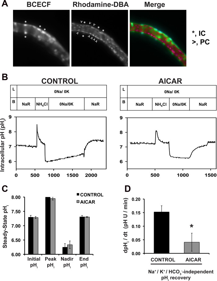
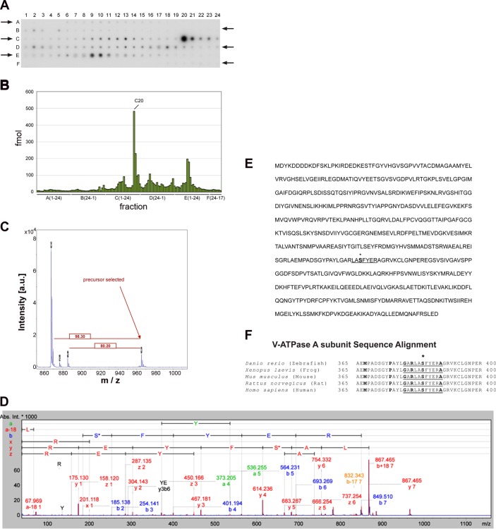
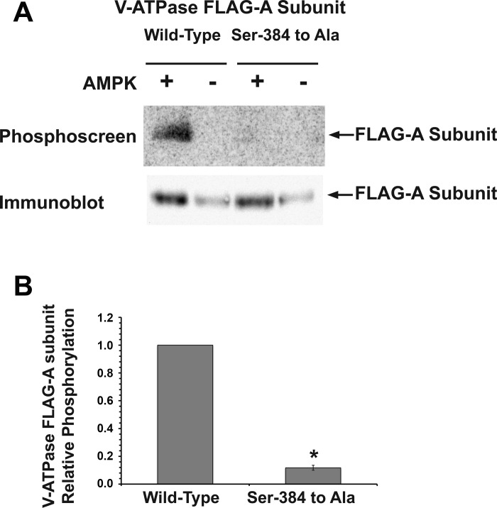
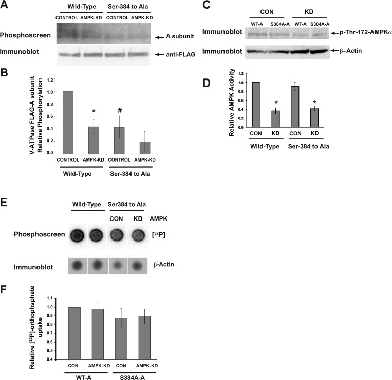
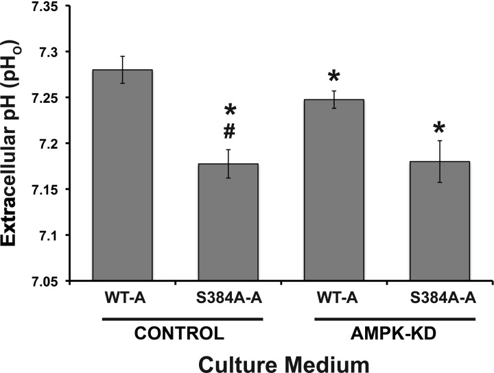
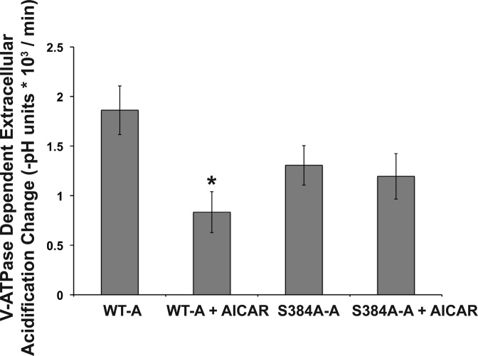
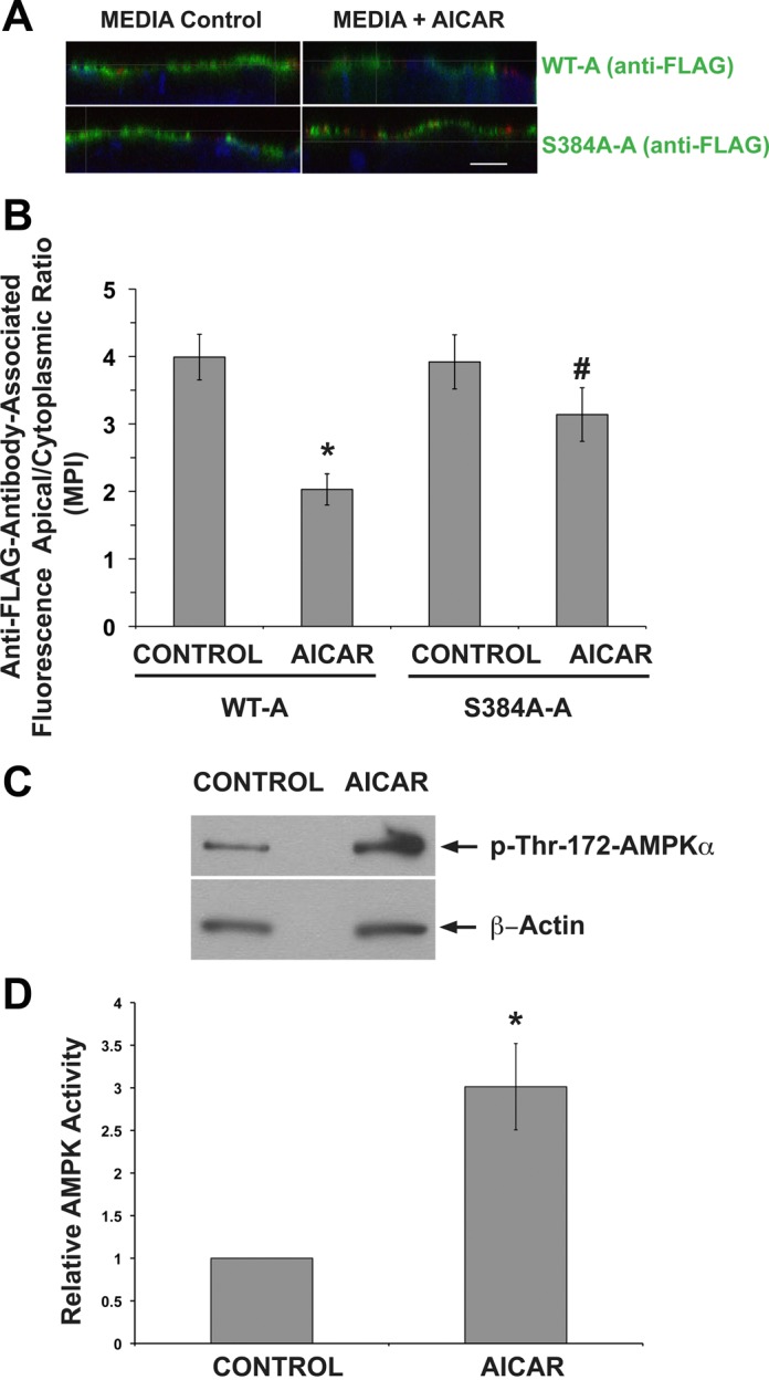
The vacuolar H(+)-ATPase (V-ATPase) in intercalated cells contributes to luminal acidification in the kidney collecting duct and nonvolatile acid excretion. We previously showed that the A subunit in the cytoplasmic V1 sector of the V-ATPase (ATP6V1A) is phosphorylated by the metabolic sensor AMP-activated protein kinase (AMPK) in vitro and in kidney cells. Here, we demonstrate that treatment of rabbit isolated, perfused collecting ducts with the AMPK activator 5-aminoimidazole-4-carboxamide-1-β-D-ribofuranoside (AICAR) inhibited V-ATPase-dependent H(+) secretion from intercalated cells after an acid load. We have identified by mass spectrometry that Ser-384 is a major AMPK phosphorylation site in the V-ATPase A subunit, a result confirmed by comparing AMPK-dependent phosphate labeling of wild-type A-subunit (WT-A) with that of a Ser-384-to-Ala A subunit mutant (S384A-A) in vitro and in intact HEK-293 cells. Compared with WT-A-expressing HEK-293 cells, S384A-A-expressing cells exhibited greater steady-state acidification of HCO3(-)-containing media. Moreover, AICAR treatment of clone C rabbit intercalated cells expressing the WT-A subunit reduced V-ATPase-dependent extracellular acidification, an effect that was blocked in cells expressing the phosphorylation-deficient S384A-A mutant. Finally, expression of the S384A-A mutant prevented cytoplasmic redistribution of the V-ATPase by AICAR in clone C cells. In summary, direct phosphorylation of the A subunit at Ser-384 by AMPK represents a novel regulatory mechanism of the V-ATPase in kidney intercalated cells. Regulation of the V-ATPase by AMPK may couple V-ATPase activity to cellular metabolic status with potential relevance to ischemic injury in the kidney and other tissues.

Keywords: AMPK; Intercalated cells; V-ATPase; kidney; mass spectrometry.


Figures

Fig. 1.
Fig. 1.
The AMPK activator 5-aminoimidazole-4-carboxamide-1-β-d-ribofuranoside (AICAR) inhibits the V-ATPase-mediated intracellular pH (pHi) recovery in rabbit isolated, perfused outer medullary collecting ducts after an acute acid load. A: fluorescence micrographs of a single microperfused rabbit outer medullary collecting duct (OMCD), each captured at a slightly different focal plane. The images show increased accumulation of the pH-sensitive dye BCECF loaded from the bath as the acetoxymethyl ester (left) in intercalated cells (IC; marked with asterisks). In the middle panel, the lectin Dolichos biflorus agglutinin coupled to rhodamine (rhodamine-DBA) decorates principal cells (PC; marked with carets). In the right panel (merge), the IC show higher fluorescence intensity (green pseudocolor), while the PC are identified by the rhodamine-DBA caps (red pseudocolor). B: representative patterns of pHi recovery observed in IC of rabbit OMCD in response to an acute in vitro acid load (NH4Cl prepulse) in the absence (left) or presence (right) of the AMPK activator AICAR (2 mM) included in the bath for 1 h before and continued throughout the experiment shown. C: under both conditions, cytosolic acidification from an initial pHi of ∼7.3 in Na+-Ringer (NaR) buffer to a nadir pHi of ∼6.3 was accomplished following a 3-min bath exposure to NH4Cl. D: partial pHi recovery was observed in the absence of Na+ and K+ (0Na/0K) and a nominal absence of HCO3/CO2 at a rate of 0.15 ± 0.02 pH U/min in control untreated OMCD, while the pHi recovery was only 0.04 ± 0.03 pH U/min in the presence of the AICAR. Values are means ± SE. Reintroduction of Na+ to the bathing solution in all tubules led to full recovery of the pHi to ∼7.3 (n = 4 OMCD in each group. *P < 0.05 vs. control).
Fig. 2.
Fig. 2.
AMP-activated protein kinase (AMPK) phosphorylates the V-ATPase A subunit at Ser-384 deduced by matrix-assisted laser desorption ionization mass spectrometry (MALDI-MS and MALDI-MSMS). FLAG-tagged wild-type V-ATPase A subunit (WT-A) was expressed in and then immunoprecipitated from HEK-293 cell lysates. Immunoprecipitated WT-A was then incubated with a substoichiometric amount of recombinant AMPK in the presence of [γ-32P]ATP. After this phosphorylation reaction, the proteins were subjected to SDS-PAGE followed by autoradiography. The band corresponding to the WT-A subunit was then excised and subjected to in-gel digestion with trypsin as described in materials and methods. A: autoradiography of a prespotted AnchorChip showing the fractionation profile of the WT-A V-ATPase subunit phosphorylated by AMPK after in-gel digestion and microfractionation. The spotting occurred from A1 to A24 and continued from B24 to B1 then C1 to C24 and D24 to D1, etc. The arrows denote the directionality of the spotting. B: analysis by densitometry of individual radioactive spots identified after the microfractionation (shown in A). The phosphorylated peptides were quantified using the appropriate standards, and the major radioactive peptide peak was eluted with fraction C20. C: characterization and identification of the V-ATPase A subunit phosphorylated peptide by MALDI-MS. The peptide of spot C20 with a mass of 965.4 Da showed predominant neutral mass losses of 80 Da (−HPO3) and 98 Da (−H3PO4). D: the MSMS fragmentation spectrum of the precursor identified the tryptic peptide to be LASFYER (with the unphosphorylated peptide having a mass of Mr 867.4 Da). E: the amino acid sequence for the mouse FLAG-tagged WT-A subunit is shown, spotlighting the highly phosphorylated peptide by AMPK (underlined C20 fraction), and Ser-384 (asterisk). F: Ser-384 (bold and asterisk) is part of a highly conserved region that appears to conform to the loose consensus AMPK target phosphorylation motif (underlined residues).
Fig. 3.
Fig. 3.
AMPK phosphorylation of the A subunit occurs at Ser-384 in vitro. A: representative phosphoscreen image (top) revealing the signal of the AMPK in vitro phosphorylated V-ATPase WT-A subunit compared with the Ser-384-to-Ala mutant. The immunoblot using an anti-FLAG antibody (bottom) confirms similar protein expression and loading of the gel for the different conditions. B: quantification of the V-ATPase A subunit phosphorylation signal normalized for protein loading as assessed by densitometry of the immunoblot. Values are means ± SE. Compared with the WT-A subunit, the phosphorylation-deficient (Ser-to-Ala) mutant showed a significant 90–95% decrease in phosphorylation by AMPK in vitro. *P < 0.05 relative to WT, unpaired t-test; n = 3.
Fig. 4.
Fig. 4.
Ser-384 is the target for AMPK-dependent phosphorylation of the V-ATPase A subunit in HEK-293 cells. Plasmids encoding FLAG-tagged WT-A or the S384A-A mutant were transfected into HEK-293 cells induced to express either an irrelevant mammalian short hairpin (sh) RNA (CONTROL) or an shRNA for AMPK-α1 knockdown (AMPK-KD) 2 days before experimentation. The cells were then incubated with [32P]orthophosphate for 2 h in the presence of a PKA inhibitor (10 μM mPKI) for the entire labeling period. Cell lysis, immunoprecipitation using an anti-FLAG antibody, SDS-PAGE, immunoblotting with anti-FLAG antibody, and exposure of the same membrane to a phosphoscreen were then performed as described in materials and methods. A: representative phosphoscreen image (top) revealing the signal of the phosphorylated A subunit in cells expressing the WT-A subunit or S384A-A subunit. The immunoblot (bottom) confirms similar protein expression and loading of the gel for the different conditions. B: quantification of the V-ATPase A subunit phosphorylation signal relative to WT-A control condition and normalized for protein expression. Values are means ± SE. *P < 0.05 relative to WT control. #P < 0.05 relative to WT control (one-tailed t-test); n = 3 replicate experiments. C: representative immunoblot analysis of cell lysates from each condition for the active form of AMPK using an antibody raised against pThr-172-AMPK-α. β-Actin was used as a loading control to normalize pThr-172-AMPK-α across conditions. D: summary of relative AMPK activity (pThr-172-AMPK-α intensity corrected to β-actin as a loading control). Values are means ± SE normalized to that of control cells expressing the WT A subunit. *P < 0.05 relative to control cells expressing WT-A. E: representative phosphoscreen image of total [32P]orthophosphate protein labeling in cellular lysates from the above experiments (top). Also shown is a representative immunoblot for the loading control β-actin (bottom). F: the relative [32P]orthophosphate signal to β-actin was not significantly different across conditions.
Fig. 5.
Fig. 5.
Expression of WT and mutant V-ATPase A subunit in HEK-293 cells modulates extracellular pH (pHo). Shown is quantification of chronic pHo from media incubated with HEK-293 cells for 28–31 h. The cells expressed either an irrelevant mammalian shRNA (Control) or an shRNA targeting knockdown of AMPK-α1 (AMPK-KD) and had been transfected with either WT-A or S384A-A subunits. *P < 0.05 relative to WT-A Control. #P < 0.05 relative to WT-A AMPK-KD; n = 4.
Fig. 6.
Fig. 6.
The Ser-384 A subunit mutant modulates bafilomycin-sensitive, V-ATPase-dependent extracellular acidification with AMPK activation in clone C cells. Clone C IC were transiently transfected with either the WT-A or S384A-A subunit. Twenty-four hours posttransfection, the cells were treated ± AICAR (2 mM) for 4 h and then pHo measurements were performed. The rate of extracellular acidification under each indicated condition was measured in a low-buffering-capacity solution before and after the addition of bafilomycin A1, a specific V-ATPase inhibitor (see materials and methods). The bafilomycin-sensitive rate of extracellular acidification [−(final buffer pH − initial buffer pH)/Δt] was obtained for cells incubated either with or without the AMPK activator AICAR (n = 5 for each transfection and treatment condition). Values are means ± SE. The transfection efficiency in each well was calculated as described in materials and methods, and no significant differences were observed across all conditions. *P < 0.01 relative to untreated cells transfected with the WT-A subunit.
Fig. 7.
Fig. 7.
The AMPK phosphorylation-deficient S384A V-ATPase A subunit mutant remains at the apical membrane of IC in response to an AMPK activator. Clone C cells were transiently transfected with either the WT-A or S384A-A subunit. A: 1 day after transfection with either the WT-A (top) or S384A-A mutant subunit (bottom), clone C cells were plated onto Transwell filters. After 4 days, the filters were incubated for 4 h in media (left) or in media with 2 mM AICAR (right). Filters were then incubated with concanavalin A coupled to CY3 (red), fixed, and immunofluorescently labeled using anti-FLAG antibody (green) and TO-PRO-3 nuclear stain (blue). Scale bar = 10 μM. B: quantification of V-ATPase-associated MPI ratio of apical region of interest (ROI-1; here the A subunit colocalizes with concanavalin A) and cytoplasmic ROI-2 (A subunit alone). This ROI-1/ROI-2 ratio under the different conditions reveals a significant AICAR-mediated inhibition of apical V-ATPase accumulation in cells expressing the WT A subunit compared with cells expressing the S384A mutant. Values are means ± SE. *P < 0.05 vs. WT-A Control. #P < 0.05 vs. WT-A AICAR; n = 20–45 cells analyzed for both conditions. C: representative immunoblots of pThr-172-AMPKα and β-actin in clone C cell lysate filters treated with media alone (left) or with media+AICAR (right). D. quantification of the ratio of pThr-172-AMPK-α to β-actin Western blot signals shown in C normalized to Control as a measure of relative AMPK activity. *P < 0.05; n = 3.
Fig. 8.
Fig. 8.
The AMPK phosphorylation-deficient S384A V-ATPase A subunit mutant coimmunoprecipitates with the V0 sector of the a subunit. Clone C cell monolayers were transiently transfected to express either the WT-A or S384A-A subunit. A: representative set of immunoblots of an immunoprecipitation experiment using anti-FLAG antibody (IP FLAG; left) followed by immunoblotting with antibodies against FLAG (top) and the V0 sector V-ATPase a subunit (bottom). Whole cell lysate samples (4.5% of total) of the transfected monolayers were also directly immunoblotted for the A subunit (top right) and the a subunit (bottom right). B: results shown are the apparent a subunit binding (coimmunoprecipitation) relative to the WT-A subunit. Values are means ± SE. Densitometric quantitation of the relevant bands and determinations of relative binding strength were performed as described in materials and methods (n = 3).

Comment in

References

    1. Al-Awqati Q, Gao XB. Differentiation of intercalated cells in the kidney. Physiology (Bethesda) 26: 266–272, 2011 - PubMed
    1. Alzamora R, Thali RF, Gong F, Smolak C, Li H, Baty CJ, Bertrand CA, Auchli Y, Brunisholz RA, Neumann D, Hallows KR, Pastor-Soler NM. PKA regulates vacuolar H+-ATPase localization and activity via direct phosphorylation of the A subunit in kidney cells. J Biol Chem 285: 24676–24685, 2010 - PMC - PubMed
    1. Bens M, Vallet V, Cluzeaud F, Pascual-Letallec L, Kahn A, Rafestin-Oblin ME, Rossier BC, Vandewalle A. Corticosteroid-dependent sodium transport in a novel immortalized mouse collecting duct principal cell line. J Am Soc Nephrol 10: 923–934, 1999 - PubMed
    1. Bhalla V, Oyster NM, Fitch AC, Wijngaarden MA, Neumann D, Schlattner U, Pearce D, Hallows KR. AMP-activated kinase inhibits the epithelial Na+ channel through functional regulation of the ubiquitin ligase Nedd4–2. J Biol Chem 281: 26159–26169, 2006 - PubMed
    1. Bouley R, Pastor-Soler N, Cohen O, McLaughlin M, Breton S, Brown D. Stimulation of AQP2 membrane insertion in renal epithelial cells in vitro and in vivo by the cGMP phosphodiesterase inhibitor sildenafil citrate (Viagra). Am J Physiol Renal Physiol 288: F1103–F1112, 2005 - PubMed

Publication types

Substances