. 2013 Aug;140(16):3403-12.
doi: 10.1242/dev.095307. Epub 2013 Jul 17.

Circulating Bmp10 acts through endothelial Alk1 to mediate flow-dependent arterial quiescence

Affiliations

Circulating Bmp10 acts through endothelial Alk1 to mediate flow-dependent arterial quiescence

Derek W Laux et al. Development. 2013 Aug.

Abstract

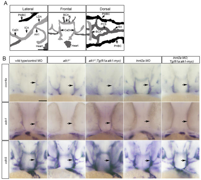
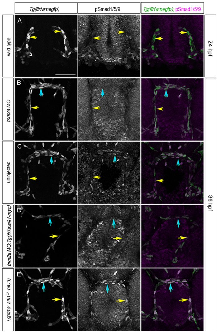
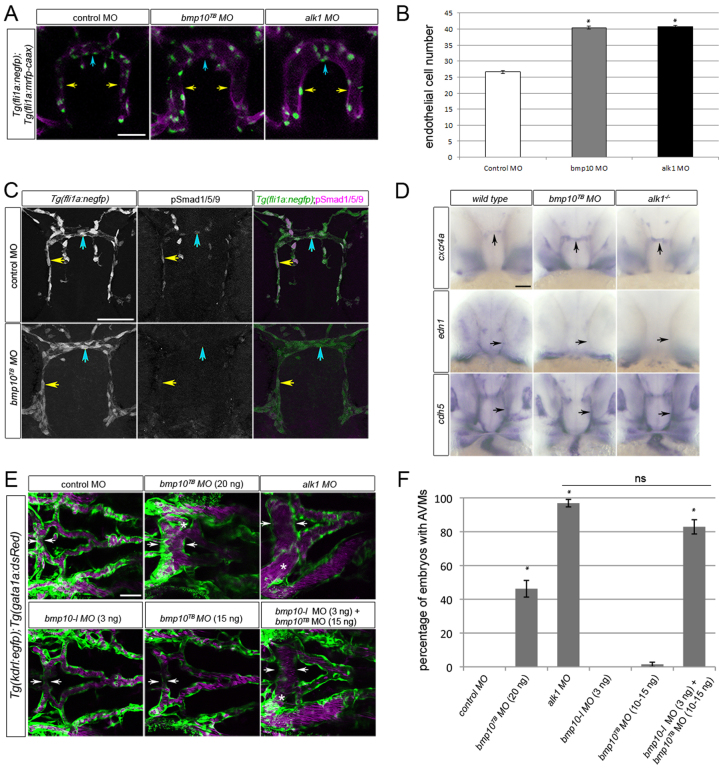
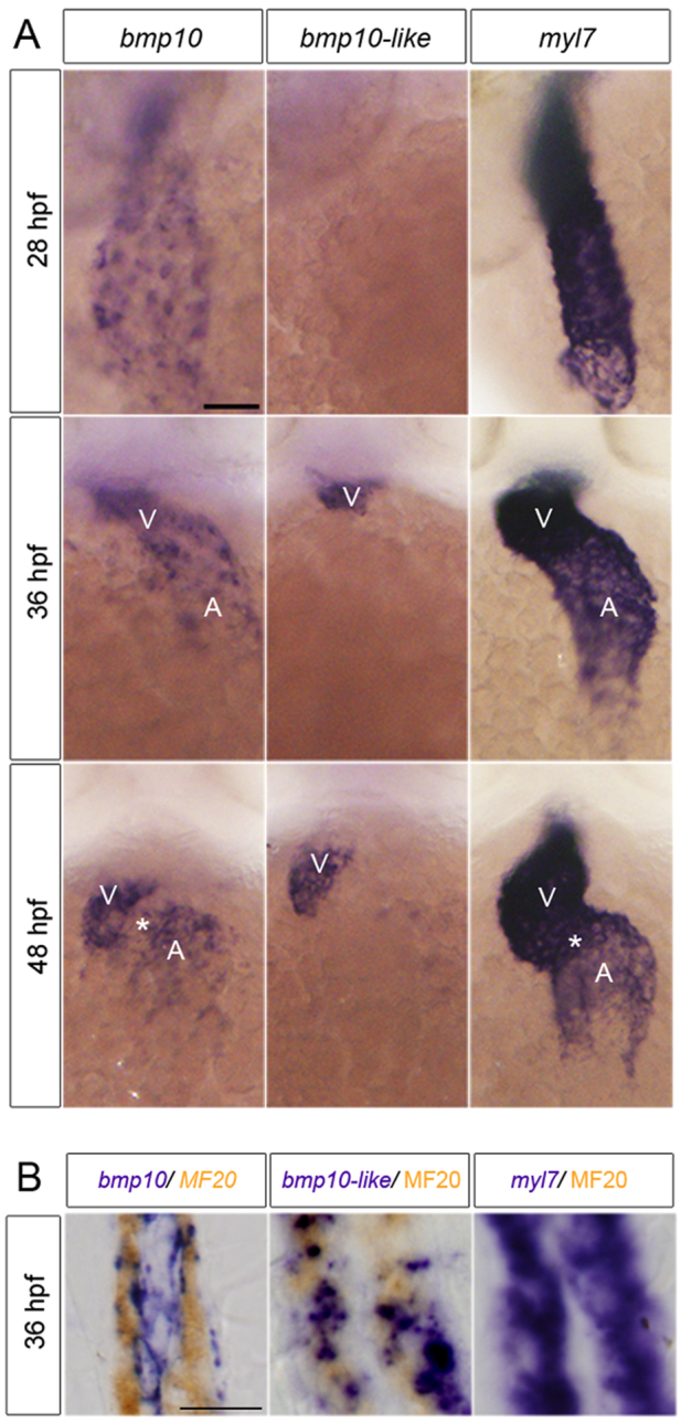
Blood flow plays crucial roles in vascular development, remodeling and homeostasis, but the molecular pathways required for transducing flow signals are not well understood. In zebrafish embryos, arterial expression of activin receptor-like kinase 1 (alk1), which encodes a TGFβ family type I receptor, is dependent on blood flow, and loss of alk1 mimics lack of blood flow in terms of dysregulation of a subset of flow-responsive arterial genes and increased arterial endothelial cell number. These data suggest that blood flow activates Alk1 signaling to promote a flow-responsive gene expression program that limits nascent arterial caliber. Here, we demonstrate that restoration of endothelial alk1 expression to flow-deprived arteries fails to rescue Alk1 activity or normalize arterial endothelial cell gene expression or number, implying that blood flow may play an additional role in Alk1 signaling independent of alk1 induction. To this end, we define cardiac-derived Bmp10 as the crucial ligand for endothelial Alk1 in embryonic vascular development, and provide evidence that circulating Bmp10 acts through endothelial Alk1 to limit endothelial cell number in and thereby stabilize the caliber of nascent arteries. Thus, blood flow promotes Alk1 activity by concomitantly inducing alk1 expression and distributing Bmp10, thereby reinforcing this signaling pathway, which functions to limit arterial caliber at the onset of flow. Because mutations in ALK1 cause arteriovenous malformations (AVMs), our findings suggest that an impaired flow response initiates AVM development.

Keywords: Alk1/Acvrl1; Arteriovenous malformation; Bmp10; Flow response; Hereditary hemorrhagic telangiectasia; Zebrafish.


Figures

Fig. 1.
Fig. 1.
Restoration of alk1 expression does not rescue cxcr4a or edn1 expression in the absence of blood flow. (A) Lateral (anterior left), frontal (anterior upwards) and dorsal (anterior left) views of the zebrafish cranial vasculature at 36 hpf. alk1-positive arteries are light gray, alk1-negative arteries are dark gray and veins are black. alk1-positive arteries are closest to the heart. AA1, aortic arch 1; BA, basilar artery; BCA, basal communicating artery; CaDI, caudal division of the internal carotid artery; ICA, internal carotid artery; LDA, lateral dorsal aorta; PCS, posterior communicating segments; PHBC, primordial hindbrain channel; PMBC, primordial midbrain channel. Scale bar: 50 μm. (B) Whole-mount in situ hybridization for cxcr4a, edn1 and cadherin 5 (cdh5, pan-endothelial control) at 36 hpf. cxcr4a and edn1 are upregulated and downregulated, respectively, in alk1 mutants (column 2) and tnnt2a morphants (column 4) compared with controls (column 1), and expression can be normalized to wild-type/control morphant levels by transgene-mediated, flow-independent alk1 expression in alk1 mutants (column 3) but not in tnnt2a morphants (column 5). cdh5 expression is not altered by alk1 or flow conditions. n≥50 for all groups. Frontal views, dorsal upwards. Arrows indicate left CaDI. Scale bar: 50 μm.
Fig. 2.
Fig. 2.
Alk1 activity is dependent on blood flow. (A-E) pSmad1/5/9 (middle column) in endothelial cell nuclei (marked by fli1a:negfp transgene, left column); in merge (right column), EGFP-expressing endothelial cell nuclei are green and pSmad1/5/9 immunofluorescence is magenta. (A) 24 hpf wild type, prior to blood flow; (B) 36 hpf tnnt2a morphant (no flow); (C) 36 hpf wild type; (D) 36 hpf tnnt2a morphant harboring a fli1a:alk1-myc transgene; (E) 36 hpf tnnt2a morphant harboring a fli1a:alk1CA-mCh transgene. Tg(fli1a:alk1CA-mCh) embryos do not have lumenized vessels. In merge (right column), EGFP-expressing endothelial cell nuclei are green, pSmad1/5/9 immunofluorescence is magenta. Yellow and blue arrows indicate endothelial cells in the caudal division of the internal carotid artery (CaDI) and basal communicating artery (BCA), respectively. 2D confocal projections of 50 μm frontal sections, dorsal upwards. Scale bar: 50 μm. See supplementary material Table S1 for fluorescence quantitation.
Fig. 3.
Fig. 3.
Knockdown of bmp10 phenocopies zebrafish alk1 mutants. (A) CaDIs (caudal divisions of the internal carotid artery; yellow arrows) and BCA (basal communicating artery; blue arrows) in 36 hpf Tg(fli1a:mrfp-caax);Tg(fli1a:negfp) embryos injected with 20 ng control, 20 ng bmp10TB or 2.5 ng alk1 morpholino. Endothelial cell membranes are magenta; nuclei are green. 2D projections of 10 optical sections (Z-step, 2 μm), frontal views, dorsal upwards. Scale bar: 50 μm. (B) Endothelial cell number in the CaDI/BCA in control, bmp10TB and alk1 morphants. n=7-10 in three independent experiments. Values are mean±s.e.m. Student’s t-test: *P<0.001. (C) pSmad1/5/9 (middle column) in endothelial cells (nuclei marked by fli1a:negfp transgene, left column) in 36 hpf control and bmp10TB morphants. In merge (right column), EGFP-expressing endothelial cell nuclei are green, pSmad1/5/9 immunofluorescence is magenta. Yellow and blue arrows indicate endothelial cells in the CaDI and BCA, respectively. 2D confocal projections of 50 μm frontal sections, dorsal upwards. Scale bar: 50 μm. See supplementary material Table S1 for fluorescence quantitation. (D) Whole-mount in situ hybridization for cxcr4a, edn1 and cdh5 (pan-endothelial control) in wild type/control morphant, bmp10TB morphant and alk1-/- at 36 hpf. Frontal views, dorsal upwards. Arrows indicate BCA (vertical) or CaDI (horizontal). Scale bar: 50 μm. (E) Cranial vasculature in 48 hpf Tg(kdrl:gfp);Tg(gata1a:dsRed) embryos injected with bmp10TB morpholino (15-20 ng, as indicated), alk1 morpholino (2.5 ng) and/or bmp10-like morpholino (3 ng). Arrows highlight width of BCA; asterisk indicates AVM. Endothelial cells are green, red blood cells magenta. 2D confocal projections, dorsal views, anterior leftwards. Scale bar: 50 μm. (F) Quantification of AVM development in 48 hpf morpholino-injected embryos. n=43-146. Values are mean±s.e.m. Student’s t-test: *P<0.001; ns, not significant.
Fig. 4.
Fig. 4.
bmp10 and bmp10-like are expressed in the heart. (A) Whole-mount in situ hybridization for bmp10, bmp10-like and myl7 at 28, 36 and 48 hpf. Ventral views, anterior upwards. Scale bar: 50 μm. V, ventricle; A, atrium. Asterisk indicates atrioventricular canal. (B) Sagittal sections (8 μm) through the heart of 36 hpf embryos co-stained with bmp10, bmp10-like or myl7 (purple) and MF20 (sarcomeric myosin; brown). Scale bar: 10 μm.
Fig. 5.
Fig. 5.
Bmp10/Alk1 lies downstream of blood flow in regulation of pSmad1/5/9, cxcr4a and edn1. (A) BRE:luciferase activity in C2C12 cells transfected with human (h) ALK1, zebrafish (z) alk1, hALK1 kinase dead mutant (R411Q), hALK3 (which does not bind BMP10) or lipofectamine (lipo), and treated with rhBMP10. Data are normalized to pTK-Renilla luciferase activity. Values are mean±s.e.m., n=3 biological replicates, each representing 3 technical replicates. (B) pSmad1/5/9 (middle column) in endothelial cells (nuclei marked by fli1a:negfp transgene, left column) of the CaDI (caudal division of the internal carotid artery) of 36 hpf tnnt2a morphants in which endothelial cell alk1 expression [Tg(fli1a:alk1-myc)], BMP10 availability (rhBMP10; 2 nl of 10 μM rhBMP10 injected into base of CaDI at 28 hpf), or both, are restored. In merge (right column), EGFP-expressing endothelial cell nuclei are green, pSmad1/5/9 immunofluorescence is magenta. Yellow arrows indicate endothelial cells in the injected CaDI. 2D confocal projections of 50 μm frontal sections, dorsal upwards. Scale bar: 10 μm. See supplementary material Table S1 for fluorescence quantitation. (C) Whole-mount in situ hybridization for cxcr4a, edn1 and cdh5 (pan-endothelial control) in 36 hpf tnnt2a morphant;Tg(fli1a:alk1-myc) embryos injected at 28 hpf into the base of the left CaDI with 2 nl tracer with or without 10 μM rhBMP10. Yellow arrow indicates injected CaDI, red arrow indicates uninjected CaDI. Asterisk indicates injection site. Frontal views, dorsal upwards; lateral views, right (uninjected) side or left (injected) side. Scale bar: 50 μm. (D) Quantitation of CaDI endothelial cell number in 36 hpf Tg(fli1a:alk1-myc) embryos left uninjected (white bars) or injected at the one-cell stage with tnnt2a morpholino (gray, black bars). At 28 hpf, tnnt2a morphants were injected in the right CaDI with 2 nl tracer (gray bars) or 2 nl 10μM rhBMP10 (black bars). tnnt2a morphants injected intravascularly with tracer exhibited increased endothelial cell number throughout the CaDI/BCA, whereas intravascular injection of rhBMP10 normalized endothelial cell number in the injected CaDI and BCA (basal communicating artery). n=10-12 per group in two independent experiments. Values are mean±s.e.m. Student’s t-test: *P<0.05; **P<0.001; ns, not significant.

References

    1. Bhullar I. S., Li Y. S., Miao H., Zandi E., Kim M., Shyy J. Y., Chien S. (1998). Fluid shear stress activation of IkappaB kinase is integrin-dependent. J. Biol. Chem. 273, 30544–30549 - PubMed
    1. Brown M. A., Zhao Q., Baker K. A., Naik C., Chen C., Pukac L., Singh M., Tsareva T., Parice Y., Mahoney A., et al. (2005). Crystal structure of BMP-9 and functional interactions with pro-region and receptors. J. Biol. Chem. 280, 25111–25118 - PubMed
    1. Bussmann J., Wolfe S. A., Siekmann A. F. (2011). Arterial-venous network formation during brain vascularization involves hemodynamic regulation of chemokine signaling. Development 138, 1717–1726 - PMC - PubMed
    1. Chang K., Weiss D., Suo J., Vega J. D., Giddens D., Taylor W. R., Jo H. (2007). Bone morphogenic protein antagonists are coexpressed with bone morphogenic protein 4 in endothelial cells exposed to unstable flow in vitro in mouse aortas and in human coronary arteries: role of bone morphogenic protein antagonists in inflammation and atherosclerosis. Circulation 116, 1258–1266 - PubMed
    1. Chen H., Shi S., Acosta L., Li W., Lu J., Bao S., Chen Z., Yang Z., Schneider M. D., Chien K. R., et al. (2004). BMP10 is essential for maintaining cardiac growth during murine cardiogenesis. Development 131, 2219–2231 - PMC - PubMed

Publication types

MeSH terms