. 2013 Jul 1;123(8):3331-3342.
doi: 10.1172/JCI69210. Online ahead of print.

Gray platelet syndrome and defective thrombo-inflammation in Nbeal2-deficient mice

Gray platelet syndrome and defective thrombo-inflammation in Nbeal2-deficient mice

Carsten Deppermann et al. J Clin Invest. .

Abstract

Platelets are anuclear organelle-rich cell fragments derived from bone marrow megakaryocytes (MKs) that safeguard vascular integrity. The major platelet organelles, α-granules, release proteins that participate in thrombus formation and hemostasis. Proteins stored in α-granules are also thought to play a role in inflammation and wound healing, but their functional significance in vivo is unknown. Mutations in NBEAL2 have been linked to gray platelet syndrome (GPS), a rare bleeding disorder characterized by macrothrombocytopenia, with platelets lacking α-granules. Here we show that Nbeal2-knockout mice display the characteristics of human GPS, with defective α-granule biogenesis in MKs and their absence from platelets. Nbeal2 deficiency did not affect MK differentiation and proplatelet formation in vitro or platelet life span in vivo. Nbeal2-deficient platelets displayed impaired adhesion, aggregation, and coagulant activity ex vivo that translated into defective arterial thrombus formation and protection from thrombo-inflammatory brain infarction following focal cerebral ischemia. In a model of excisional skin wound repair, Nbeal2-deficient mice exhibited impaired development of functional granulation tissue due to severely reduced differentiation of myofibroblasts in the absence of α-granule secretion. This study demonstrates that platelet α-granule constituents are critically required not only for hemostasis but also thrombosis, acute thrombo-inflammatory disease states, and tissue reconstitution after injury.


Figures
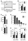
Figure 1
Figure 1. Nbeal2–/– mice are macrothrombocytopenic and lack platelet α-granules.
(A) Analysis of Nbeal2 mRNA in bone marrow (b) and thymus (t) in wild-type (+/+) and Nbeal2–/– (–/–) mice. Gapdh mRNA served as loading control. (B) Spleen to body weight ratio was analyzed in 6-week-old and 6-month-old mice. Values are mean ± SD (n = 4). (C) Peripheral platelet counts and mean platelet volume (MPV) in wild-type and Nbeal2–/– mice. Values are mean ± SD (n = 7). (D) Platelet life span as measured by injection of DyLight 488 α-GPIX. (E) Determination of MK numbers per visual field (294 × 221 μm) in H&E-stained spleen and bone marrow sections of 6-week-old and 6-month-old mice. Values are mean ± SD (n = 4). *P < 0.05; **P < 0.01; ***P < 0.001.
Figure 2
Figure 2. Analysis of α-granule content in Nbeal2–/– platelets.
(A) Representative transmission electron microscopy images of resting wild-type and Nbeal2–/– platelets. Note the lack of α-granules in Nbeal2–/– platelets. AG, α-granules; DG, δ-granules; M, mitochondria; V, vacuoles. Scale bar: 1 μm. (B) Nbeal2–/– platelet ultrastructure. Scale bar: 0.5 μm (left); 0.25 μm (right). (C) Analysis of filamentous actin structure (red) and VWF (green) localization in spread (30 minutes on fibrinogen) wild-type and Nbeal2–/– platelets by confocal microscopy. Arrows indicate residual VWF in Nbeal2–/– platelets. Scale bar: 7.5 μm. (D) ELISA of VWF and fibrinogen content in resting wild-type and Nbeal2–/– platelets. Data are presented as ΔOD 450 nm–OD 620 nm of 4 mice per group, normalized to Vwf–/– or PBS as control, and are representative of 3 individual experiments.
Figure 3
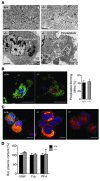
Figure 3. Paucity of α-granules but unaltered ultrastructure in Nbeal2–/– MKs.
(A) Representative transmission electron microscopy images of mature bone marrow MKs. Normally developed demarcation membrane system with α-granules in a wild-type mouse (top left). Scale bar: 1 μm. The demarcation membrane system continues to be seen in Nbeal2–/– MKs (top right) but with a deficiency of α-granules and a markedly increased number of vacuoles in Nbeal2–/– MKs. Scale bar: 1 μm. Note the presence of a leukocyte within the cytoplasm of the mutant MK (bottom left). Scale bar: 3 μm. Protrusions from the MK cytoplasm and developing proplatelets could be clearly seen in Nbeal2–/– MKs (bottom right). Scale bar: 1 μm. VS, vascular sinus. (B) Analysis of actin (red) and tubulin-containing (green) structures in fetal liver cell–derived (FLC-derived) proplatelet-forming MKs by confocal microscopy. Nuclei were counterstained with DAPI (blue). Proplatelet formation was unaltered in FLC-derived MKs. Results are quantified as the percentage of proplatelet-forming MKs per visual field ± SD from ≥ 7 samples per group. Scale bar: 50 μm. (C) Distribution of VWF (green) in FLC- and bone marrow–derived MKs. Note absence of VWF staining in some Nbeal2–/– MKs and the accumulation of VWF in other mutant cells, in contrast to a uniform distribution in wild-type MKs. The actin cytoskeleton was visualized using phalloidin (red). Scale bar: 25 μm. (D) ELISA assay of plasma VWF, fibrinogen, and PF4 content. Data are presented as ΔOD 450 nm–OD 620 nm of 5 mice per group.
Figure 4
Figure 4. Agonist responses of Nbeal2–/– platelets in suspension.
(A) Flow cytometric analysis of Nbeal2–/– platelets shows decreased degranulation-dependent P-selectin exposure but normal activation of αIIbβ3 integrin (binding of JON/A-PE) upon stimulation with the indicated agonists. (B) Recruitment of integrin αIIbβ3 to the platelet surface upon stimulation with different agonists was assessed using the JON7-FITC antibody. Results in A and B are expressed as MFI ± SD (top) and mean relative fluorescence intensities ± SD (bottom) (n = 4 mice per group) and are representative of 3 independent experiments. (C) Impaired aggregation of Nbeal2–/– platelets in response to different agonists. Aggregation traces (recording time = 10 minutes) representative of 2 independent experiments are depicted. (D) Normal ATP release from platelets activated by thrombin and collagen, as assessed by luciferase activity. U46, U46619; Thr, thrombin; RC, rhododcytin; CVX, convulxin; rest, resting. *P < 0.05; **P < 0.01; ***P < 0.001.
Figure 5
Figure 5. Nbeal2–/– platelets show defective adhesion, thrombus formation, and PS exposure under flow.
(A) Severely impaired adhesion and thrombus formation of Nbeal2–/– platelets on collagen under flow at a shear rate of 1,700 s–1. Representative phase-contrast (bright field [BF]) and fluorescence images are shown as well as mean surface coverage and relative platelet deposition, as measured by integrated fluorescent intensity (IFI) per mm2 ± SD (n = 4 mice per group). Scale bar: 50 μm. (B) Representative phase-contrast (BF) and fluorescence images of Nbeal2–/– platelets stained with Annexin A5–DyLight 488 perfused over a collagen-coated surface at a shear rate of 1,000 s–1. Scale bar: 65 μm. (C) Procoagulant index represents the ratio of Annexin A5–positive cells to surface coverage (n = 5 mice per group). (D) Highly diluted washed platelets were stimulated with 1 μg/ml convulxin, 20 μg/ml CRP, or 20 μg/ml CRP plus 0.1 U/ml thrombin, and the percentage of Annexin A5–positive cells was determined by flow cytometry (n = 5). Data are representative of 3 independent experiments. *P < 0.05; ***P < 0.001.
Figure 6
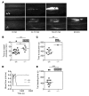
Figure 6. Severely impaired arterial thrombus formation and hemostasis in Nbeal2–/– mice.
(A and B) Thrombus formation in small mesenteric arterioles was induced by topical application of 20% FeCl3. (A) Representative images. (B) Time to stable vessel occlusion is depicted. Each symbol represents 1 arteriole. (C) In an aorta injury model, the blood flow was monitored for 30 minutes or until complete occlusion occurred. (D) Representative blood flow curves. (E) Tail bleeding times of wild-type and Nbeal2–/– mice. (C and E) Each symbol represents 1 individual mouse. *P < 0.05; **P < 0.01; ***P < 0.001.
Figure 7
Figure 7. Nbeal2–/– mice are protected from cerebral ischemia.
Infarct volumes and functional outcome 24 hours after focal cerebral ischemia in wild-type and Nbeal2–/– mice, as well as wild-type mice reconstituted with wild-type and Nbeal2–/– bone marrow, were investigated in a murine model of ischemic stroke. Mice were subjected to 60 minutes of tMCAO. (A) Representative images of 3 coronal sections stained with 2,3,5-triphenyltetrazolium chloride 24 hours after tMCAO. (B) Brain infarct volumes (n ≥ 6) were measured by planimetry. Results represent mean ± SD. (C) Bederson score and (D) grip test determined 24 hours after tMCAO. Each symbol represents 1 individual mouse. *P < 0.05; **P < 0.01.
Figure 8
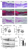
Figure 8. Impaired dermal repair due to reduced myofibroblast activity in wounds of Nbeal2-deficient mice and severely diminished mature and pro–TGF-β in Nbeal2-deficient platelets.
(AC) Representative sections of wounds at 7 days after injury. Wounds of Nbeal2-deficient mice display (A) reduced development of granulation tissue but similar epithelial wound closure (H&E staining), (B) reduced amounts of collagenous granulation tissue (central darker area in picrosirius red stain), (C) and severely diminished myofibroblast numbers per wound. Scale bar: 500 μm. (D) Area of granulation tissue is significantly reduced in mutants (n = 19) versus that in wild-type mice (n = 15). (E) Area occupied by αSMA-positive myofibroblasts is significantly diminished in mutants (n = 20) versus that in controls (n = 16). (F) Reduced amounts of mature TGF-β monomer (12 kDa) and of pro–TGF-β monomer (50 kDa) in lysates from Nbeal2-deficient versus wild-type platelets (corresponding to 6 × 10⁶ platelets each). (G) Densitometric evaluation of TGF-β signals (12 + 50 kDa), normalized to β-actin signals. n = 4 for each genotype. *P < 0.05; ***P < 0.001.

References

    1. Furie B, Furie BC. Mechanisms of thrombus formation. N Engl J Med. 2008;359(9):938–949. doi: 10.1056/NEJMra0801082. - DOI - PubMed
    1. Jackson SP. Arterial thrombosis--insidious, unpredictable and deadly. Nat Med. 2011;17(11):1423–1436. doi: 10.1038/nm.2515. - DOI - PubMed
    1. Bhatt DL, Topol EJ. Scientific and therapeutic advances in antiplatelet therapy. Nat Rev Drug Discovery. 2003;2(1):15–28. doi: 10.1038/nrd985. - DOI - PubMed
    1. Blair P, Flaumenhaft R. Platelet α-granules: basic biology and clinical correlates. Blood Rev. 2009;23(4):177–189. doi: 10.1016/j.blre.2009.04.001. - DOI - PMC - PubMed
    1. Nurden A. Platelets, inflammation and tissue regeneration. Thromb Haemost. 2011;105(6):S13–S33. doi: 10.1160/THS10-11-0720. - DOI - PubMed