. 2013 Jul 1;123(8):3446-3458.
doi: 10.1172/JCI67484. Online ahead of print.

Neutrophil extracellular traps sequester circulating tumor cells and promote metastasis

Neutrophil extracellular traps sequester circulating tumor cells and promote metastasis

Jonathan Cools-Lartigue et al. J Clin Invest. .

Abstract

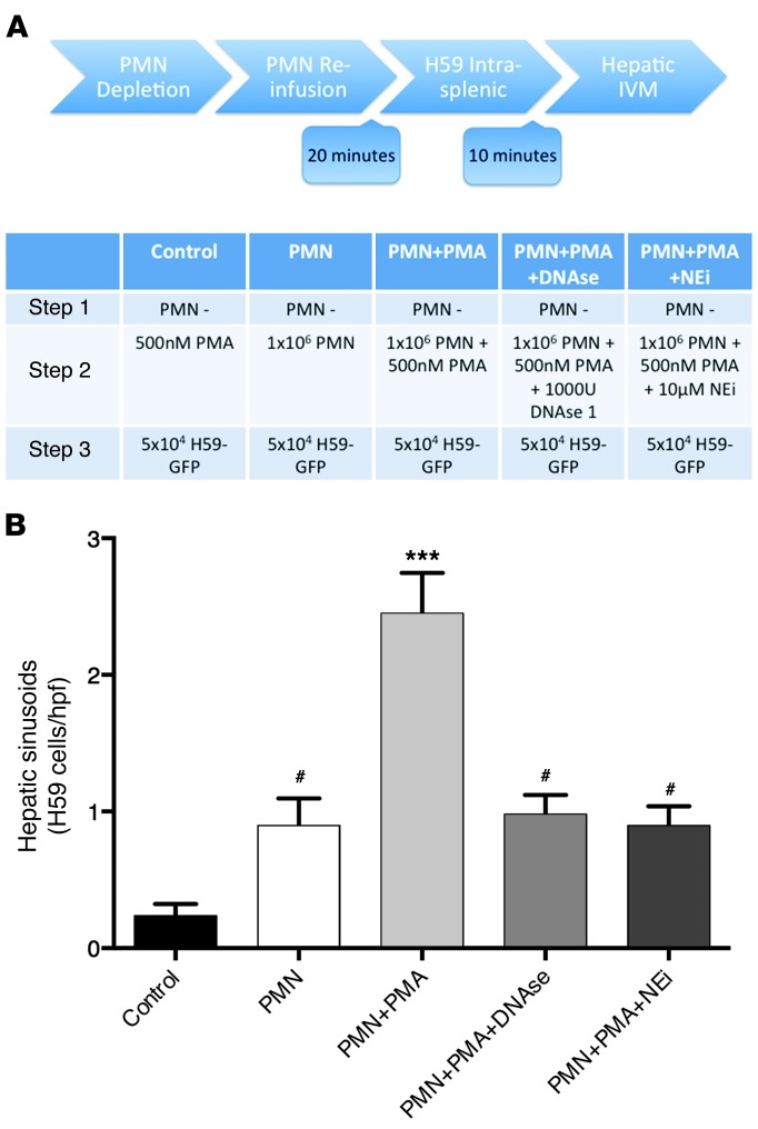
The majority of patients with cancer undergo at least one surgical procedure as part of their treatment. Severe postsurgical infection is associated with adverse oncologic outcomes; however, the mechanisms underlying this phenomenon are unclear. Emerging evidence suggests that neutrophils, which function as the first line of defense during infections, facilitate cancer progression. Neutrophil extracellular traps (NETs) are extracellular neutrophil-derived DNA webs released in response to inflammatory cues that trap and kill invading pathogens. The role of NETs in cancer progression is entirely unknown. We report that circulating tumor cells become trapped within NETs in vitro under static and dynamic conditions. In a murine model of infection using cecal ligation and puncture, we demonstrated microvascular NET deposition and consequent trapping of circulating lung carcinoma cells within DNA webs. NET trapping was associated with increased formation of hepatic micrometastases at 48 hours and gross metastatic disease burden at 2 weeks following tumor cell injection. These effects were abrogated by NET inhibition with DNAse or a neutrophil elastase inhibitor. These findings implicate NETs in the process of cancer metastasis in the context of systemic infection and identify NETs as potential therapeutic targets.


Figures

Figure 1
Figure 1. CLP results in widespread deposition of extracellular DNA, which colocalizes with neutrophils and expresses neutrophil-derived granules.
(A) Under anesthesia, murine livers were externalized and imaged using SD-IVM, permitting real-time visualization of neutrophil trafficking and DNA extrusion. Intravascular administration of E-fluor 660 anti-GR1, Sytox Green, Alexa Fluor 555 anti-H2AX, or anti-NE was used to visualize polymorphonuclear neutrophils (PMN, blue), DNA (green), H2AX (red), and NE (yellow), respectively. DNA is visualized adjacent to PMN within hepatic sinusoids. DNA stains positive for histone H2AX and NE, in keeping with what has been described for NETs (48). Scale bars: 40 μm. Extracellular DNA was quantified within (B) hepatic sinusoids and (C) pulmonary capillaries by measuring total area of fluorescence per hpf over 5 hpf. Data are represented as relative area of fluorescence compared with sham. CLP was associated with increased amounts of extracellular DNA compared with sham. Neutrophil depletion or systemic administration of DNAse 1 or NEi after CLP results in decreased extracellular DNA staining compared with CLP alone. Data are presented as mean ± SEM from n = 3–5 mice per group. ***P < 0.001, *P < 0.05 versus sham as determined by 1-way ANOVA with Tukey’s HSD post-hoc analysis. See also Supplemental Figures 1 and 2.
Figure 2
Figure 2. Systemic sepsis promotes the development of gross metastasis, which is attenuated by systemic administration of inhibitors of NET formation.
Mice were subjected to CLP in order to induce sepsis. Intrasplenic injection of H59 Lewis lung cancer cell lines was performed 24 hours later. Administration of DNAse 1 intramuscularly or a NEi per os was started 24 hours prior to CLP and continued daily for 14 days. At 14 days, mice were sacrificed, and the number of gross hepatic metastases was quantified. (A) Representative images of hepatic nodules after necropsy in mice subjected to sham surgery, CLP, CLP with daily DNAse 1 administration, and CLP with daily NEi administration. (B) CLP resulted in a significant increase in the number of gross metastatic nodules compared with sham. Treatment with DNAse or NEi after CLP resulted in a significant decrease in the number of gross metastases. Data are presented as mean ± SEM from n = 5 mice per group. *P < 0.05, **P < 0.01 versus CLP. #P < 0.001 versus sham as determined by 1-way ANOVA with Tukey’s HSD post-hoc analysis.
Figure 3
Figure 3. Tumor cell adhesion after CLP is augmented by trapping within neutrophil-derived extracellular DNA.
In order to demonstrate that tumor cells become embedded within NETs, SD-IVM was performed permitting visualization of (A) hepatic sinusoids in living mice and (B) pulmonary capillaries ex vivo within 10 minutes. Images shown in A and B represent a single 3-dimensional reconstruction of confocal z-stacks (10- to 20-μm thickness,1-μm intervals), rotated 180° from an inferior to a superior perspective. In both liver and lungs, tumor cells (red) were found to arrest within extracellular chromatin (Alexa Fluor 555 anti-histone H2AX [green]) adjacent to neutrophils (E-fluor 660 anti-GR1 [blue]). Scale bars: 20 μm. See also Supplemental Video 1 and Supplemental Figure 3. (C) Quantification of adhesion of H59 cells within hepatic sinusoids. 3 × 10⁴ cells were injected via the spleen 24 hours after CLP or sham surgery. Adhesion was increased after CLP compared with sham. Systemic administration of DNAse 1 or NEi starting 1 day prior to CLP abolished this increase. (D) Quantification of B16 melanoma cells within pulmonary capillaries. 1 × 10⁶ cells were injected via tail vein 24 hours after CLP or sham. Adhesion was increased after CLP compared with sham and was abolished by administration of DNAse 1 or NEi starting 1 day prior to CLP. Quantification was performed by counting the number of cells per hpf in 8 to 10 hpf (×20) per experiment. Data are presented as mean ± SEM from n = 5 mice per group. ***P < 0.001 versus sham, DNAse, and NEi as determined by 1-way ANOVA with Tukey’s HSD post-hoc analysis.
Figure 4
Figure 4. NET production by neutrophils is sufficient to increase tumor cell adhesion within hepatic sinusoids.
(A) A schematic representation of the experimental design is depicted. All mice used were subjected to neutrophil depletion via intraperitoneal injection of anti-GR1 (150 μg). Twenty-four hours later, 1 × 10⁶ bone marrow–derived neutrophils (or control buffer) from syngeneic mice were reinfused via the intrasplenic route. This was followed by injection of 3 × 10⁴ H59-GFP cells into the spleen 20 minutes later. Ten minutes later, epifluorescence microscopy was used for quantification of adherent cells. IVM, intravital microscopy. (B) Quantification of arrested tumor cells within hepatic sinusoids 10 minutes following intrasplenic injection of H59-GFP cells. Injection of 500 nM PMA without neutrophils prior to tumor cell injection resulted in low levels of tumor cell adhesion. Infusion of unstimulated neutrophils increased tumor cell adhesion above control, but this was significantly lower than when neutrophils were pretreated with 500 nM PMA. This increase is abrogated if PMA treatment of neutrophils occurs in the presence of NEi (10 μm) or DNAse 1 (1,000 U) prior to reinfusion. Adherent cells were quantified by counting the number of cells per hpf in 8 to 10 hpf (×20) per experiment. Data are presented as mean ± SEM from n = 4–5 mice per group. Significance was determined using 1-way ANOVA with Tukey’s HSD post-hoc analysis. ***P < 0.0001, #P < 0.05 compared with control.
Figure 5
Figure 5. NETs trap both human and murine tumor cells in vitro.
(A) Under static conditions, H59 and A549 cells demonstrate increased adhesion to neutrophil monolayers stimulated with PMA (800 nM) compared with unstimulated neutrophils. Addition of DNAse 1 (1,000 U) or pretreatment of neutrophils with NEi (5 μM) results in levels of adhesion comparable to control. (B) Tumor cells were perfused over neutrophil monolayers at shear rates of 1 dyne/cm/s–1. H59 and A549 cells demonstrate increased adhesion to neutrophils after stimulation with PMA (800 nM) compared with controls. This was abrogated by addition of DNAse 1 (1,000 U) or pretreatment of neutrophils with NEi (5 μM). Data are presented as mean ± SEM from n = 2–4 separate experiments. ***P < 0.01 versus control, DNAse, and NEi. #P < 0.05 versus control. Significance was determined using 1-way ANOVA with Tukey’s HSD post-hoc analysis. (C) Confocal imaging reveals that after PMA stimulation, A549 cells (red) become trapped within webs of extracellular DNA (green) in proximity to neutrophils (blue). (D) Scanning electron microscopy of A549- and PMA-stimulated neutrophils (800 nM) (original magnification, ×3,000). After PMA stimulation, neutrophils flatten (white arrow) and extrude strands consistent with NETs. Strands encompass a cluster of adherent tumor cells (black arrow). (E) Scanning electron microscopy demonstrates that NETs are in direct contact with adherent A549 tumor cells (original magnification, ×3,500). Scale bars: 40 μm (confocal microscopy); 5 μm (electron microscopy). See also Supplemental Figures 2 and 3.
Figure 6
Figure 6. CLP promotes the development of micrometastases, which is attenuated by treatment with DNAse 1 and NEi.
H59 cells were injected via the spleen 24 hours after CLP. (A) Quantification of micrometastatic foci demonstrated increased numbers of tumor islands at 48 hours after intrasplenic injection in mice subjected to CLP compared with those after sham surgery. Systemic administration of DNAse 1 or NEi after CLP is associated with decreased micrometastases compared with CLP alone. (B) Representative images of tumor cells trapped within hepatic sinusoidal spaces in mice subjected to sham surgery, CLP, and NEi after CLP at 24 and 48 hours following intrasplenic injection of 5 × 10⁵ H59 cells. Scale bars: 40 μm. (C) To further demonstrate that adherent tumor cells persist and are able to grow in this model of sepsis, individual tumor cells (as depicted in B) were quantified within the liver at 24 and 48 hours after intrasplenic injection. Quantification of individual tumor cells within hepatic sinusoids reveals increased numbers at both 24 and 48 hours after intrasplenic injection in mice subjected to CLP compared with sham. Systemic administration of NEi after CLP resulted in decreased numbers of tumor cells at both 24 and 48 hours compared with CLP alone. Data were acquired using n = 3–5 mice per group. Quantification was performed by counting the number of cells per 5–10 hpf per experiment (×20). *P < 0.05, ***P < 0.0001 compared with control, DNAse, and sham. Significance was determined using 1-way ANOVA with Tukey’s HSD post-hoc analysis.
Figure 7
Figure 7. Migration and invasion of A549 cells in vitro is promoted in the presence of intact NETs.
In the presence of neutrophils exposed to PMA, (A) A549 cell migration through 8-μm PET membranes and (B) invasion through endothelial monolayers was increased compared with A549 cells alone, A549 cells in the presence of unstimulated neutrophils, and A549 cells stimulated with PMA (25 nM) in the absence of neutrophils. This phenotype was reversed back to control levels after addition of DNAse 1 (1,000 U) or pretreatment of neutrophils with NEi (5 μm). Data are presented as mean ± SEM from n = 2–4 separate experiments. **P < 0.01, ***P < 0.001 versus A549 alone. #P < 0.05 versus A549 plus neutrophils and PMA. Significance was determined using 1-way ANOVA with post-hoc multiple comparisons with Tukey’s HSD post-hoc analysis.

References

    1. Parkin DM, Bray F, Ferlay J, Pisani P. Global cancer statistics, 2002. CA Cancer J Clin. 2005;55(2):74–108. doi: 10.3322/canjclin.55.2.74. - DOI - PubMed
    1. Mountain CF. Revisions in the international system for staging lung cancer. Chest. 1997;111(6):1710–1717. doi: 10.1378/chest.111.6.1710. - DOI - PubMed
    1. McDonald B, Spicer J, Giannais B, Fallavollita L, Brodt P, Ferri LE. Systemic inflammation increases cancer cell adhesion to hepatic sinusoids by neutrophil mediated mechanisms. Int J Cancer. 2009;125(6):1298–1305. doi: 10.1002/ijc.24409. - DOI - PubMed
    1. van Hagen P, et al. Preoperative chemoradiotherapy for esophageal or junctional cancer. N Engl J Med. 2012;366(22):2074–2084. doi: 10.1056/NEJMoa1112088. - DOI - PubMed
    1. Khan O, Goh S, Byrne B, Somers S, Mercer S, Toh S. Long-term outcomes of extended proximal gastrectomy for oesophagogastric junctional tumours. World J Surg. 2011;35(10):2245–2251. doi: 10.1007/s00268-011-1235-z. - DOI - PubMed