. 2013 Aug;123(8):3564-76.
doi: 10.1172/JCI67521. Epub 2013 Jul 25.

Transcription factor EGR1 directs tendon differentiation and promotes tendon repair

Affiliations

Transcription factor EGR1 directs tendon differentiation and promotes tendon repair

Marie-Justine Guerquin et al. J Clin Invest. 2013 Aug.

Abstract

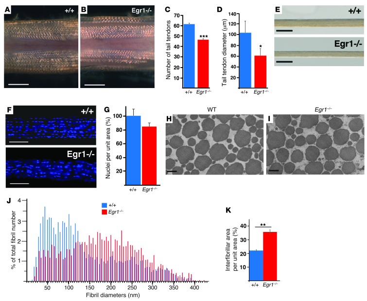
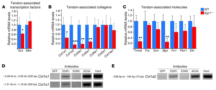
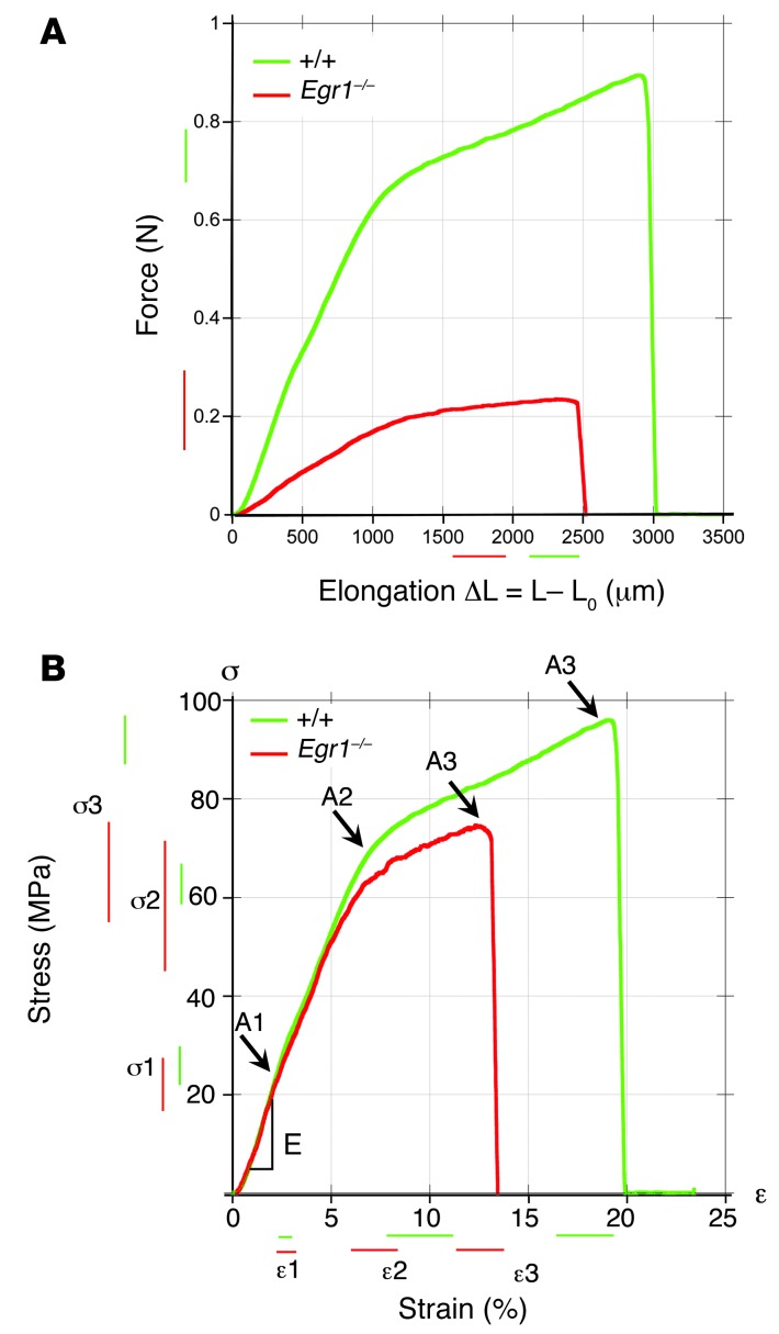
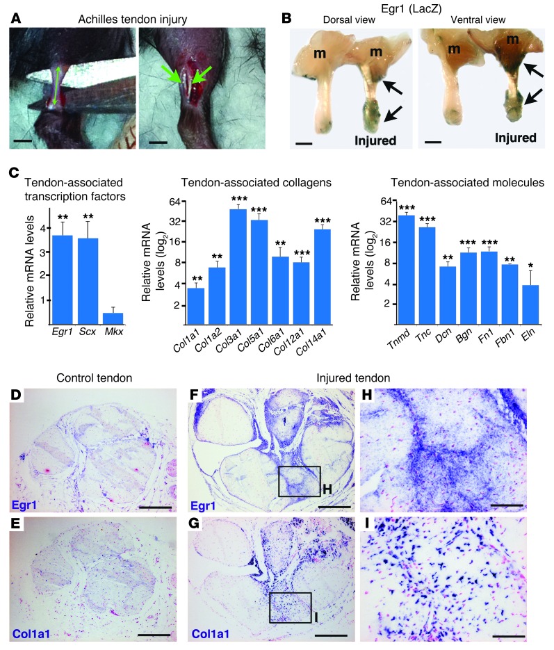
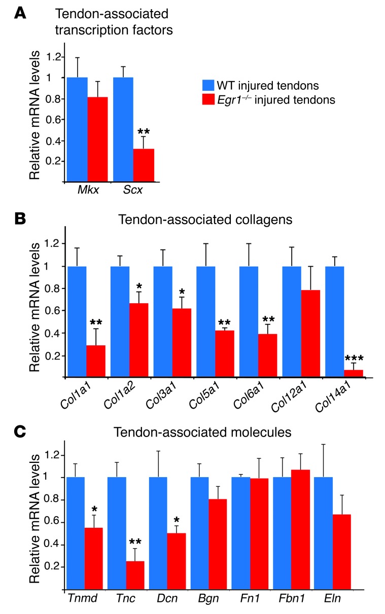
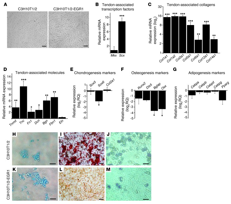
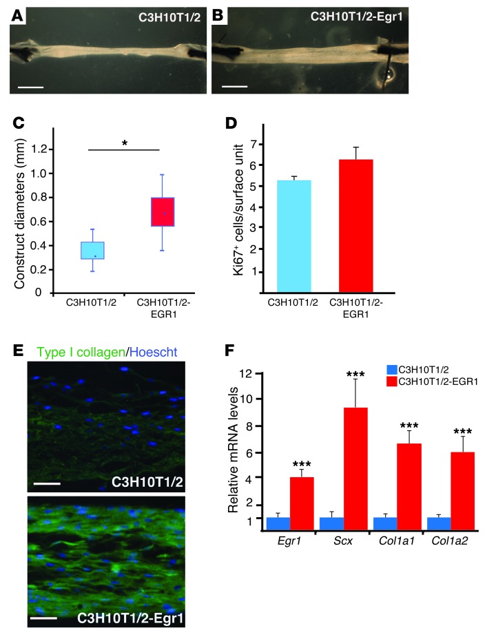
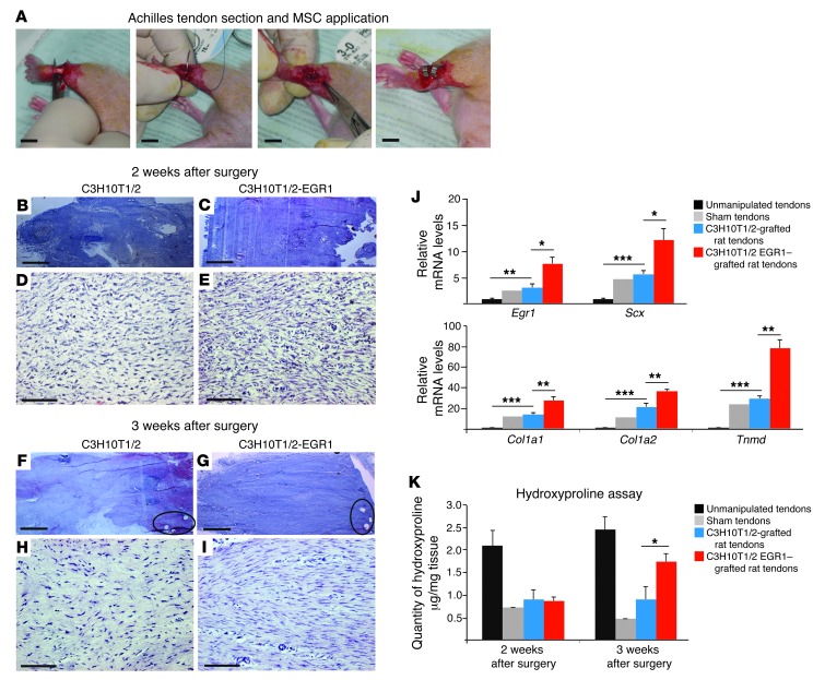
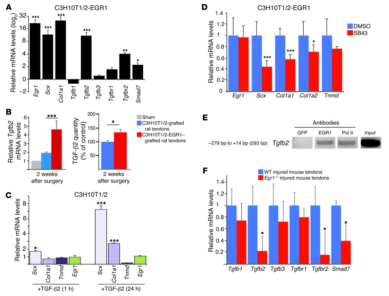
Tendon formation and repair rely on specific combinations of transcription factors, growth factors, and mechanical parameters that regulate the production and spatial organization of type I collagen. Here, we investigated the function of the zinc finger transcription factor EGR1 in tendon formation, healing, and repair using rodent animal models and mesenchymal stem cells (MSCs). Adult tendons of Egr1-/- mice displayed a deficiency in the expression of tendon genes, including Scx, Col1a1, and Col1a2, and were mechanically weaker compared with their WT littermates. EGR1 was recruited to the Col1a1 and Col2a1 promoters in postnatal mouse tendons in vivo. Egr1 was required for the normal gene response following tendon injury in a mouse model of Achilles tendon healing. Forced Egr1 expression programmed MSCs toward the tendon lineage and promoted the formation of in vitro-engineered tendons from MSCs. The application of EGR1-producing MSCs increased the formation of tendon-like tissues in a rat model of Achilles tendon injury. We provide evidence that the ability of EGR1 to promote tendon differentiation is partially mediated by TGF-β2. This study demonstrates EGR1 involvement in adult tendon formation, healing, and repair and identifies Egr1 as a putative target in tendon repair strategies.


Figures

Figure 1
Figure 1. Multiscale analysis of tendons from adult Egr1–/– mice.
(A and B) Macroscopic views showing tail tendons of mouse tails close to the body from 2-month-old WT (A) and Egr1–/– (B) mice. (C) Number of tail tendons counted at the same level (close to the body) from 2- to 3-month-old WT and Egr1–/– mutant mice. (D) Tail tendon diameters from 2- to 3-month-old WT and Egr1–/– mice. (E) Examples of isolated tail tendons from WT and Egr1–/– mice. (F) Hoechst staining of individual tail tendons from WT and Egr1–/– mice. (G) Number of nuclei per unit area in tail tendon sections from 2- to 3-month-old age-matched WT and Egr1–/– mice. Numbers of nuclei were normalized to those in WT mice. (H and I) Electron microscopic analysis of tail tendons from 2-month-old WT and Egr1–/– mice. (J) Histograms showing the frequencies of fibril diameters from WT versus Egr1–/– mice. For WT mice, the mean of the diameters = 143.89 nm; SD = 87.63. For Egr1–/–, the mean of the diameters = 183.73 nm; SD = 84.88. (K) Interfibrillar area (per unit area) of tail tendons from WT and Egr1–/– mice. The error bars represent the SEM. *P < 0.05; **P < 0.01; ***P < 0.001. Scale bars: 1 mm (A and B); 100 μm (E and F); 200 nm (H and I).
Figure 2
Figure 2. Molecular analyses of tendons from Egr1–/– mutant mice.
(AC) qRT-PCR analyses of tendon gene expression in Egr1–/– versus WT mice. The following tendon markers were analyzed: the tendon-associated transcription factors, Scx and Mkx (A); the tendon-associated collagens, Col1a1, Col1a2, Col3a1, Col5a1, Col6a1, Col12a1, and Col14a1; (B) and the tendon-associated molecules, Tnmd, Tnc, Dcn, Bgn, Fn1, Fbn1, and Eln (C). mRNA levels of tendon markers in WT tendons were normalized to 1. Error bars represent SEM. *P < 0.05; **P < 0.01; ***P < 0.001, unpaired Student’s t tests. (D and E) ChIP assays were performed on tendons from postnatal mice with antibodies against EGR1 and EGR2 or ACH4 (acetylated histone H4) as a positive control, or with GFP antibody as a negative control. ChIP products were analyzed by PCR to study the interaction of EGR1 with the tendon regulatory regions of mouse Col1a1 and Col1a2 promoters. Primers targeting a 230-bp region flanking the tendon-specific elements TSE1 and TSE2 and targeting a 330-bp region within the tendon-specific deletion identified DNA regions immunoprecipitated by EGR1 (D). Primers targeting a 73-bp fragment of the Col1a2 promoter identified DNA regions immunoprecipitated by EGR1 (E).
Figure 3
Figure 3. Biomechanical properties of tail tendons deficient for Egr1.
(A) Typical force-elongation curves for WT (green) and Egr1–/– (red) mouse tail tendons. (B) Typical stress-strain curves for WT (green) and Egr1–/– (red) mouse tail tendons. These curves can be characterized by the slope E (Young’s modulus) of the linear regime at low strain, by the coordinates (ε1, σ1) and (ε2, σ2) of the 2 successive changes of slopes A1 and A2, and finally, by the coordinates (ε3, σ3) of the rupture point A3. The position and length of the bars next to the axes represent the means and SDs of these coordinates for all animals, averaged over all the tendons tested for a single animal.
Figure 4
Figure 4. Upregulation of tendon gene expression following injury.
(A) Longitudinal sections parallel to the axis of Achilles tendons were made in adult mice to create a mouse model for tendon injury. (B) We observed increased LacZ expression (reflecting Egr1 expression) in Achilles tendons 1 week after tendon injury in Egr1+/– mice. m, muscle. (C) qRT-PCR analyses of tendon-associated gene expression in injured tendons versus noninjured tendons 1 week after injury. The following tendon markers were analyzed: the tendon-associated transcription factors, Scx and Mkx; the tendon-associated collagens, Col1a1, Col1a2, Col3a1, Col5a1, Col6a1, Col12a1, and Col14a1; and the tendon-associated molecules, Tnmd, Tnc, Dcn, Bgn, Fn1, Fbn1, and Eln. mRNA levels of noninjured tendons were normalized to 1. Error bars represent SEM. *P < 0.05; **P < 0.01; ***P < 0.001, paired Student’s t tests. (DI) Adjacent transverse sections of noninjured (D and E) and injured (FI) Achilles tendons were hybridized with Egr1 (D, F, and H) or Col1a1 (E, G, and I) probes (blue labeling). Three weeks after injury, Egr1 and Col1a1 expression was upregulated in injured tendons at the site of injury compared with control (noninjured) tendons. Scale bars: 1 mm (A and B); 200 μm (DG); 50 μm (H and I).
Figure 5
Figure 5. Egr1 is required for normal tendon gene response following mouse tendon injury.
qRT-PCR analyses of tendon-associated gene expression in Egr1–/– injured tendons versus WT injured tendons 1 week after injury. The following tendon markers were analyzed: the tendon-associated transcription factors, Scx and Mkx (A); the tendon-associated collagens, Col1a1, Col1a2, Col3a1, Col5a1, Col6a1, Col12a1, and Col14a1 (B); and the tendon-associated molecules, Tnmd, Tnc, Dcn, Bgn, Fn1, Fbn1, and Eln (C). mRNA levels of injured tendons from WT mice were normalized to 1. Error bars represent SEM. *P < 0.05; **P < 0.01; ***P < 0.001, paired Student’s t tests.
Figure 6
Figure 6. EGR1-producing C3H10T1/2 cells increase the expression of tendon markers and lose their capacity to differentiate into bone and fat lineages.
(A) EGR1-producing C3H10T1/2 cells showed a fibroblastic phenotype compared with C3H10T1/2 cells. EGR1-producing C3H10T1/2 cells displayed increased mRNA expression levels of Scx (B), of the all the tendon-associated collagens, Col1a1, Col1a2, Col3a1, Col5a1, Col6a1, Col12a1, and Col14a1 (C), and of the tendon-associated molecules, Tnmd, Tnc, Fn1, Dcn, Bgn, and Fbn1 (D), but no change in Mkx (B) or Eln (D) expression levels compared with C3H10T1/2 cells. Relative expression levels of mRNAs for the markers associated with chondrogenic (E), osteogenic (F), and adipogenic (G) differentiation were not increased in EGR1-producing C3H10T1/2 cells compared with C3H10T1/2 cells. Relative mRNA expression levels for Sox6 (chondrogenesis) (E), Bglap, and Osx (osteogenesis) (F) were downregulated in the presence of EGR1 compared with control C3H10T1/2 cells. Error bars represent SEM. *P < 0.05; **P < 0.01; ***P < 0.001, unpaired Student’s t tests. (HM) C3H10T1/2 (HJ) and C3H10T1/2-EGR1 (KM) cells were subjected to chondrocyte (H and K), osteocyte (I and L), and adipocyte (J and M) differentiation. In contrast to C3H10T1/2 cells showing full differentiation into the 3 lineages (HJ), C3H10T1/2-EGR1 cells showed a minimal capacity to differentiate into osteocytes (L) and adipocytes (M), but appeared to retain their capacity to differentiate into chondrocytes (K). Scale bars: 200 μm (A, HM).
Figure 7
Figure 7. EGR1 promotes the formation of tendon-like structures from C3H10T1/2 cells.
(A and B) C3H10T1/2 (A) or C3H10T1/2-EGR1 (B) fibrin gel constructs. The 3-week-old tendon-like constructs made with C3H10T1/2-EGR1 cells were larger than those made with C3H10T1/2 cells. (C) Diameters were significantly larger in the presence of Egr1 compared with those of controls. (D) There was no significant increase in cell proliferation based on Ki67+ cell numbers in C3H10T1/2-EGR1 tendon constructs compared with C3H10T1/2 tendon constructs. (E) Longitudinal sections of C3H10T1/2 or C3H10T1/2-EGR1 fibrin gel constructs were immunostained with type I collagen antibody. (F) An increase in the expression levels of Scx, Col1a1, and Col1a2 transcripts was observed in tendon-like constructs from C3H10T1/2-EGR1 cells compared with control constructs from C3H10T1/2 cells. *P < 0.05; ***P < 0.001, unpaired Student’s t tests. Scale bars: 1 mm (A and B); 250 μm (E).
Figure 8
Figure 8. EGR1 promotes the formation of tendon-like tissues in a rat model for tendon injury.
(A) Achilles tendons of adult nude rats were separated from the plantaris and soleus tendons. A total transverse section of the Achilles tendon was created, and both ends were immediately sutured back together with surgical sutures. C3H10T1/2 or C3H10T1/2-EGR1 cells were then implanted in the injured/sutured tendon. C3H10T1/2- and C3H10T1/2-EGR1–grafted tendons were morphologically and histologically analyzed by H&E staining of sections along the axis of the tendon, 2 (BE) and 3 (FI) weeks after manipulation. (F and G) Holes left by the suture points are indicated by ellipses. All the longitudinal sections (BI) are orientated bone to the left and muscle to the right. (J) qRT-PCR analyses of tendon gene expression in nonmanipulated tendons, sham tendons (with no cell application), C3H10T1/2-grafted tendons, and C3H10T1/2-EGR1–grafted tendons 2 weeks after the operation. mRNA levels of nonmanipulated tendons were normalized to 1. (K) Collagen quantity was assessed using a hydroxyproline assay in nonmanipulated tendons, sham tendons, C3H10T1/2-grafted tendons, and C3H10T1/2-EGR1–grafted tendons 2 and 3 weeks postoperation. Error bars represent SD (J and K). *P < 0.05; **P < 0.01; ***P < 0.001, unpaired Student’s t tests. Scale bars: 1 cm (A); 1 mm (B, C, F, and G); 100 μm (D, E, H, and I).
Figure 9
Figure 9. Link between Egr1 and TGF-β signaling pathway components during tendon cell differentiation in postnatal tendons and injured tendons.
(A) EGR1-producing C3H10T1/2 cells displayed increased mRNA expression levels of the TGF-β signaling pathway components, Tgfb2, Tgfbr2, and Smad7, compared with control C3H10T1/2 cells. mRNA levels for each gene in the control C3H10T1/2 cells were established at 1. (B) Tgfb2 mRNA levels and TGF-β2 quantity were determined in manipulated rat tendons grafted with C3H10T1/2-EGR1 versus control C3H10T1/2 cells 2 weeks postoperation. (C) Application of human recombinant TGF-β2 in C3H10T1/2 cells led to an increase in Scx and Col1a1 expression, while that of Egr1 and Tnmd was not induced 1 or 24 hours after TGF-β2 exposure. (D) Application of a specific TGF-β inhibitor SB43 on C3H10T1/2-EGR1 cells diminished the mRNA expression levels of Scx and Col1a1 genes. mRNA levels of C3H10T1/2-EGR1 cells treated with DMSO were normalized to 1. (E) ChIP assays were performed on tendons from postnatal mice with antibodies against EGR1. ChIP products were analyzed by PCR. Primers targeting a 293-bp fragment of the Tgfb2 promoter identified DNA regions immunoprecipitated by EGR1. (F) qRT-PCR analyses of TGF-β pathway components in Egr1–/– injured tendons versus WT injured tendons 1 week after injury. mRNA levels of injured tendons from Egr1–/– and WT mice were normalized to those of Gapdh in each experiment. For qRT-PCR analyses, the error bars represent SEM. *P < 0.05; **P < 0.01; ***P < 0.001.

Comment in

Similar articles

Cited by

References

    1. Butler DL, et al. Functional tissue engineering for tendon repair: A multidisciplinary strategy using mesenchymal stem cells, bioscaffolds, and mechanical stimulation. J Orthop Res. 2008;26(1):1–9. doi: 10.1002/jor.20456. - DOI - PubMed
    1. Kadler KE, Holmes DF, Trotter JA, Chapman JA. Collagen fibril formation. Biochem J. 1996;316(pt 1):1–11. - PMC - PubMed
    1. Silver FH, Freeman JW, Seehra GP. Collagen self-assembly and the development of tendon mechanical properties. J Biomech. 2003;36(10):1529–1553. doi: 10.1016/S0021-9290(03)00135-0. - DOI - PubMed
    1. Schweitzer R, et al. Analysis of the tendon cell fate using Scleraxis, a specific marker for tendons and ligaments. Development. 2001;128(19):3855–3866. - PubMed
    1. Murchison ND, et al. Regulation of tendon differentiation by scleraxis distinguishes force-transmitting tendons from muscle-anchoring tendons. Development. 2007;134(14):2697–2708. doi: 10.1242/dev.001933. - DOI - PubMed

Publication types

MeSH terms