. 2013 Aug;123(8):3624-33.
doi: 10.1172/JCI69746. Epub 2013 Jul 25.

Somitic disruption of GNAS in chick embryos mimics progressive osseous heteroplasia

Affiliations

Somitic disruption of GNAS in chick embryos mimics progressive osseous heteroplasia

Dana M Cairns et al. J Clin Invest. 2013 Aug.

Erratum in

  • J Clin Invest. 2013 Nov;123(11):4981

Abstract

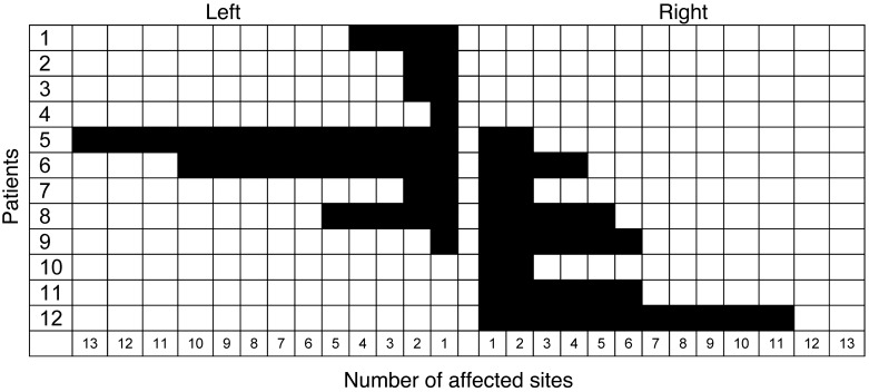
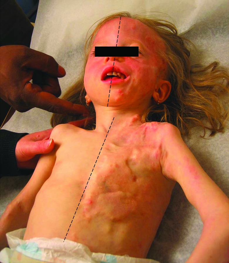
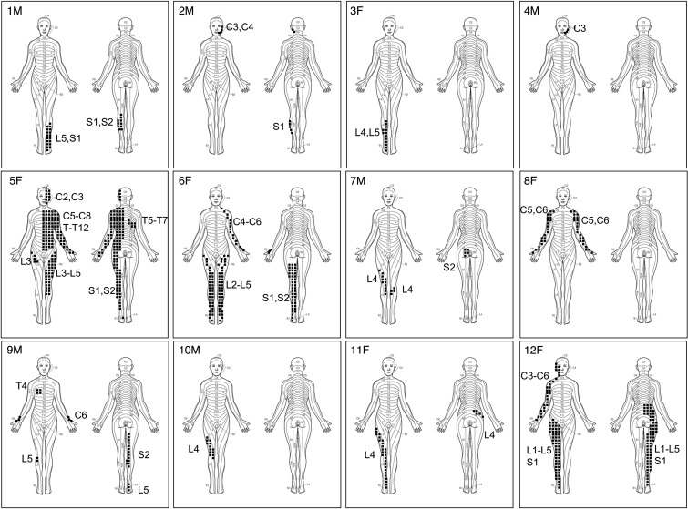
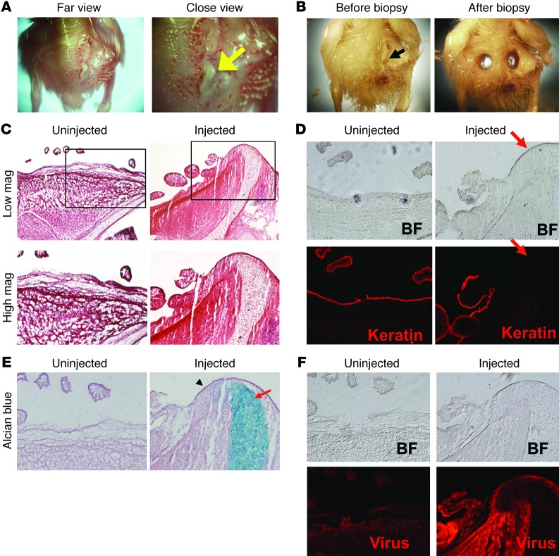
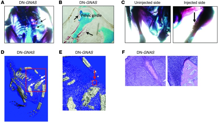
Progressive osseous heteroplasia (POH) is a rare developmental disorder of heterotopic ossification (HO) caused by heterozygous inactivating germline mutations in the paternal allele of the GNAS gene. Interestingly, POH lesions have a bewildering mosaic distribution. Using clinical, radiographic, and photographic documentation, we found that most of the 12 individuals studied had a lesional bias toward one side or the other, even showing exclusive sidedness. Most strikingly, all had a dermomyotomal distribution of HO lesions. We hypothesized that somatic mutations in a progenitor cell of somitic origin may act on a background of germline haploinsufficiency to cause loss of heterozygosity at the GNAS locus and lead to the unilateral distribution of POH lesions. Taking advantage of the chick system, we examined our hypothesis by mimicking loss of heterozygosity of GNAS expression using dominant-negative GNAS that was introduced into a subset of chick somites, the progenitors that give rise to dermis and muscle. We observed rapid ectopic cartilage and bone induction at the axial and lateral positions in a unilateral distribution corresponding to the injected somites, which suggests that blocking GNAS activity in a targeted population of progenitor cells can lead to mosaic ectopic ossification reminiscent of that seen in POH.


Figures

Figure 1
Figure 1. Lateralization of POH lesions.
The number of affected anatomic sites was scored as the presence of HO in any of 30 regional body areas. Patients are ranked in order of percent sidedness, from the leftmost to the rightmost. Note the exclusive lateralization in 7 of the 12 patients (83%) examined.
Figure 2
Figure 2. Sidedness in POH.
Left-sided lesions of HO on the head, face, chest, and abdomen of patient 5 did not cross the midline.
Figure 3
Figure 3. Dermomyotomal distribution of HO in POH.
Stippled areas depict lesion formation along the indicated dermomyotomes of patients 1–12. F, female; M, male.
Figure 4
Figure 4. Ectopic expression of DN-GNAS increased mineralization of adipose-derived stromal cells.
(A) RT-PCR analysis of PPARG and ALPL in adipose-derived stromal cells after culturing for 5, 7, and 14 days in DMEM with 10% FBS. Data represent mean ± SD. (B) ALPL activity assay in adipose-derived stromal cells after 5 days of culture. (C) Alizarin red staining of adipose-derived stromal cells after 14 days of culture. Original magnification, ×10 (B and C).
Figure 5
Figure 5. DN-GNAS viral expression in the somites induces skin protrusions.
(A) “Hardened” outgrowth (arrow) in the lumbosacral region of the embryo on the DN-GNAS–injected side. Note the shiny appearance of the outgrowth, which was devoid of feather structures. (B) The outgrowth on the DN-GNAS–injected side (arrow) was excised for histological analysis. (C) Histology showing DN-GNAS–induced extension of the pelvic girdle through the dermis; (D) absence of the epithelial cell marker pan-keratin (arrow) in areas near the outgrowth; (E) and extension of cartilage elements of the pelvic girdle (Alcian blue [arrow]; the area immediately left of the cartilage element was devoid of staining for Alcian blue as well as pan-keratin [arrowhead]). (F) Immunohistochemistry detection of viral coat protein GAG. Original magnification, ×1 (A and B); ×10 (C, low mag); ×20 (C, high mag, and DF).
Figure 6
Figure 6. DN-GNAS expression in the somites causes skeletal abnormalities.
(A) Whole-mount Alcian blue images of retrovirus GFP and DN-GNAS coinjected embryos. GFP encoding avian retrovirus vector RCAS-B was coinjected with DN-GNAS encoding retrovirus RCAS-A to allow visualization of viral expression. Thickening of the cartilaginous ribs (arrow) as well as the pelvic girdle (arrowhead) was observed. DN-GNAS virus injection alone yielded the same results as DN-GNAS coinjection with GFP virus. (B) Alcian blue and Alizarin red staining showing a cartilage nodule in the forelimb of DN-GNAS–injected side (arrow), while the contralateral side showed uniform mineralization. (C) An ectopic cartilage element was present in the hindlimb of the DN-GNAS–injected side (arrow) of a representative embryo. Note that the mineralized femur was also thicker on the injected side (asterisk) than on the contralateral side. (D) μCT analysis of GFP-injected embryos, indicating normal mineralization. (E) A DN-GNAS–injected embryo showing altered mineralization during endochondral bone formation in the ribs (arrowheads). (F) A DN-GNAS–injected embryo showing thickened mineralized ribs after endochondral bone formation (arrows). Original magnification, ×0.71 (A, low mag); ×2.0 (A, high mag); ×1.6 (B); ×1.25 (C).
Figure 7
Figure 7. DN-GNAS expression in somites induces HO independently of the skeletal elements.
(A) Alcian blue and Alizarin red staining showing patches of Alizarin red–positive tissues in between the ribs and the pelvic girdle (arrows). (B) Alcian blue and Alizarin red staining of sections of the protruding pelvic girdle. Note islands of Alizarin red stained areas outside the cartilaginous pelvic girdle (arrows). (C) Alcian blue and Alizarin red staining showing patches of mineralized tissues in the musculature of the hindlimb (arrow). (D) μCT analysis showing HO (arrows). (E) Enlarged images showing islands of mineralized zones. (F) H&E staining of heterotopic bone from the boxed region (red arrow) in E. Original magnification, ×1.6 (A and C); ×20 (B); ×10 (F).

References

    1. Shore EM, Kaplan FS. Inherited human diseases of heterotopic bone formation. Nat Rev Rheumatol. 2010;6(9):518–527. doi: 10.1038/nrrheum.2010.122. - DOI - PMC - PubMed
    1. Kaplan FS, et al. Progressive osseous heteroplasia: a distinct developmental disorder of heterotopic ossification. Two new case reports and follow-up of three previously reported cases. J Bone Joint Surg Am. 1994;76(3):425–436. - PubMed
    1. Shore EM, et al. Paternally inherited inactivating mutations of the GNAS1 gene in progressive osseous heteroplasia. N Engl J Med. 2002;346(2):99–106. doi: 10.1056/NEJMoa011262. - DOI - PubMed
    1. Adegbite NS, Xu M, Kaplan FS, Shore EM, Pignolo RJ. Diagnostic and mutational spectrum of progressive osseous heteroplasia (POH) and other forms of GNAS-based heterotopic ossification. Am J Med Genet A. 2008;146A(14):1788–1796. - PMC - PubMed
    1. Kaplan FS, Shore EM. Progressive osseous heteroplasia. J Bone Miner Res. 2000;15(11):2084–2094. doi: 10.1359/jbmr.2000.15.11.2084. - DOI - PubMed

Publication types

Substances