. 2013 Jul 17;5(194):194ra92.
doi: 10.1126/scitranslmed.3005795.

Overcoming preexisting humoral immunity to AAV using capsid decoys

Affiliations

Overcoming preexisting humoral immunity to AAV using capsid decoys

Federico Mingozzi et al. Sci Transl Med. .

Abstract

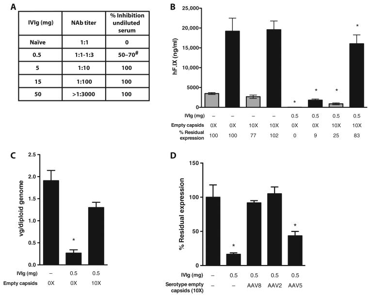
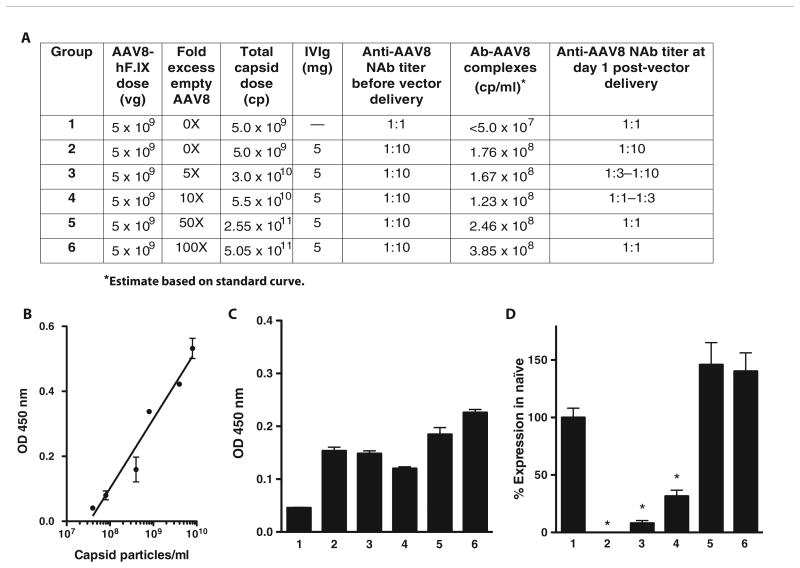
Adeno-associated virus (AAV) vectors delivered through the systemic circulation successfully transduce various target tissues in animal models. However, similar attempts in humans have been hampered by the high prevalence of neutralizing antibodies to AAV, which completely block vector transduction. We show in both mouse and nonhuman primate models that addition of empty capsid to the final vector formulation can, in a dose-dependent manner, adsorb these antibodies, even at high titers, thus overcoming their inhibitory effect. To further enhance the safety of the approach, we mutated the receptor binding site of AAV2 to generate an empty capsid mutant that can adsorb antibodies but cannot enter a target cell. Our work suggests that optimizing the ratio of full/empty capsids in the final formulation of vector, based on a patient's anti-AAV titers, will maximize the efficacy of gene transfer after systemic vector delivery.


Conflict of interest statement

Competing interests: F.M., X.M.A., G.M.P., J.F.W., and K.A.H. are inventors in a pending patent on the formulation of AAV vectors for highly efficient transgene delivery (U.S. Provisional Patent Application Serial No. 61/682,019). J.F.W. and K.A.H. have consulted for companies developing AAV-based gene therapeutics. All other authors declare no competing interests.

Figures

Fig. 1
Fig. 1. AAV empty capsids prevent vector neutralization by anti-AAV NAbs in vivo in mice
(A) Anti-AAV8 antibody analysis in naïve mice injected intraperitoneally with PBS only or mice passively immunized intraperitoneally with 0.5, 5, 15, or 50 mg of IVIg (n = 5 per group). The analysis was performed 24 hours after immunization. “NAb titer” represents the reciprocal serum dilution at which <50% inhibition of the reporter vector signal was measured in the NAb assay. “% Inhibition undiluted serum” represents the inhibition of the reporter gene signal observed when undiluted test serum was mixed with an equal volume of a solution containing the reporter vector. #, range. (B) Male C57BL/6 mice (n = 5 per group) were injected intraperitoneally with 0.5 mg of IVIg (resulting in a NAb titer of 1:1 to 1:3) or injected with PBS (−). After 24 hours, animals received either 1 × 10⁹ vg (gray bars) or 5 × 10⁹ vg (black bars) of an AAV8-F.IX vector alone (0X) or formulated with a 10X excess of AAV8 empty capsids [1 × 10¹⁰ capsid particles (gray bars) or 5 × 10¹⁰ capsid particles (black bars)]. hF.IX transgene levels in plasma at week 8 after vector delivery are shown as averages; error bars, SEM. *P < 0.05 versus naïve mice (two-tailed, unpaired t test). (C) Vector gene copy number in mouse livers collected at week 12 after AAV8-F.IX gene transfer at a vector dose of 5 × 10⁹ vg per animal. Results are shown as average copy number of five livers. Error bars, SEM. Experimental groups are the same as shown in (B). *P < 0.05 versus naïve mice (two-tailed, unpaired t test). (D) Empty capsids from alternate AAV serotypes protect AAV8-F.IX vector from NAb neutralization. Male C57BL/6 mice (n = 5 per group) were passively immunized with 0.5 mg of IVIg or injected with PBS (−) intraperitoneally. Twenty-four hours after, animals received 5 × 10⁹ vg of an AAV8-F.IX vector alone (−) or formulated with a 10X excess of AAV8, AAV2, or AAV5 empty capsids. The percent residual expression is calculated relative to the F.IX transgene plasma levels in naïve animals receiving the AAV-F.IX vector only. *P < 0.05 versus naïve mice (two-tailed, unpaired t test).
Fig. 2
Fig. 2. Addition of empty capsid in defined amounts based on pretreatment NAb titers can overcome transduction barrier posed by anti-AAV antibodies
(A to D) Percent residual F.IX transgene expression in mice immunized with 0.5 mg (A), 5.0 mg (B), 15 mg (C), or 50 mg (D) of IVIg and injected 24 hours later with 5 × 10⁹ vg per mouse of AAV8-F.IX vector alone or vector formulated in excess empty capsids as indicated in the x axes. The percent residual expression is calculated relative to the F.IX transgene product plasma levels in naïve animals (No IVIg) receiving the AAV-F.IX vector alone. Results are shown as average residual expression (n = 5 animals per group); error bars represent the SEM. *P < 0.05 versus naïve mice (two-tailed, unpaired t test).
Fig. 3
Fig. 3. Detection of antibody-capsid immune complexes after vector AAV delivery
(A) Outline of experimental groups and summary of results. Mice (n = 5 per group) received 5 × 10⁹ vg of AAV8-F.IX 24 hours after the administration of 5 mg of IVIg (groups 2 to 6) or saline (group 1, naïve control) intraperitoneally. Vector was formulated in increasing amounts of AAV8 empty capsids, from 0X to 100X. Twenty-four hours after vector administration, plasma was collected and assayed for immune complexes and anti-AAV8 NAb titers. (B) Standard curve used for the quantification of immune complexes prepared with known amounts of AAV8 capsid particles incubated with IVIg for 1 hour at 37°C. Each sample was tested in duplicate. (C) Detection of immune complexes after AAV vector delivery. An increasing amount of immune complexes is detectable at increasing capsid doses. Each sample was tested in triplicate. (D) F.IX transgene expression levels 4 weeks after vector administration. At the same AAV8-F.IX vector dose, increasing amounts of F.IX expression are measured at higher empty capsid doses and when circulating antibody-capsid immune complexes are detected. F.IX levels are expressed as percent of F.IX levels in naïve mice. *P < 0.05 versus group 1 (two-tailed, unpaired t test). vg, vector genomes; cp, capsid particles; OD, optical density. Error bars represent the SEM.
Fig. 4
Fig. 4. Safety and efficacy of coadministration of AAV8-F.IX vectors and AAV8 empty capsids in rhesus macaques
(A) Plasma hF.IX levels in male rhesus macaques receiving an AAV8-hF.IX vector at doses of 1 × 10¹² or 2 × 10¹² vg/kg. Animals received the AAV8-hF.IX vector in PBS (1001, 1002, 3001, 3002, and 5001) or formulated in excess of empty AAV8 capsids (2001, 4001, 4002, and 6001). Results are shown as average hF.IX levels in plasma at weeks 4 to 12 (weekly measurements). Error bars represent the SEM. Statistical analysis of hF.IX expression levels was performed with two-tailed, unpaired t test. (B) Antibody-capsid circulating immune complexes measured after vector administration. Serum samples were collected at baseline and at days 1, 2, 14, 21, and 28 after vector delivery and assayed for the presence of immune complexes. Results are shown as optical densities (OD). Error bars represent the SD of the average of triplicate testing. When detectable, immune complexes are present only briefly after vector delivery.
Fig. 5
Fig. 5. The AAV585/8 capsid mutant effectively protects AAV vector neutralization by NAbs and does not infect cells
(A) Male C57BL/6 mice (n = 5 per group) were passively immunized with 0.5 mg of IVIg (+) or injected with PBS (−). Twenty-four hours later, animals received 5 × 10⁹ vg of an AAV8-F.IX vector alone (0X) or formulated with a 10X or 1000X excess of AAV585/8 empty capsids (10X AAV585/8 and 1000X AAV585/8, respectively) or 1000X excess of AAV8 empty capsids (1000X AAV8). Results are shown as percent of expression of F.IX in naïve mice. Error bars represent the SEM. *P < 0.05 versus vector alone in naïve animals (two-tailed, unpaired t test). (B) Naïve (−) or passively immunized mice (+) were given a 5 × 10⁹ vg (AAVdj) or 5 × 10¹⁰ vg (AAV2, AAV5, and AAV6) of vectors expressing F.IX. Vector was given alone (black bars) or formulated in a 10X excess of AAV585/8 empty capsids (red bars). Results are shown as percent of expression of F.IX in naïve mice. Error bars represent the SEM. *P < 0.05 versus vector alone in naïve animals (two-tailed, unpaired t test). (C to H) AAV2 (C to E) and AAV585/8 (F to H) vector internalization in HHL5 human hepatocytes. Cells were treated for 4 hours at a multiplicity of infection (MOI) of 1 × 10⁵ with the AAV vectors and subsequently intracellularly stained with the anti-AAV2 monoclonal antibody A20, which binds equally both AAV2 and AAV585/8 (fig. S5E). The gates in (C) and (F) indicate the percent of AAV signal localized intracellularly; the gates in (D) and (G) indicate the percent of the AAV signal localized in early endosomes; the gates in (E) and (H) indicate the percent of AAV signal localized in the nuclei. (I) CTL assay with HHL5 human hepatocytes. Target cells were transduced overnight at increasing MOIs of AAV2 (black line) or AAV585/8 (red line) vectors and incubated with human leukocyte antigen–matched AAV-specific effector cells at an effector/target ratio of 10:1. Percent cytotoxicity is calculated after background subtraction relative to the maximum cell lysis obtained by treating targets with Triton X-100. Error bars, SEM of triplicate readings.

References

    1. Nathwani AC, Tuddenham EG, Rangarajan S, Rosales C, McIntosh J, Linch DC, Chowdary P, Riddell A, Pie AJ, Harrington C, O'Beirne J, Smith K, Pasi J, Glader B, Rustagi P, Ng CY, Kay MA, Zhou J, Spence Y, Morton CL, Allay J, Coleman J, Sleep S, Cunningham JM, Srivastava D, Basner-Tschakarjan E, Mingozzi F, High KA, Gray JT, Reiss UM, Nienhuis AW, Davidoff AM. Adenovirus-associated virus vector–mediated gene transfer in hemophilia B. N Engl J Med. 2011;365:2357–2365. - PMC - PubMed
    1. Manno CS, Pierce GF, Arruda VR, Glader B, Ragni M, Rasko JJ, Ozelo MC, Hoots K, Blatt P, Konkle B, Dake M, Kaye R, Razavi M, Zajko A, Zehnder J, Rustagi PK, Nakai H, Chew A, Leonard D, Wright JF, Lessard RR, Sommer JM, Tigges M, Sabatino D, Luk A, Jiang H, Mingozzi F, Couto L, Ertl HC, High KA, Kay MA. Successful transduction of liver in hemophilia by AAV-factor IX and limitations imposed by the host immune response. Nat Med. 2006;12:342–347. - PubMed
    1. Li H, Haurigot V, Doyon Y, Li T, Wong SY, Bhagwat AS, Malani N, Anguela XM, Sharma R, Ivanciu L, Murphy SL, Finn JD, Khazi FR, Zhou S, Paschon DE, Rebar EJ, Bushman FD, Gregory PD, Holmes MC, High KA. In vivo genome editing restores haemostasis in a mouse model of haemophilia. Nature. 2011;475:217–221. - PMC - PubMed
    1. Arruda VR, Stedman HH, Haurigot V, Buchlis G, Baila S, Favaro P, Chen Y, Franck HG, Zhou S, Wright JF, Couto LB, Jiang H, Pierce GF, Bellinger DA, Mingozzi F, Nichols TC, High KA. Peripheral transvenular delivery of adeno-associated viral vectors to skeletal muscle as a novel therapy for hemophilia B. Blood. 2010;115:4678–4688. - PMC - PubMed
    1. Boutin S, Monteilhet V, Veron P, Leborgne C, Benveniste O, Montus MF, Masurier C. Prevalence of serum IgG and neutralizing factors against adeno-associated virus (AAV) types 1, 2, 5, 6, 8, and 9 in the healthy population: Implications for gene therapy using AAV vectors. Hum Gene Ther. 2010;21:704–712. - PubMed

Publication types