. 2013 Oct 3;122(14):2338-45.
doi: 10.1182/blood-2012-12-470971. Epub 2013 Jul 17.

Mouse extraembryonic arterial vessels harbor precursors capable of maturing into definitive HSCs

Affiliations

Mouse extraembryonic arterial vessels harbor precursors capable of maturing into definitive HSCs

Sabrina Gordon-Keylock et al. Blood. .

Abstract

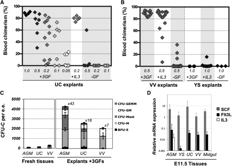
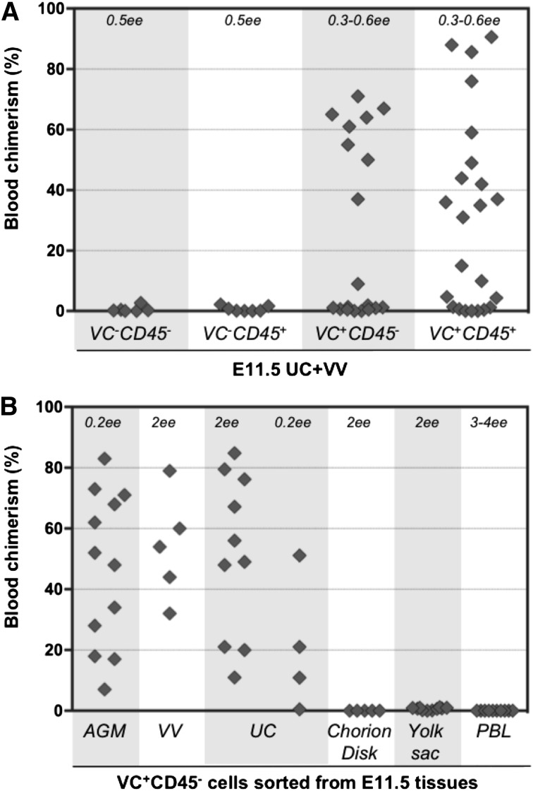
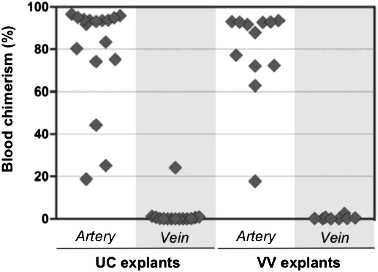
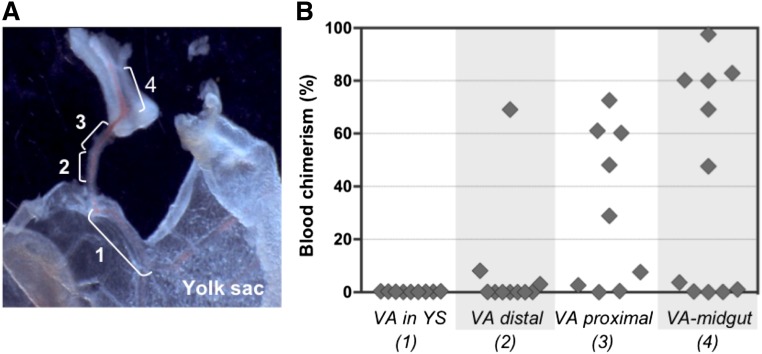
During mouse development, definitive hematopoietic stem cells (dHSCs) emerge by late E10.5 to E11 in several hematopoietic sites. Of them, the aorta-gonad-mesonephros (AGM) region drew particular attention owing to its capacity to autonomously initiate and expand dHSCs in culture, indicating its key role in HSC development. The dorsal aorta contains characteristic hematopoietic clusters and is the initial site of dHSC emergence, where they mature through vascular endothelial (VE)-cadherin(+)CD45(-)CD41(low) (type 1 pre-HSCs) and VE-cadherin(+)CD45(+) (type 2 pre-HSCs) intermediates. Although dHSCs were also found in other embryonic niches (placenta, yolk sac, and extraembryonic vessels), attempts to detect their HSC initiating potential have been unsuccessful to date. Extraembryonic arterial vessels contain hematopoietic clusters, suggesting that they develop HSCs, but functional evidence for this has been lacking. Here we show that umbilical cord and vitelline arteries (VAs), but not veins, contain pre-HSCs capable of maturing into dHSCs in the presence of exogenous interleukin 3, although in fewer numbers than the AGM region, and that pre-HSC activity in VAs increases with proximity to the embryo proper. Our functional data strongly suggest that extraembryonic arteries can actively contribute to adult hematopoiesis.


Figures

Figure 1
Figure 1
Definitive HSCs in E11.5 UC and VV are enriched in the VE-cad+CD45+ fraction. (A) UC and VV were pooled and sorted on the basis of VE-cadherin and CD45 expression. (B) Four sorted cell populations were transplanted directly into irradiated adult recipients (8 e.e. per mouse), and the level of donor-derived engraftment in PBL was assessed 13 to 14 weeks posttransplantation (n = 6 independent experiments).
Figure 2
Figure 2
Pre-HSC potential is revealed in explant cultures of UC and VV. (A-B) Direct transplants. E11.5 UC or VV were separated on dissection and transplanted individually into irradiated recipients, and the level of donor-derived PBL chimerism (%) was determined 14 weeks posttransplantation. Only 5 out of 30 UCs transplanted into recipients and 5 out of 22 VV recipients were repopulated. (C-D) Transplantation of explant cultures. UC or VV explants were cultured for 4 days with GF (IL-3 + SCF + Flt3l) and transplanted separately into irradiated mice as follows: 1 UC explant per 2 recipients (8 explants in total) and 0.2 to 0.5 VV explants per 1 recipient. Note that after culture all UC and VV explants contained HSC activity. M, recipient mouse.
Figure 3
Figure 3
Dissection of extraembryonic vessels. (A) E11.5 wild-type embryo, showing position of VV and UC. (B) Dissection of UC. Separation of artery and vein is shown. (C) Dissection of VV. YS and midgut loop were removed. White dotted lines indicate points of dissection to isolate vessels. Original magnification ×8 (A), ×40 (B), and ×32 (C). CD, chorion disk; D, distal; Em, embryo proper; FL, fetal liver; P, proximal; PL, placenta.
Figure 4
Figure 4
Expansion of HSC and progenitor numbers in the presence of GFs. (A) In the absence of GFs, HSCs are only maintained in UC explants but show 56-fold increase with GFs (IL-3 + SCF + Flt3L). Addition of IL-3 alone was found to be sufficient to support HSC expansion; 0.05 to 1 e.e. was injected per recipient, as indicated, and engraftment was assessed 12 to 14 weeks posttransplantation in PBL. (B) VV or YSs were cultured for 4 days in the presence of the 3 GFs, with IL-3 only, or without GFs and injected into irradiated recipients (at 0.5 VV per mouse and 1 YS per mouse). Engraftment was assessed 12 to 14 weeks posttransplantation in PBL (n = 5 independent experiments). (C) CFU-C progenitor numbers increase after 4 days of explant culture in the presence of 3 GFs (n = 2-5 independent experiments). Values refer to fold increase in CFU-C in explants compared with their uncultured counterparts. (D) Levels of IL-3, SCF, and Flt3l expression were assessed by qRT-PCR relative to expression of the TBP housekeeping gene (n = 3-8 independent RNA preparations, representing up to 10 pooled tissues each).
Figure 5
Figure 5
Type 1 pre-HSCs are present in UC and VV. (A) Four populations sorted based on VE-cadherin and CD45 expression were coaggregated with OP9 cells and transplanted after 4 days of culture (with IL-3 + SCF + Flt3l). The level (%) of donor engraftment in PBL was assessed 14 weeks after transplantation (n = 7 independent experiments). HSC activity correlated with VE-cad+CD45+ and VE-cad+CD45 fractions. (B) Type 1 pre-HSCs (VE-cad+CD45) from different embryonic tissues were coaggregated with OP9 cells and transplanted after 4 days of culture. Note that VV and UC contain type 1 pre-HSCs.
Figure 6
Figure 6
Pre-HSC activity is associated with extraembryonic arteries, but not veins. Arteries and veins subdissected from UC and VV were cultured separately for 4 days with GFs (IL-3 + SCF + Flt3l) and transplanted into irradiated recipients (1 e.e. per mouse). Donor engraftment was assessed 12 to 14 weeks posttransplantation (n = 5 independent experiments).
Figure 7
Figure 7
Proximal-distal gradient of pre-HSC activity in the VA. (A) Subdissection scheme of the VA: 1, portion of VA associated with YS; 2, distal VA portion; 3, proximal VA portion; 4, portion of VA associated with midgut. Original magnification ×32. (B) VA subsections were transplanted into irradiated recipients after 4 days of culture (1 e.e. per mouse). Donor engraftment was assessed 14 weeks posttransplantation (n = 2 independent experiments).

Similar articles

Cited by

References

    1. Zeigler BM, Sugiyama D, Chen M, Guo Y, Downs KM, Speck NA. The allantois and chorion, when isolated before circulation or chorio-allantoic fusion, have hematopoietic potential. Development. 2006;133(21):4183–4192. - PubMed
    1. Corbel C, Salaün J, Belo-Diabangouaya P, Dieterlen-Lièvre F. Hematopoietic potential of the pre-fusion allantois. Dev Biol. 2007;301(2):478–488. - PubMed
    1. Palis J, Robertson S, Kennedy M, Wall C, Keller G. Development of erythroid and myeloid progenitors in the yolk sac and embryo proper of the mouse. Development. 1999;126(22):5073–5084. - PubMed
    1. Cumano A, Ferraz JC, Klaine M, Di Santo JP, Godin I. Intraembryonic, but not yolk sac hematopoietic precursors, isolated before circulation, provide long-term multilineage reconstitution. Immunity. 2001;15(3):477–485. - PubMed
    1. Medvinsky A, Dzierzak E. Definitive hematopoiesis is autonomously initiated by the AGM region. Cell. 1996;86(6):897–906. - PubMed

Publication types