. 2013 Aug 29;500(7464):603-7.
doi: 10.1038/nature12321. Epub 2013 Jul 17.

Mitotic trigger waves and the spatial coordination of the Xenopus cell cycle

Affiliations

Mitotic trigger waves and the spatial coordination of the Xenopus cell cycle

Jeremy B Chang et al. Nature. .

Abstract

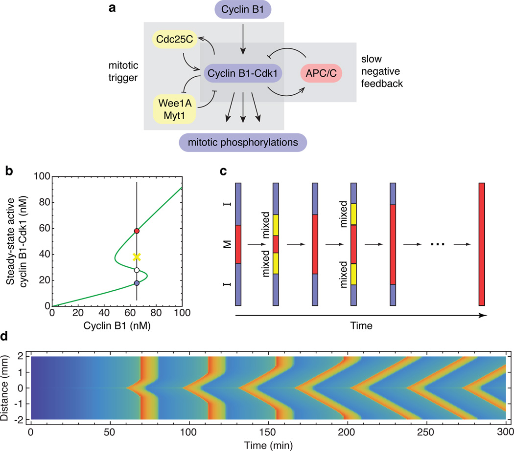
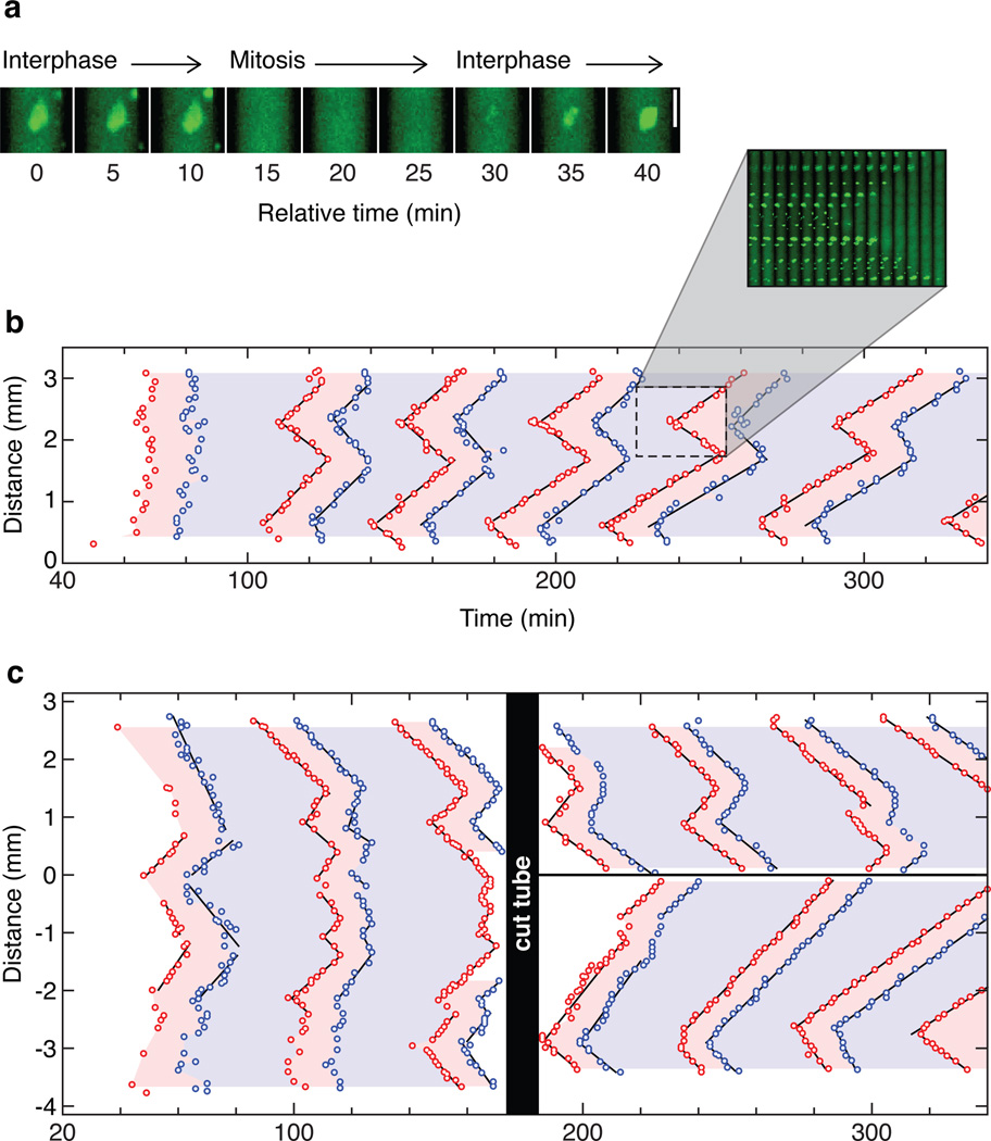
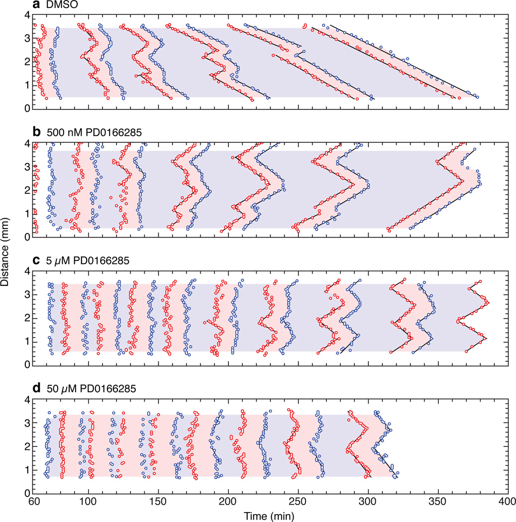
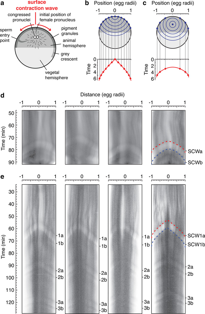
Despite the large size of the Xenopus laevis egg (approximately 1.2 mm diameter), a fertilized egg rapidly proceeds through mitosis in a spatially coordinated fashion. Mitosis is initiated by a bistable system of regulatory proteins centred on Cdk1 (refs 1, 2), raising the possibility that this spatial coordination could be achieved through trigger waves of Cdk1 activity. Using an extract system that performs cell cycles in vitro, here we show that mitosis does spread through Xenopus cytoplasm via trigger waves, propagating at a linear speed of approximately 60 µm min(-1). Perturbing the feedback loops that give rise to the bistability of Cdk1 changes the speed and dynamics of the waves. Time-lapse imaging of intact eggs argues that trigger waves of Cdk1 activation are responsible for surface contraction waves, ripples in the cell cortex that precede cytokinesis. These findings indicate that Cdk1 trigger waves help ensure the spatiotemporal coordination of mitosis in large eggs. Trigger waves may be an important general mechanism for coordinating biochemical events over large distances.


Figures

Figure 1
Figure 1. Trigger waves in Cdk1 activation
a, Schematic view of the Cdk1-APC/C circuit. b, Modeled steady-state response of Cdk1 to cyclin B1, with parameters based on experimental studies,. At intermediate concentrations of cyclin, the response is bistable. The stable low (interphase) and high (mitotic) Cdk1 activity steady-states corresponding to one such cyclin concentration (65 nM) are shown as blue and red circles, respectively. The white circle denotes the unstable steady state, and the yellow X denotes an intermediate level of Cdk1 activity that would be attracted to the mitotic (red) steady state. c, Schematic view of the propagation of Cdk1 activity up and down a one-dimensional tube through successive rounds of mixing and conversion. d, Modeled propagation of Cdk1 activity in a one-dimensional tube. It is assumed that cyclin B1 is synthesized at a uniform rate everywhere in the tube, but in a 5 µm region in the middle of the tube, the concentration of Cdc25C is 50% higher than in the rest of the tube, allowing Cdk1 to become activated earlier. Cyclin B1-Cdk1 activity is denoted by the color scale (blue is low, red is high). Numerical solution of the PDEs was carried out using Mathematica 9.0 (Wolfram) as described in the Supplementary Materials. See also Supplementary Fig. 1.
Figure 2
Figure 2. Rapid, linear propagation of mitotic entry and exit through Xenopus cytoplasm
a, An example of nuclear envelope breakdown and nuclear envelope reformation in an extract with added sperm chromatin and GFP-NLS. b, The timing of mitotic entrance and exit in a 3 mm section of a Teflon tube submerged in mineral oil. Each data point represents the time and position at which an individual nucleus underwent nuclear envelope breakdown (red points) or nuclear envelope re-formation (blue points). The pink and blue regions of the plot denote mitosis and interphase, respectively. Time is measured relative to when the extract was warmed to room temperature. The inset shows frames from the video in montage form. c, Trigger waves vs. phase waves. The tube was cut under mineral oil at 160 min. See also Supplementary Fig. 2.
Figure 3
Figure 3. The Wee1/Myt1 inhibitor PD0166285 accelerates the trigger waves
a–d, Mitotic entrance and exit waves in extracts treated with DMSO or one of three concentrations of PD0166285. See also Supplementary Fig. 3.
Figure 4
Figure 4. Surface contraction waves in intact Xenopus eggs
a, Schematic view of the anatomy of a fertilized egg just before the onset of mitosis. Adapted from ref. . b, c, Expected propagation of surface contraction waves if they were due to a spherical wave of Cdk1 activation spreading from a point source. An equation for the spreading of the waves (Eq S1) is derived in the Supplementary Materials. Panel B assumes the point source is at the animal pole. Panel C assumes the point source is halfway between the animal pole and the center of the cell. d–e, Kymographs depicting surface contraction waves, indicated by transitions from light to dark or dark to light, in four fertilized eggs (d) and four parthenogenetically-activated eggs (e). The red and blue dashed curves are fits of the experimental data to the Eq S1. See also Supplementary Fig. 4.

References

    1. Pomerening JR, Sontag ED, Ferrell JE., Jr Building a cell cycle oscillator: hysteresis and bistability in the activation of Cdc2. Nature Cell Biol. 2003;5:346–351. - PubMed
    1. Sha W, et al. Hysteresis drives cell-cycle transitions in Xenopus laevis egg extracts. Proc Natl Acad Sci U S A. 2003;100:975–980. - PMC - PubMed
    1. Novak B, Tyson JJ. Numerical analysis of a comprehensive model of M-phase control in Xenopus oocyte extracts and intact embryos. J Cell Sci. 1993;106:1153–1168. - PubMed
    1. Hara K. Cinematographic observation of "surface contraction waves" (SCW) during the early cleavage of axolotl eggs. Wilhelm Roux' Archiv. 1971;167:183–186. - PubMed
    1. Hara K, Tydeman P, Kirschner M. A cytoplasmic clock with the same period as the division cycle in Xenopus eggs. Proc Natl Acad Sci U S A. 1980;77:462–466. - PMC - PubMed

Publication types

MeSH terms