. 2013 Oct;14(7):427-33.
doi: 10.1038/gene.2013.36. Epub 2013 Jul 18.

Identification of a KIR antisense lncRNA expressed by progenitor cells

Affiliations

Identification of a KIR antisense lncRNA expressed by progenitor cells

P W Wright et al. Genes Immun. 2013 Oct.

Abstract

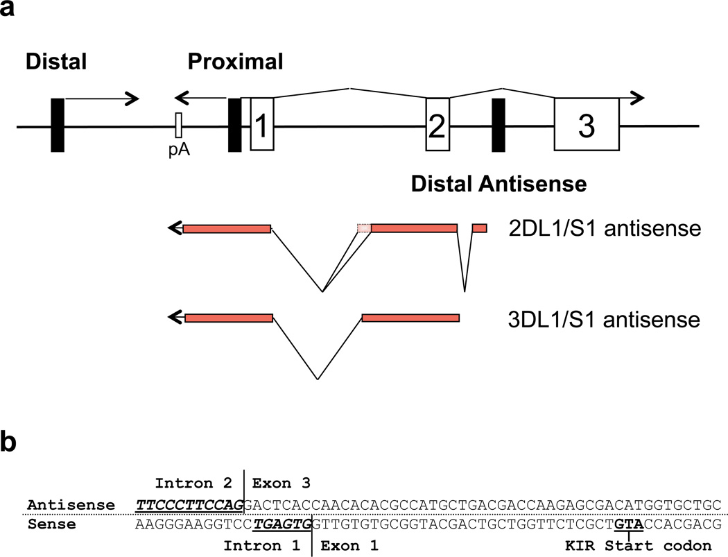
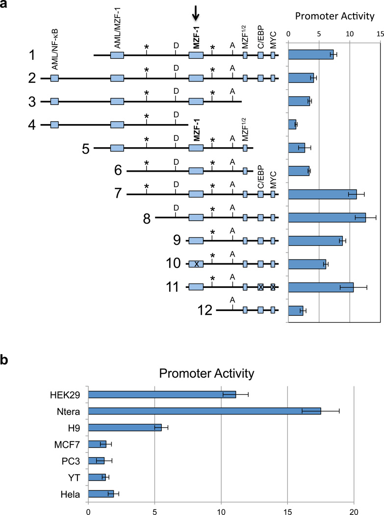
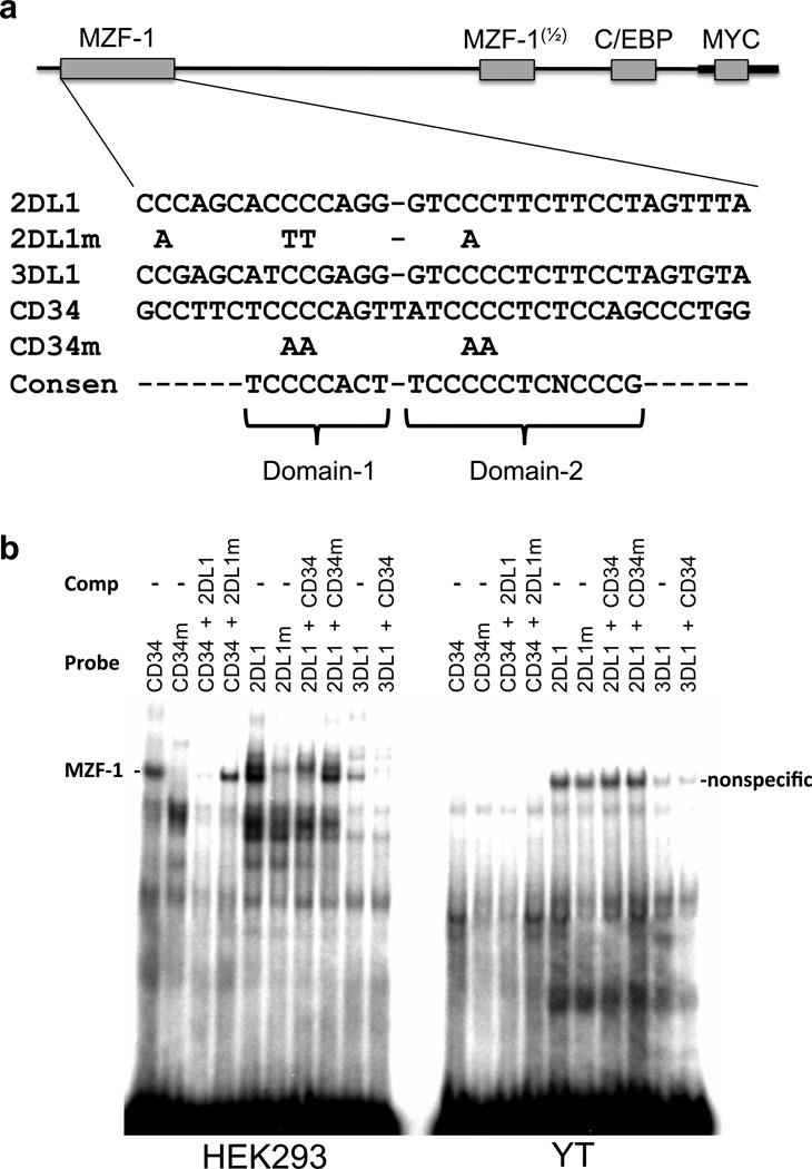
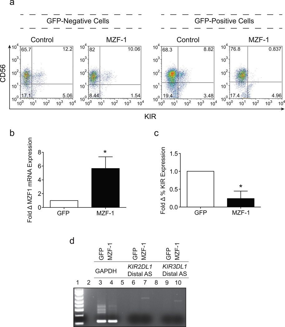
Human NK cells express cell surface class I MHC receptors (killer cell immunoglobulin-like receptor, KIR) in a probabilistic manner. Previous studies have shown that a distal promoter acts in conjunction with a proximal bidirectional promoter to control the selective activation of KIR genes. We report here the presence of an intron 2 promoter in several KIR genes that produce a spliced antisense transcript. This long noncoding RNA (lncRNA) transcript contains antisense sequence complementary to KIR-coding exons 1 and 2 as well as the proximal promoter region of the KIR genes. The antisense promoter contains myeloid zinc finger 1 (MZF-1)-binding sites, a transcription factor found in hematopoietic progenitors and myeloid precursors. The KIR antisense lncRNA was detected only in progenitor cells or pluripotent cell lines, suggesting a function that is specific for stem cells. Overexpression of MZF-1 in developing NK cells led to decreased KIR expression, consistent with a role for the KIR antisense lncRNA in silencing KIR gene expression early in development.


Conflict of interest statement

Conflict of interest

The authors declare no conflict of interest.

Figures

Figure 1
Figure 1
Identification of KIR distal antisense transcripts. (a) A schematic of the organization of the 5’ region of the KIR genes is shown. Black rectangles represent promoter elements, and numbered rectangles represent exons. Lines represent KIR transcripts with their orientation indicated by arrows. The exon structure of the KIR2DL1/S1 and KIR3DL1/S1 distal antisense transcripts is indicated below. The additional exon sequence found in the KIR2DL1 alternative transcript (GenBank: GQ422373) is indicated by the dotted rectangle. (b) The nucleotide sequence of the region containing the KIR2DL1 distal antisense intron 2/exon 3 splice junction and the exon 1/intron 1 splice junction of the KIR2DL1 coding transcript is shown. The antisense strand is shown on top with the complementary coding sense strand shown on the bottom. Consensus splice acceptor (antisense) and splice donor (sense) sequences are underlined in bold.
Figure 2
Figure 2
In vitro analysis of the KIR distal antisense promoter. (a) The left panel is a schematic representation of KIR2DL1 intron 2 fragments shown in an antisense orientation. Potential transcription factor binding sites are indicated by the labeled shaded rectangles. The central MZF-1 site is indicated by the bold arrow. Rectangles containing an “x” represent constructs wherein the respective site has been mutated. The position of the KIR2DL1 and KIR3DL1 distal antisense transcription start sites are indicated by vertical lines labeled with an asterisk. The MYC-binding site indicated at the 3’ end of the distal antisense promoter region overlaps with the exon 2/intron 2 junction on the sense strand. The boundaries of the first KIR2DL1 antisense intron are indicated by vertical lines labeled D (splice donor) and A (splice acceptor). The right panel shows the luciferase activity of pGL3 constructs containing the fragments depicted on the left. Constructs were transfected into HEK293 cells and relative luciferase activity was determined 48 hours post-transfection. Values represent the mean, and error bars indicate the standard deviation of at least 3 independent experiments. (b) Analysis of KIR2DL1 distal promoter activity (construct 7 in a) in various cell lines is shown. Values represent the mean and error bars indicate the standard deviation of at least 3 independent experiments.
Figure 3
Figure 3
EMSA analysis of the putative MZF-1 binding sites of KIR2DL1 and KIR3DL1. (a) A schematic of the core promoter fragment is shown, with the shaded rectangles indicating potential transcription factor binding sites. The expanded sequence underneath shows the oligonucleotide probes from the predicted KIR2DL1 and KIR3DL1 MZF-1 binding sites compared with the known MZF-1 binding site in the human CD34 gene. The consensus nucleotides for MZF-1 binding are shown below (Consen). Brackets indicate the sequences bound by the first and second DNA-binding domains of MZF-1. Nucleotide substitutions present in mutated KIR2DL1 (2DL1m) and CD34 (CD34m) probes are shown below the respective sequences. (b) EMSA analysis of HEK293 (left panel) and YT (right panel) nuclear extracts using the oligonucleotide probes shown in (a). Unlabeled oligonucleotides used for cold competition are indicated (Comp), as are the labeled probes (Probe). The position of a HEK293 cell-specific MZF-1 complex (MZF-1 -) is indicated next to the left panel, and a non-specific complex formed in YT cells is indicated next to the right panel (nonspecific).
Figure 4
Figure 4
Enforced expression of MZF-1 decreases KIR expression in developing NK cells. (a) FACS analysis of day 21 in vitro differentiated cord blood NK cells transduced with either a control GFP expressing lentiviral vector or a lentiviral vector expressing GFP-IRES-MZF-1. Cells gated on the GFP−ve subset are shown on the left, and cells gated on the GFP+ve subset are shown on the right. Cells were stained with a cocktail of APC-conjugated NCAM16.2 (CD56) and PE-conjugated DX9 (KIR3DL1), EB6 (KIR2DL1/S1) and GL183 (KIR2DL2/S2/L3). (b) The average fold increase in MZF-1 mRNA expression in cells over-expressing MZF-1 relative to control GFP cells calculated from 3 independent donors. Differences in MZF-1 mRNA expression were statistically significant (p=0.0189) as determined by two-tailed Student’s t test. (c) The average fold reduction of KIR expression in cells over-expressing MZF-1 relative to control GFP cells calculated from 3 independent donors. Differences in KIR expression were statistically significant (p=0.0274) as determined by two-tailed Student’s t test. (d) PCR analysis of GAPDH (Lanes 3 & 4), KIR2DL1 distal antisense transcript (Lanes 6 & 7) and KIR3DL1 distal antisense transcript (Lanes 9 & 10) in sorted GFP and MZF-1 over-expressing day 21 in vitro-derived NK cells. Data is representative of 3 donors tested.

References

    1. Trinchieri G. Biology of natural killer cells. Adv Immunol. 1989;47:187–376. - PMC - PubMed
    1. Lanier LL. NK cell recognition. Annu Rev Immunol. 2005;23:225–274. - PubMed
    1. Carrington M, Norman P. The KIR Gene Cluster. Bethesda, MD: National Library of Medicine (US), NCBI; 2001.
    1. Anderson SK, Ortaldo JR, McVicar DW. The ever-expanding Ly49 gene family: repertoire and signaling. Immunol Rev. 2001;181:79–89. - PubMed
    1. Parham P. MHC class I molecules and KIRs in human history, health and survival. Nat Rev Immunol. 2005;5:201–214. - PubMed

Publication types

MeSH terms