. 2013 Sep;24(17):2609-19.
doi: 10.1091/mbc.E13-02-0106. Epub 2013 Jul 17.

A complex of Cox4 and mitochondrial Hsp70 plays an important role in the assembly of the cytochrome c oxidase

Affiliations

A complex of Cox4 and mitochondrial Hsp70 plays an important role in the assembly of the cytochrome c oxidase

Lena Böttinger et al. Mol Biol Cell. 2013 Sep.

Abstract

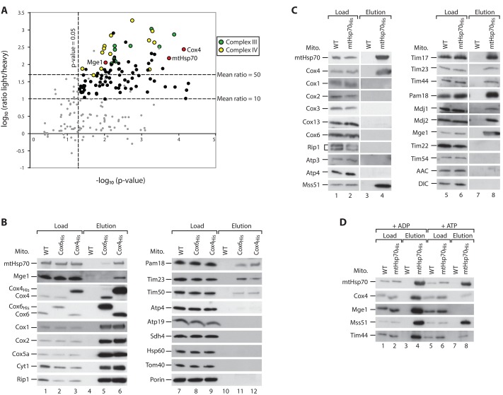
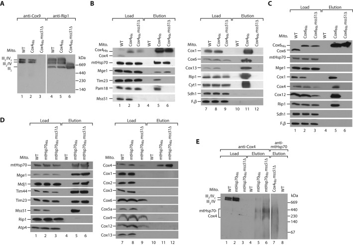
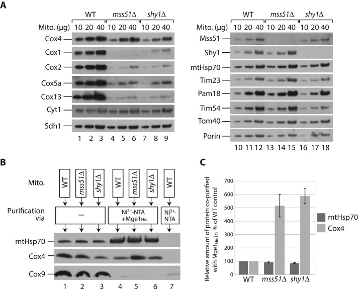
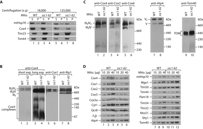
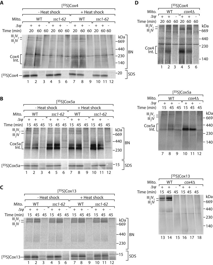
The formation of the mature cytochrome c oxidase (complex IV) involves the association of nuclear- and mitochondria-encoded subunits. The assembly of nuclear-encoded subunits like cytochrome c oxidase subunit 4 (Cox4) into the mature complex is poorly understood. Cox4 is crucial for the stability of complex IV. To find specific biogenesis factors, we analyze interaction partners of Cox4 by affinity purification and mass spectroscopy. Surprisingly, we identify a complex of Cox4, the mitochondrial Hsp70 (mtHsp70), and its nucleotide-exchange factor mitochondrial GrpE (Mge1). We generate a yeast mutant of mtHsp70 specifically impaired in the formation of this novel mtHsp70-Mge1-Cox4 complex. Strikingly, the assembly of Cox4 is strongly decreased in these mutant mitochondria. Because Cox4 is a key factor for the biogenesis of complex IV, we conclude that the mtHsp70-Mge1-Cox4 complex plays an important role in the formation of cytochrome c oxidase. Cox4 arrests at this chaperone complex in the absence of mature complex IV. Thus the mtHsp70-Cox4 complex likely serves as a novel delivery system to channel Cox4 into the assembly line when needed.


Figures

FIGURE 1:
FIGURE 1:
Cox4 interacts with mtHsp70 and Mge1 in an ATP-sensitive manner. (A) Yeast cells were labeled with heavy (H) isotopes [15N213C6]lysine/[15N413C6]arginine (wild type) or their corresponding light (L) [14N12C]-containing variants (Cox4His strain). Mitochondria were isolated, lysed, and subjected to affinity purification. Elution fractions were mixed and analyzed by quantitative mass spectrometry. The mean log10(L/H) ratio and −log10 p values of three independent experiments were determined and plotted against each other. Red dots, Cox4, mtHsp70, and Mge1; yellow, subunits of complex IV; green, subunits of complex III; black, other proteins; gray, unspecific proteins (see Supplemental Table S1). (B) Isolated mitochondria from wild-type (WT), Cox6His, and Cox4His yeast strains were lysed in digitonin and incubated with Ni2+-NTA agarose. Bound complexes were eluted with imidazole, and samples were analyzed by SDS–PAGE and Western blotting. Load, 3%; elution, 100%. (C, D) Wild-type (WT) and mtHsp70His mitochondria were analyzed as described for B. Where indicated, lysis and purification of the samples were performed in the presence of 5 mM MgCl2 and 5 mM ADP or ATP. Load, 3%; elution, 100%. AAC, ADP/ATP carrier; DIC, dicarboxylate carrier.
FIGURE 2:
FIGURE 2:
Cox4 binds independently of the respiratory chain supercomplexes to mtHsp70-Mge1. (A) Mitochondria from wild-type (WT), Cox4His, and Cox4His mss51Δ cells were lysed in digitonin and analyzed by blue native electrophoresis and Western blotting. III, complex III; IV, complex IV of mitochondrial respiratory chain. (B) Wild-type (WT), Cox4His, and Cox4His mss51Δ mitochondria were lysed in digitonin and incubated with Ni2+-NTA agarose. Bound complexes were eluted with imidazole, and samples were analyzed by SDS–PAGE and Western blotting. Load, 3%; elution, 100%. F1β, β-subunit of F1Fo-ATP synthase. (C) Wild-type (WT), Cox6His, and Cox6His mss51Δ mitochondria were analyzed as described in B. (D) Wild-type (WT), mtHsp70His, and mtHsp70His mss51Δ mitochondria were analyzed as described in B. (E) Wild-type (WT), mtHps70His, mtHsp70His mss51Δ, and Cox4His mss51Δ mitochondria were lysed in digitonin and incubated with Ni2+-NTA agarose. Bound complexes were eluted with imidazole, and samples were analyzed by blue native electrophoresis and Western blotting. Load, 3%; elution, 100%.
FIGURE 3:
FIGURE 3:
Cox4 arrests at mtHsp70-Mge1 in the absence of mature complex IV. (A) Wild-type (WT), mss51Δ, and shy1Δ mitochondria were lysed under denaturing conditions and analyzed by SDS–PAGE and Western blotting. (B) Wild-type (WT), mss51Δ, and shy1Δ mitochondria were lysed in digitonin and incubated with a noncoated or Mge1His-coated Ni2+-NTA matrix. Bound proteins were eluted with imidazole, and samples were analyzed by SDS–PAGE and Western blotting. Load (lanes 1–3), 3%; elution (lanes 4–7), 100%. (C) Quantification of three independent experiments like the one shown in B. Amount of coeluted Cox4 or mtHsp70, respectively, relative to the corresponding total mitochondrial protein content in the load fractions. Values for wild-type (WT) mitochondria were set to 100%. Error bars, SEM (n = 3).
FIGURE 4:
FIGURE 4:
A temperature-sensitive mutant of mtHsp70 (ssc1-62) is specifically defective in binding to Cox4. (A) Serial dilutions of wild-type (WT) and ssc1-62 yeast cells were spotted onto agar plates containing glucose (YPD) or glycerol (YPG) as carbon source. Plates were incubated at the indicated temperatures. (B) Isolated mitochondria from WT and ssc1-62 mutant cells were lysed in digitonin and incubated with anti-mtHsp70 antibodies coupled to protein A–Sepharose. Bound proteins were eluted with glycine, pH 2.5, and analyzed by SDS–PAGE and Western blotting. Load, 3%; elution, 100%. (C) Isolated mitochondria from WT and ssc1-62–mutant cells were lysed in digitonin and incubated with Mge1His-coated or uncoated Ni2+-NTA. Bound proteins were eluted with imidazole and analyzed by SDS–PAGE and Western blotting. Load, 3%; elution, 100%. (D) 35S-labeled Su9-DHFR (left) or F1β (right) was imported into WT and ssc1-62 mitochondria for the indicated time points with or without previous in vitro heat shock. In control reactions, the membrane potential (Δψ) was dissipated. Nonimported F1β precursor proteins were removed by treatment with proteinase K. Samples were subjected to SDS–PAGE and digital autoradiography. m, mature; p, precursor.
FIGURE 5:
FIGURE 5:
Respiratory chain supercomplexes are affected in ssc1-62 mitochondria. (A) Mitochondria from wild-type (WT) and ssc1-62 cells were lysed in digitonin and subjected to centrifugation at 18,000 × g or 125,000 × g, respectively. Supernatant (S) and pellet (P) fractions were separated and analyzed by SDS–PAGE and Western blotting. (B) Mitochondria isolated from WT and ssc1-62 cells grown under permissive conditions were lysed in digitonin and analyzed by blue native electrophoresis followed by Western blotting. III, complex III; IV, complex IV of mitochondrial respiratory chain. (C) WT and ssc1-62 cells were grown under permissive conditions and shifted to 37°C for 32 h. Mitochondria were isolated, and protein complexes were analyzed by blue native electrophoresis followed by Western blotting. TOM, translocase of outer membrane; V, complex V (F1Fo-ATP synthase). (D) Wild-type and ssc1-62 cells were grown under grown under permissive conditions and shifted to 37°C for 32 h. Isolated mitochondria were lysed under denaturing conditions and analyzed by SDS–PAGE and Western blotting.
FIGURE 6:
FIGURE 6:
mtHsp70-Mge1-Cox4 stimulates assembly of the respiratory supercomplexes. The 35S-labeled precursors of Cox4 (A), Cox5a (B), or Cox13 (C) were imported into wild-type (WT) or ssc1-62 mitochondria for the indicated time points with or without previous in vitro heat shock. In control reactions the membrane potential (Δψ) was dissipated. Samples were either lysed in digitonin and subjected to blue native electrophoresis (BN; top), or treated with proteinase K, denatured, and subjected to SDS–PAGE (bottom), followed by digital autoradiography. III, complex III; IV, complex IV of mitochondrial respiratory chain. (D) The 35S-labeled precursors of Cox4 (top), Cox5a (middle), or Cox13 (bottom) were imported into isolated mitochondria from WT and cox4Δ cells for the indicated time points in presence or absence of Δψ. Samples were lysed in digitonin and subjected to blue native electrophoresis followed by digital autoradiography. III, complex III; IV, complex IV of mitochondrial respiratory chain. Int., assembly intermediate.

References

    1. Baker MJ, Frazier AE, Gulbis JM, Ryan MT. Mitochondrial protein-import machinery: correlating structure with function. Trends Cell Biol. 2007;17:456–464. - PubMed
    1. Barrientos A, Zambrano A, Tzagoloff A. Mss51p and Cox14p jointly regulate mitochondrial Cox1p expression in Saccharomyces cerevisiae. EMBO J. 2004;23:3472–3482. - PMC - PubMed
    1. Böttinger L, Horvath SE, Kleinschroth T, Hunte C, Daum G, Pfanner N, Becker T. Phosphatidylethanolamine and cardiolipin differentially affect the stability of mitochondrial respiratory chain supercomplexes. J Mol Biol. 2012;423:677–686. - PMC - PubMed
    1. Brandner K, Mick DU, Frazier AE, Taylor RD, Meisinger C, Rehling P. Taz1, an outer mitochondrial membrane protein, affects stability and assembly of inner membrane protein complexes: implications for Barth syndrome. Mol Biol Cell. 2005;16:5202–5214. - PMC - PubMed
    1. Burbulla LF, et al. Dissecting the role of the mitochondrial chaperone mortalin in Parkinson's disease: functional impact of disease-related variants on mitochondrial homeostasis. Hum Mol Genet. 2010;19:4437–4452. - PMC - PubMed

Publication types

MeSH terms

LinkOut - more resources