

Three-dimensional evaluation of upper airway following rapid maxillary expansion: a CBCT study
- PMID: 23865825
- PMCID: PMC8673812
- DOI: 10.2319/012313-71.1
Three-dimensional evaluation of upper airway following rapid maxillary expansion: a CBCT study
Abstract
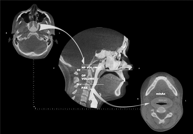
Objectives: To evaluate, by using cone beam computed tomography, the skeletal, dental, oropharyngeal (OP) airway volume, and nasal passage (NP) volume changes that occur after rapid maxillary expansion (RME).
Materials and methods: Two groups were selected, each with 35 patients (15 males, 20 females), an RME group (mean age, 14.02 ± 1.46 years) and a control group (mean age, 14.10 ± 1.44 years). The RME group consisted of patients with maxillary constriction who were treated with Hyrax palatal expanders, and the control group comprised age- and sex-matched patients who underwent comprehensive orthodontic treatment without the use of a rapid maxillary expander.
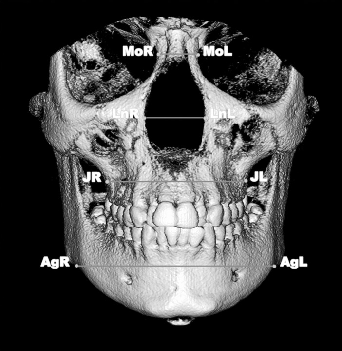
Results: All of the transverse skeletal (medial orbital width, lateral nasal width, maxillary width, and mandibular width) and interdental (intermolar, interpremolar, and intercanine) parameters were significantly enlarged in the RME group. A statistically significant increase in airway variables was seen in both groups between pretreatment (T0) and final records (T1). The mean increase of NP airway volume for the RME group (1719.9 ± 1510.7 mm(3)) was twofold compared with the control group (813.6 ± 1006.7 mm(3)), and no intergroup significant difference was found for the OP volume.
Conclusions: Rapid maxillary expansion creates a significant increase in nasal passage airway volume but no significant change in the oropharyngeal airway volume.
Figures
References
-
- Angell EC. Treatment of irregularities of the permanent or adult teeth. Dental Cosmos. 1860;1:540–544.
-
- Haas AJ. Rapid expansion of the maxillary dental arch and nasal cavity by opening the mid palatal suture. Angle Orthod. 1961;31:73–89.
-
- Haas AJ. The treatment of maxillary deficiency by opening the midpalatal suture. Angle Orthod. 1965;35:200–217. - PubMed
-
- Wertz RA. Changes in nasal airflow incident to rapid maxillary expansion. Angle Orthod. 1968;38:1–11. - PubMed
-
- Johal A, Conaghan C. Maxillary morphology in obstructive sleep apnea: a cephalometric and model study. Angle Orthod. 2004;74:648–656. - PubMed
Publication types
MeSH terms
LinkOut - more resources
Full Text Sources
Other Literature Sources
Miscellaneous
