. 2013 Jul 18:11:69.
doi: 10.1186/1477-7827-11-69.

Superovulation induces defective methylation in line-1 retrotransposon elements in blastocyst

Affiliations

Superovulation induces defective methylation in line-1 retrotransposon elements in blastocyst

Xing-Wei Liang et al. Reprod Biol Endocrinol. .

Abstract

Background: Series of epigenetic events happen during preimplantation development. Therefore assistant reproduction techniques (ART) have the potential to disrupt epigenetic regulation during embryo development. The purpose of this study was to investigate whether defects in methylation patterns in blastocyst due to superovulation originate from abnormal expression of Dnmts.

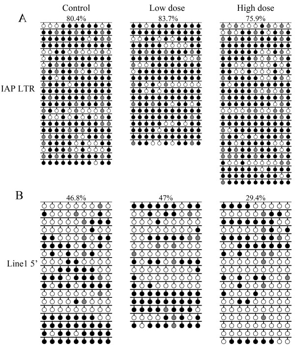
Methods: Low- (6 IU) and high- (10 IU) dosage of PMSG was used to stimulate the female mice. The metaphase II(MII) oocytes, zygotes and blastocyst stage embryos were collected. Global methylation and methylation at H3K9 in zygote, and methylation at repeated sequence Line 1 and IAP in blastocysts were assayed. In addition, expression of Dnmts was examined in oocytes and zygotes.

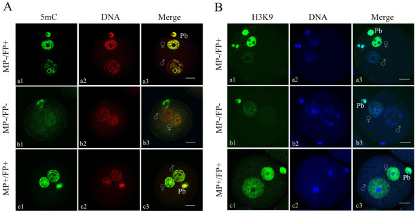
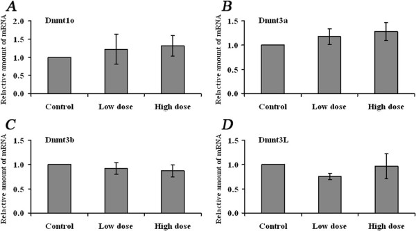
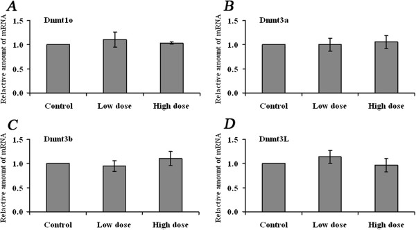
Results: Global DNA methylation and methylation at H3K9 in zygotes derived from females after low- or high-dosage hormone treatment were unaltered compared to that in controls. Moreover, DNA methylation at IAP in blastocysts was also unaffected, regardless of hormone dosage. In contrast, methylation at Line1 decreased when high-dose hormone was administered. Unexpectedly, expression of Dnmt3a, Dnmt3b, Dnmt3L as well as maintenance Dnmt1o in oocytes and zygotes was not disrupted.

Conclusions: The results suggest that defects in embryonic methylation patterns do not originate from the disruption of Dnmt expression.


Figures

Figure 1
Figure 1
Distribution patterns of global DNA methylation (A) and methylation at histone H3K9 (B) in pronucleus-stage mouse zygotes. The zygotes were stained with 5mC-specific antibody (green) and counterstained with DAPI (red), or stained for H3K9 dimethylation (green) and counterstained with Hoechst 33342 (blue). MP-/FP + indicates absence of a signal in male pronuclei and presence of a signal in female pronuclei; MP-/FP- indicates that both pronuclei had a very weak signal or lacked a signal; and MP+/FP + indicates both pronuclei had a signal. The polar body (Pb) was always positively stained. MP: male pronucleus; FP: female pronucleus; (+) positive signal; (−) negative or weak signal.
Figure 2
Figure 2
IAP and Line1 DNA methylation profiles in blastocysts. Sodium bisulfite sequencing was used to examine the DNA methylation patterns of (A) IAP LTR and (B) Line1 5′ end sequences in blastocyst DNA. Blastocysts were obtained from control, low- and high-dose hormone treatment groups. Target sequences were amplified, cloned and sequenced. Open circles, unmethylated CpGs; black circles, methylated CpGs; gray circles, not analyzable/mutated CpG site. Each row represents an individual sequenced clone. Only black and white circles were analyzed. The percentage of methylated CpGs (black circles/(black + white circles)) is indicated.
Figure 3
Figure 3
Expression of Dnmts in MII oocytes derived from control and superovulated females. qRT-PCR was applied to evaluate the mRNA abundance of A) Dnmt1o, B) Dnmt3a, C) Dnmt3b and D) Dnmt3L in MII oocytes derived from control, low- (6 IU) and high-dose hormone treatment groups, respectively. Two reference genes, Gapdh and Atp5, were used for each sample. Data were normalized to the average levels of the two reference genes. The fold change was calculated using the comparative CT method. Data are presented as mean ± SD.
Figure 4
Figure 4
Expression of Dnmts in zygotes derived from control and superovulated females. qRT-PCR was applied to evaluate the mRNA abundance of A) Dnmt1o, B) Dnmt3a, C) Dnmt3b and D) Dnmt3L in zygotes derived from control, low- (6 IU) and high-dose hormone treatment groups, respectively. Data were normalized to the average levels of Gapdh and Atp5 for each sample. The fold change was calculated using the comparative CT method. Data are presented as mean ± SD.

Similar articles

Cited by

References

    1. Roberts RM. Embryo culture conditions: what embryos like best. Endocrinology. 2005;146:2140–2041. doi: 10.1210/en.2005-0221. - DOI - PubMed
    1. Gosden R, Trasler J, Lucifero D, Faddy M. Rare congenital disorders, imprinted genes, and assisted reproductive technology. Lancet. 2003;361:1975–1977. doi: 10.1016/S0140-6736(03)13592-1. - DOI - PubMed
    1. van Montfoort AP, Hanssen LL, de Sutter P, Viville S, Geraedts JP, de Boer P. Assisted reproduction treatment and epigenetic inheritance. Hum Reprod Update. 2012;18:171–197. doi: 10.1093/humupd/dmr047. - DOI - PMC - PubMed
    1. Ma JY, Liang XW, Schatten H, Sun QY. Active DNA demethylation in mammalian preimplantation embryos: new insights and new perspectives. Mol Hum Reprod. 2012;18:333–340. doi: 10.1093/molehr/gas014. - DOI - PubMed
    1. Bestor TH. The DNA methyltransferases of mammals. Hum Mol Genet. 2000;9:2395–2402. doi: 10.1093/hmg/9.16.2395. - DOI - PubMed

Publication types

MeSH terms

LinkOut - more resources