. 2014;23(10):1255-66.
doi: 10.3727/096368913X670200. Epub 2013 Jul 17.

Transplantation of human fetal-derived neural stem cells improves cognitive function following cranial irradiation

Affiliations

Transplantation of human fetal-derived neural stem cells improves cognitive function following cranial irradiation

Munjal M Acharya et al. Cell Transplant. 2014.

Abstract

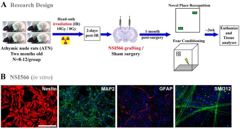
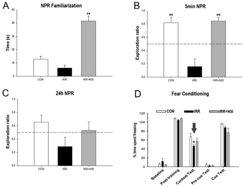
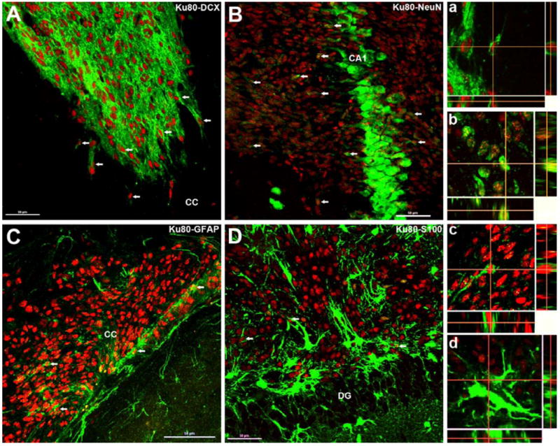
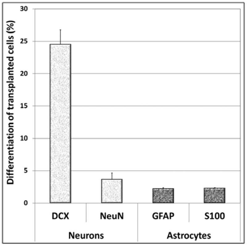
Treatment of central nervous system (CNS) malignancies typically involves radiotherapy to forestall tumor growth and recurrence following surgical resection. Despite the many benefits of cranial radiotherapy, survivors often suffer from a wide range of debilitating and progressive cognitive deficits. Thus, while patients afflicted with primary and secondary malignancies of the CNS now experience longer local regional control and progression-free survival, there remains no clinical recourse for the unintended neurocognitive sequelae associated with their cancer treatments. Multiple mechanisms contribute to disrupted cognition following irradiation, including the depletion of radiosensitive populations of stem and progenitor cells in the hippocampus. We have explored the potential of using intrahippocampal transplantation of human stem cells to ameliorate radiation-induced cognitive dysfunction. Past studies demonstrated the capability of cranially transplanted human embryonic (hESCs) and neural (hNSCs) stem cells to functionally restore cognition in rats 1 and 4 months after cranial irradiation. The present study employed an FDA-approved fetal-derived hNSC line capable of large scale-up under good manufacturing practice (GMP). Animals receiving cranial transplantation of these cells 1 month following irradiation showed improved hippocampal spatial memory and contextual fear conditioning performance compared to irradiated, sham surgery controls. Significant newly born (doublecortin positive) neurons and a smaller fraction of glial subtypes were observed within and nearby the transplantation core. Engrafted cells migrated and differentiated into neuronal and glial subtypes throughout the CA1 and CA3 subfields of the host hippocampus. These studies expand our prior findings to demonstrate that transplantation of fetal-derived hNSCs improves cognitive deficits in irradiated animals, as assessed by two separate cognitive tasks.


Conflict of interest statement

Authors declare no conflicts of interest.

Figures

Figure 1
Figure 1
Schematics of research design. (A) Two month old athymic nude rats received 10Gy head-only γ-irradiation were transplanted two days later with human-fetal derived neural stem cells (NSI566). At 1-month post-transplantation surgery, animals were administered a novel place recognition and a fear conditioning tasks. Three weeks later, after cognitive testing, animals were euthanized for immunohistochemical analysis. Non-irradiated control and irradiated animals receiving sterile hibernation buffer served as sham surgery groups. (B) Immunocytochemical analysis of NSI566 stained with DAPI shows strong expression and co-localization of the multipotent marker (Nestin, red). In vitro differentiated NSIs express both neuronal (green, neurofilament proteins MAP2 and SMI312) and astrocytic (red, GFAP) markers. (Scale bars: Nestin, 10 μm; MAP2, SMI312 and GFAP, 50 μm).
Figure 2
Figure 2
Human fetal-derived neural stem cell transplantation improved radiation-induced cognitive impairments at 1-month post-transplantation. For the novel place recognition task (NPR), animals were first familiarized with two identical objects in specific spatial locations in an open field arena, and total time spent exploring both identical objects was assessed. Following a 5-min retention interval, animal were placed in the same arena with one object moved to a novel spatial location. (A) Analysis of total time spent exploring both objects during the initial familiarization phase of NPR task revealed NSI-transplanted (IRR+NSI) animals explore more than controls- (CON) and irradiated-sham (IRR) groups (P’s<0.001, post hoc). Exploration ratios were calculated as, timenovel/timenovel+timefamiliar, for the first minute of 5min (B) and 24h (C) test sessions in NPR task. (B) In 5min test phase, IRR animals spent a significantly lower proportion of time exploring the novel place (P’s<0.001 vs. CON, and vs. IRR+NSI, post hoc), while CON and IRR+NSI animals did not differ. IRR animals did not spent more time exploring the novel place than expected by chance (dashed line at 50%). (C) 24h after the initial familiarization phase, animals were presented with the same two objects, with one moved to a new spatial location. None of the groups spent more time exploring the novel place than expected by chance. For the context and cued fear-conditioning task (D), baseline freezing levels were established using a series of 5 tone-shock pairings (post-training bars, D), and all groups showed increased post-training freezing behavior. 24h hour later, a context test was administered, and the IRR group spent significantly less time freezing compared to CON, while CON and IRR+NSI did not differ (P=0.014, post hoc comparisons, indicated by arrow). 48h after the initial training phase, the context was changed, which resulted in a substantial reduction in freezing behavior in all groups (pre-cue bars, D). Further, freezing levels were restored in all groups following the tone sound (cue test bars, D), indicating intact amygdala function in all groups Data are presented as Means + 1 SEM. P values were derived from FPLSD post hoc comparisons. *, P=0.014 indicates significant difference versus CON and IRR+NSI, and **, P=0.001 indicates significant difference versus IRR animals.
Figure 3
Figure 3
Survival and location of transplanted NSIs. At ~ 2 months post-transplantation, NSI are located near the injection site (dual labeled cells displayed as white, Nt, needle track; Tc, transplant core, A-E, 5 to 60× magnification) and CA1 and corpus callosum (CC) areas. Transplanted NSI (green) did not show extensive migration patterns in the host hippocampus (dentate gyrus, DG; dentate hilus DH, CA3 subfields). Transplanted NSIs were detected by human specific nuclear antigen (Ku80, green) and counterstained with nuclear dye (TOTO-3, pink). Insert (E) represents orthogonal reconstruction of confocal Z-stacks. (Scale bars: A-B, 100 μm; C, 50 μm; D, 20 μm; E, 10 μm and E-insert, 5 μm).
Figure 4
Figure 4
Differentiation of transplanted NSIs in the irradiated hippocampus. At ~ 2 months post-transplantation, Ku80 positive (human specific nuclear antigen, red) NSIs differentiated into immature (doublecortin, DCX, A and a, green) and mature neurons (neuron specific nuclear antigen, NeuN, B and b, green) as visualized by dual labeling of neuron-specific markers with Ku80 (red). A similar pattern of differentiation was observed for immature (glial fibrillary acidic protein, GFAP, C and c, green) and mature (S100 protein, D and d, green) astrocytes. Arrows indicate representative dual-labeled NSI transplant-derived cells (A-D). Confocal Z-stack orthogonal reconstructions of dual-labeled cells are shown for each neuronal (NeuN, a; DCX, b) and astrocytic (GFAP, c; S100, d) phenotypes. DG, dentate gyrus; CC, corpus callosum. (Scale bars: A-D, 50 μm and a-d, 10 μm).
Figure 5
Figure 5
Engrafted NSIs differentiated into neuronal and astrocytic phenotypes 2-months after irradiation and transplantation. The majority of NSI graft-derived cells (Ku80+) differentiated into immature neurons (doublecortin, DCX). Transplanted NSI also differentiated into mature (S100β+) and immature (GFAP+) astrocytes, though minimal oligodendrocytic differentiation was observed at this time-point (not shown). The data is represented as the Mean ± S.E.M. of 4 independent observations for each marker.

References

    1. Abayomi OK. Pathogenesis of cognitive decline following therapeutic irradiation for head and neck tumors. Acta Oncol. 2002;41(4):346–351. - PubMed
    1. Abayomi OK. Pathogenesis of irradiation-induced cognitive dysfunction. Acta Oncol. 1996;35(6):659–663. - PubMed
    1. Acharya MM, Christie LA, Lan ML, Donovan PJ, Cotman CW, Fike JR, Limoli CL. Rescue of radiation-induced cognitive impairment through cranial transplantation of human embryonic stem cells. Proc Natl Acad Sci U S A. 2009;106(45):19150–19155. - PMC - PubMed
    1. Acharya MM, Christie LA, Lan ML, Giedzinski E, Fike JR, Rosi S, Limoli CL. Human neural stem cell transplantation ameliorates radiation-induced cognitive dysfunction. Cancer Res. 2011;71(14):4834–4845. - PMC - PubMed
    1. Acharya MM, Christie LA, Lan ML, Limoli CL. Comparing the functional consequences of human stem cell transplantation in the irradiated rat brain. Cell Transplant. 2013;22(1):55–64. - PMC - PubMed

Publication types

MeSH terms

Substances