. 2013 Jul 17;2(1):15.
doi: 10.1186/2047-9158-2-15.

Biological function of Presenilin and its role in AD pathogenesis

Affiliations

Biological function of Presenilin and its role in AD pathogenesis

Shuting Zhang et al. Transl Neurodegener. .

Abstract

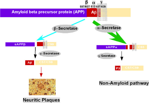
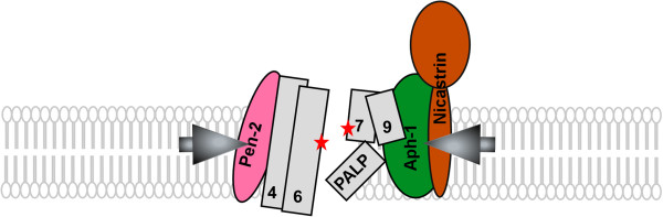
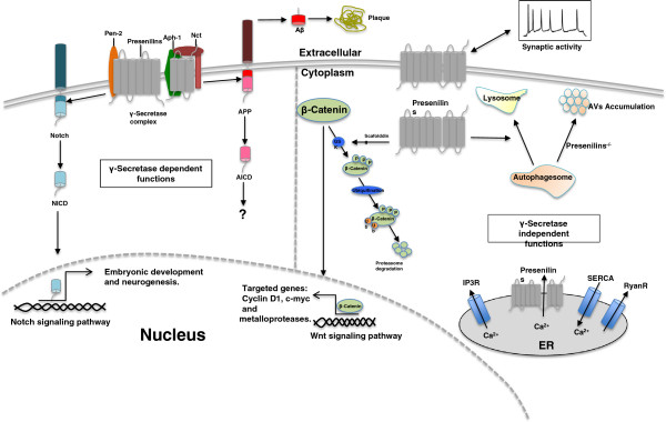
Presenilins (PSs) are the catalytic core of γ-secretase complex. However, the mechanism of FAD-associated PS mutations in AD pathogenesis still remains elusive. Here we review the general biology and mechanism of γ-secretase and focus on the catalytic components - presenilins and their biological functions and contributions to the AD pathogenesis. The functions of presenilins are divided into γ-secretase dependent and γ-secretase independent ones. The γ-secretase dependent functions of presenilins are exemplified by the sequential cleavages in the processing of APP and Notch; the γ-secretase independent functions of presenilins include stabilizing β-catenin in Wnt signaling pathway, regulating calcium homeostasis and their interaction with synaptic transmission.


Figures

Figure 1
Figure 1
APP processing pathways. Under the physiological conditions, the majority of APP undergoes the non-amyloidogenic pathway. The α-secretase processes APP within the Aβ domain to generate C83 and this cleavage abolishes Aβ generation. In the amyloidogenic pathway, β-secretase processes APP at Asp1 site to generate C99 fragment, which is the substrate for γ-secretase for Aβ generation.
Figure 2
Figure 2
Presenilin 1 structure. This diagram shows the amino acid sequence of PS1 and the distribution of the FAD-associated mutations. Blue circles represent the FAD-associated mutations and red circles indicate the two catalytic active aspartates.
Figure 3
Figure 3
γ-secretase complex and the formation of the catalytic pore of PS1. PS1 transmembrane domains (TMDs) are shown as columns with numbers. Without the assistance of other subunits, PS1 forms a relatively open pore structure within the membrane. Upon the binding of subunits, the PALP motif moves to the proximity of the catalytic center, and the catalytic structure is activated by the structural changes in TMDs of PS1.
Figure 4
Figure 4
Presenilin functions. This diagram divides the functions of presenilins into γ-secretase-dependent and γ-secretase-independent ones by the vertical dash line. The γ-secretase activity of presenilins are exemplified by APP processing and Notch processing. Other γ-secretase independent functions of presenilins include stabilizing β-catenin in Wnt signaling pathway, regulating calcium homeostasis and its interaction with synaptic transmission. APP, amyloid β precursor protein; AICD, APP intracellular domain; NICD, Notch intracellular domain; Nct, Nicastrin; IP3, Inositol trisphosphate receptor; SERCA, Sarco/endoplasmic reticulum Ca2+−ATPase; Ryan R, Ryanodine receptor; GSK, Glycogen synthase kinase.

References

    1. Goldgaber D, Lerman MI, McBride OW, Saffiotti U, Gajdusek DC. Characterization and chromosomal localization of a cDNA encoding brain amyloid of Alzheimer’s disease. Science. 1987;235:877–880. doi: 10.1126/science.3810169. - DOI - PubMed
    1. Kang J, Lemaire HG, Unterbeck A, Salbaum JM, Masters CL, Grzeschik KH, Multhaup G, Beyreuther K, Muller-Hill B. The precursor of Alzheimer’s disease amyloid A4 protein resembles a cell-surface receptor. Nature. 1987;325:733–736. doi: 10.1038/325733a0. - DOI - PubMed
    1. Robakis NK, Ramakrishna N, Wolfe G, Wisniewski HM. Molecular cloning and characterization of a cDNA encoding the cerebrovascular and the neuritic plaque amyloid peptides. Proc Natl Acad Sci U S A. 1987;84:4190–4194. doi: 10.1073/pnas.84.12.4190. - DOI - PMC - PubMed
    1. St George-Hyslop PH, Tanzi RE, Polinsky RJ, Haines JL, Nee L, Watkins PC, Myers RH, Feldman RG, Pollen D, Drachman D. et al.The genetic defect causing familial Alzheimer’s disease maps on chromosome 21. Science. 1987;235:885–890. doi: 10.1126/science.2880399. - DOI - PubMed
    1. Tanzi RE, Gusella JF, Watkins PC, Bruns GA, St George-Hyslop P, Van Keuren ML, Patterson D, Pagan S, Kurnit DM, Neve RL. Amyloid beta protein gene: cDNA, mRNA distribution, and genetic linkage near the Alzheimer locus. Science. 1987;235:880–884. doi: 10.1126/science.2949367. - DOI - PubMed