Review
. 2013 Dec;23(6):1057-64.
doi: 10.1016/j.conb.2013.06.006. Epub 2013 Jul 15.

'Sealing off the CNS': cellular and molecular regulation of blood-brain barriergenesis

Affiliations
Review

'Sealing off the CNS': cellular and molecular regulation of blood-brain barriergenesis

Julie A Siegenthaler et al. Curr Opin Neurobiol. 2013 Dec.

Abstract

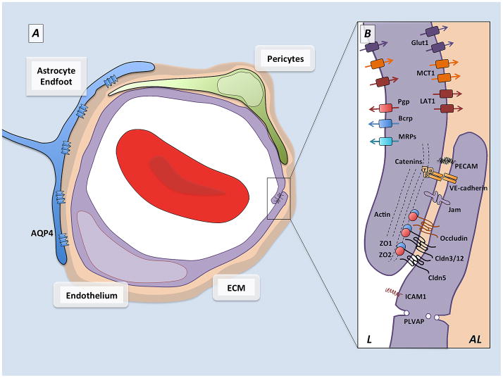
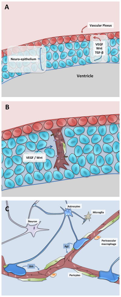
From their initial ingression into the neural tube to the established, adult vascular plexus, blood vessels within the CNS are truly unique. Covered by a virtually continuous layer of perivascular cells and astrocytic endfeet and connected by specialized cell-cell junctional contacts, mature CNS blood vessels simultaneously provide nutritive blood flow and protect the neural milieu from potentially disruptive or harmful molecules and cells flowing through the vessel lumen. In this review we will discuss how the CNS vasculature acquires blood-brain barrier (BBB) properties with a specific focus on recent work identifying the cell types and molecular pathways that orchestrate barriergenesis.


Figures

Figure 1
Figure 1. Schematic representation of the neurovascular unit
A) Cellular components of the blood-brain barrier. Capillaries in the central nervous system are made up of endothelial cells (purple) which form the walls of the blood vessels, and their ablumenal surface is incompletely covered by a pericytes (green) which are embedded in the vascular extracellular matrix. Astrocytes (blue), a major glial cell population, extend cellular processes whose endfeet ensheath the blood vessels. Between the astrocytes and the vascular tube are two layers of extracellular matrix, the vascular extracellular matrix secreted by the endothelial cells and pericytes, and the glial matrix secreted by astrocytes. B) Barrier components of the blood-brain barrier. Many of the properties of the BBB are manifested within the endothelial cells that make up the walls of the vessels. The endothelial cells are held together by tight junctions (TJs) which create tight paracellular barrier, and polarize the cells creating distinct lumenal (L) and ablumenal (AL) membrane compartments. The TJs are made up of transmembrane molecules including claudin family members, occludin and JAMs which are linked to the cytoskeleton and adherens junctions by cytoplasmic adaptors including ZO-1 and ZO-2. The endothelial cells undergo extremely low rates of transcytosis, mediated by low levels of PLVAP, limiting the transcellular movement of molecules and ions. These endothelial cells also express polarized transporters that determine the movement of many solutes across the endothelial cells. These include lumenal efflux transporters, such as Pgp and BCRP, which use ATP hydrolysis to actively transport a variety of small molecule substrates into the blood, as well as solute carriers such as Glut1, MCT1, and LAT1 which deliver specific nutrients (glucose, lactate and amino acids respectively) into the CNS. In addition endothelial cells express low levels of leukocyte adhesion molecules, including Icam1, which correlates with the low levels of CNS immune surveillance. These properties allow CNS endothelial cells to tightly regulate the movement of ions, molecules, and cells between the blood and the brain.
Figure 2
Figure 2. Schematic representation of BBB development
A) During early development, endothelial cell progenitors (brown) form a vascular plexus surrounding the developing neural tissue (blue). B) Angiogenic sprouts, consisting of endothelial tip and stalk cells, invade the neural tissue under the influence of general angiogenic factors (VEGF) as well as CNS-specific angiogenic factors (Wnt). These initial sprouts recruit pericytes (green) and are observed to have ‘tight’ properties including tight junction proteins as well as Glut1 expression, as well as ‘leaky’ properties including high rates of transcytosis and expression of leukocyte adhesion molecules (LAMs) such as Icam1. C) Over the next several days and weeks under the influence of pericyte-derived factors, neural derived factors and astrocyte derived factors, the endothelial cell tight junctions mature, the cells start to increase expression of efflux transporters and lower their transcytosis and expression of LAMs.

References

    1. Stewart P, Hayakawa K. Early ultrastructural changes in blood-brain barrier vessels of the rat embryo. Brain Res Dev Brain Res. 1994;78:25–34. - PubMed
    1. Bauer H, Bauer H, Lametschwandtner A, Amberger A, Ruiz P, Steiner M. Neovascularization and the appearance of morphological characteristics of the blood-brain barrier in the embryonic mouse central nervous system. Brain Res Dev Brain Res. 1993;75:269–278. - PubMed
    1. Daneman R, Zhou L, Kebede AA, Barres BA. Pericytes are required for blood-brain barrier integrity during embryogenesis. Nature. 2010;468:562–566. Using a pericyte-deficient mouse model, showed that perictyes were required for certain aspects of pre-natal barriergenesis including supression of endothelial trancytosis and LAMs. Provided a developmental timeline of BBB characteristics in the mouse. - PMC - PubMed
    1. Nakao T, Ishizawa A, Ogawa R. Observations of vascularization in the spinal cord of mouse embryos, with special reference to development of boundary membranes and perivascular spaces. Anat Rec. 1988;221:663–677. - PubMed
    1. Wakai S, Hirokawa N. Development of the blood-brain barrier to horseradish peroxidase in the chick embryo. Cell Tissue Res. 1978;195:195–203. - PubMed

LinkOut - more resources