

Neuroimaging standards for research into small vessel disease and its contribution to ageing and neurodegeneration
- PMID: 23867200
- PMCID: PMC3714437
- DOI: 10.1016/S1474-4422(13)70124-8
Neuroimaging standards for research into small vessel disease and its contribution to ageing and neurodegeneration
Abstract
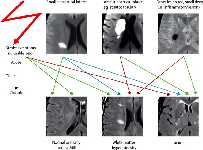
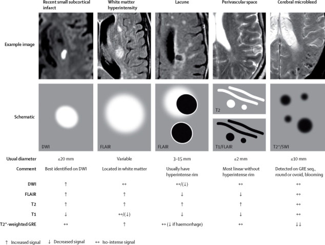
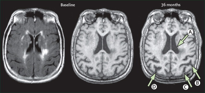
Cerebral small vessel disease (SVD) is a common accompaniment of ageing. Features seen on neuroimaging include recent small subcortical infarcts, lacunes, white matter hyperintensities, perivascular spaces, microbleeds, and brain atrophy. SVD can present as a stroke or cognitive decline, or can have few or no symptoms. SVD frequently coexists with neurodegenerative disease, and can exacerbate cognitive deficits, physical disabilities, and other symptoms of neurodegeneration. Terminology and definitions for imaging the features of SVD vary widely, which is also true for protocols for image acquisition and image analysis. This lack of consistency hampers progress in identifying the contribution of SVD to the pathophysiology and clinical features of common neurodegenerative diseases. We are an international working group from the Centres of Excellence in Neurodegeneration. We completed a structured process to develop definitions and imaging standards for markers and consequences of SVD. We aimed to achieve the following: first, to provide a common advisory about terms and definitions for features visible on MRI; second, to suggest minimum standards for image acquisition and analysis; third, to agree on standards for scientific reporting of changes related to SVD on neuroimaging; and fourth, to review emerging imaging methods for detection and quantification of preclinical manifestations of SVD. Our findings and recommendations apply to research studies, and can be used in the clinical setting to standardise image interpretation, acquisition, and reporting. This Position Paper summarises the main outcomes of this international effort to provide the STandards for ReportIng Vascular changes on nEuroimaging (STRIVE).
Copyright © 2013 Elsevier Ltd. All rights reserved.
Figures
References
-
- Norrving B. Lacunar infarcts: no black holes in the brain are benign. Pract Neurol. 2008;8:222–228. - PubMed
-
- Pantoni L. Cerebral small vessel disease: from pathogenesis and clinical characteristics to therapeutic challenges. Lancet Neurol. 2010;9:689–701. - PubMed
-
- Dichgans M, Zietemann V. Prevention of vascular cognitive impairment. Stroke. 2012;43:3137–3146. - PubMed
-
- Brayne C, Richardson K, Matthews FE. Neuropathological correlates of dementia in over-80-year-old brain donors from the population-based Cambridge City over-75s cohort (CC75C) study. J Alzheimers Dis. 2009;18:645–658. - PubMed
Publication types
MeSH terms
Grants and funding
LinkOut - more resources
Full Text Sources
Other Literature Sources
Medical
