. 2013 Aug;123(8):3498-509.
doi: 10.1172/JCI68127. Epub 2013 Jul 8.

Collagen VII plays a dual role in wound healing

Affiliations

Collagen VII plays a dual role in wound healing

Alexander Nyström et al. J Clin Invest. 2013 Aug.

Abstract

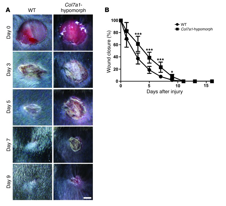
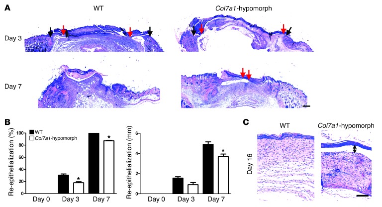
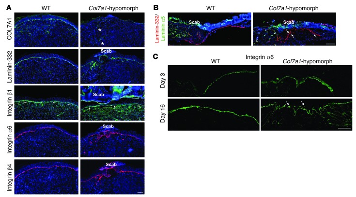
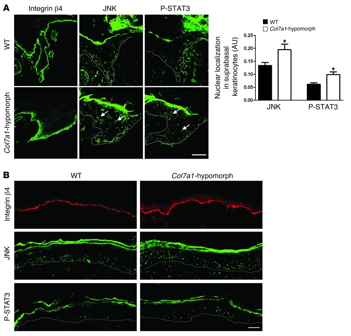
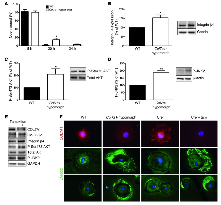
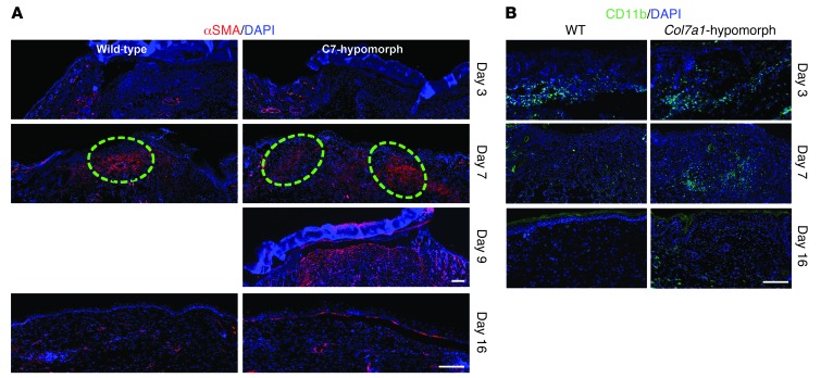
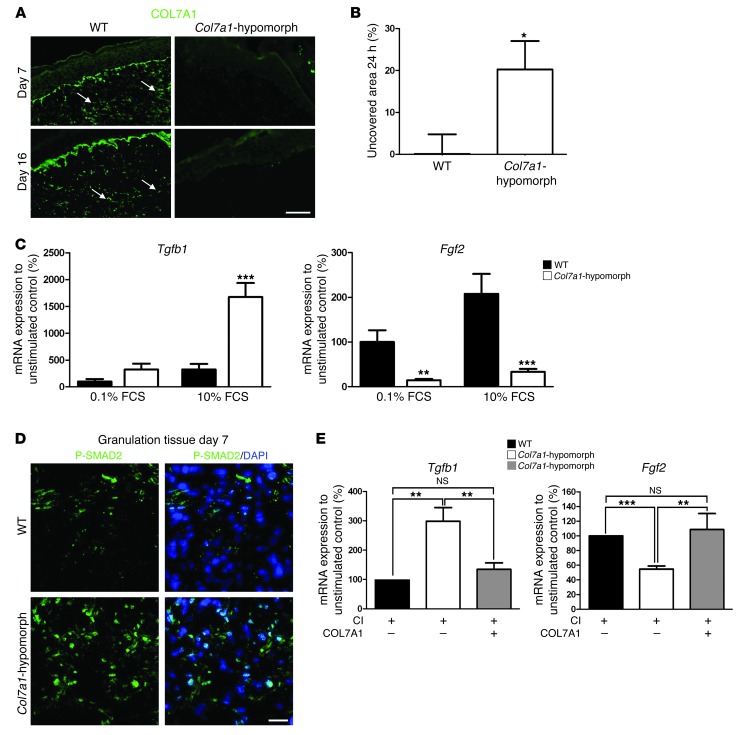
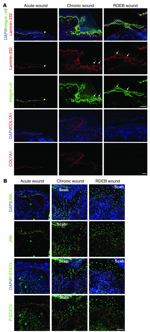
Although a host of intracellular signals is known to contribute to wound healing, the role of the cell microenvironment in tissue repair remains elusive. Here we employed 2 different mouse models of genetic skin fragility to assess the role of the basement membrane protein collagen VII (COL7A1) in wound healing. COL7A1 secures the attachment of the epidermis to the dermis, and its mutations cause a human skin fragility disorder coined recessive dystrophic epidermolysis bullosa (RDEB) that is associated with a constant wound burden. We show that COL7A1 is instrumental for skin wound closure by 2 interconnected mechanisms. First, COL7A1 was required for re-epithelialization through organization of laminin-332 at the dermal-epidermal junction. Its loss perturbs laminin-332 organization during wound healing, which in turn abrogates strictly polarized expression of integrin α6β4 in basal keratinocytes and negatively impacts the laminin-332/integrin α6β4 signaling axis guiding keratinocyte migration. Second, COL7A1 supported dermal fibroblast migration and regulates their cytokine production in the granulation tissue. These findings, which were validated in human wounds, identify COL7A1 as a critical player in physiological wound healing in humans and mice and may facilitate development of therapeutic strategies not only for RDEB, but also for other chronic wounds.


Figures

Figure 1
Figure 1. Delayed wound closure in Col7a1-hypomorphic mice.
(A) Closure of 6-mm punch biopsy wounds on the back skin of wild-type and Col7a1-hypomorphic mice over time. Scale bar: 2 mm. (B) Quantification of the wound area showed significantly delayed gross wound closure between days 3 and 9. n = 14 wounds per group; values represent mean ± SD. *P < 0.05; ***P < 0.001.
Figure 2
Figure 2. Loss of Col7a1 delays re-epithelialization.
(A) H&E staining of wounds in wild-type and Col7a1-hypomorphic mice at days 3, 7, and 16. Arrows indicate wound width (black arrows, initial wound border; red arrows, epithelial front). The original wound size was similar, but re-epithelialization was clearly protracted in Col7a1-hypomorphic wounds. Scale bar: 500 μm. (B) Quantification of percent re-epithelialization (left) and migration distance of the epithelial tongue (right). COL7A1 loss caused a significant delay in re-epithelialization. n ≥ 3 wounds; values represent mean ± SD. *P < 0.05. (C) Wounds after 16 days stained with H&E; note the detached epidermis in the Col7a1-hypomorphic wound (double-headed arrow). Note also the changes in the granulation tissue (see also Figure 6 and Supplemental Figure 12). Scale bar: 100 μm.
Figure 3
Figure 3. Loss of COL7A1 alters laminin-332 deposition and integrin α6β4 distribution in healing epidermis.
(A) 7-day wounds stained for laminin-332 and its integrin receptors. Laminin-332 deposition and integrin α6β4 distribution was altered in Col7a1-hypomorphic wounds. Asterisks show autofluorescence from red blood cells trapped in capillaries; arrows point to integrin β1 at the DEJZ. Scale bar: 100 μm. (B) Epidermal tongue in 3-day wounds stained for laminin-332 (red) and the laminin α5 chain (green). Laminin-332 deposition was irregular and patchy in Col7a1-hypomorphic wounds (arrows), in contrast to the distinct linear signal of the laminin α5 chain. Scale bar: 50 μm. (C) 3- and 16-day-old wounds stained for integrin α6. Arrows indicate the patchy suprabasal integrin α6 expression in 16-day-old Col7a1-hypomorpic wounds. Scale bar: 50 μm.
Figure 4
Figure 4. Abnormal activation of the laminin-332/integrin α6β4 signaling axis in Col7a1-hypomorphic wound epidermis.
(A) Epidermal tongue of 3-day-old wild-type and Col7a1-hypomorphic wounds stained for integrin β4, JNK, and phospho-STAT3. Note the abundant suprabasal presence of nuclear JNK and phospho-STAT3 in Col7a1-hypomorphic wounds (arrows), indicative of suprabasal activation. Quantification of staining is also shown (n ≥ 3; values represent mean ± SD). *P < 0.05. Scale bar: 50 μm. (B) Middle area of 7-day-old wild-type and Col7a1-hypomorphic wounds stained for integrin β4 (red), JNK (green), and phospho-STAT3 (green). Suprabasal activation persisted in older Col7a1-hypomorphic wounds. Scale bar: 50 μm.
Figure 5
Figure 5. Mechanisms of delayed wound closure in Col7a1-hypomorphic keratinocytes.
(A) In in vitro assays, wound closure by keratinocytes was assessed at 8, 20, and 24 hours. At 20 and 24 hours, Col7a1-hypomorphic keratinocytes closed wounds at a reduced rate. n = 6; values represent mean ± SEM. *P < 0.05. (BD) Western blotting of wild-type and Col7a1-hypomorphic keratinocyte protein lysates revealed increased expression of integrin β4 subunit (B) and increased phosphorylation of Ser473 AKT (C) and JNK2 (D) in cultured Col7a1-hypomorphic keratinocytes. Densitometric quantification of keratinocyte isolations is also shown. n ≥ 3; values represent mean ± SD. *P < 0.05; **P < 0.01. (E) Western blots on cell lysates from tamoxifen-induced Col7a1 knockout mouse keratinocytes. Keratinocytes from the same isolation were divided into 2 pools: one was treated with 4-OH tamoxifen to induce Col7a1 knockout, the other received only DMSO and served as control. The changes in Col7a1-hypomorphic keratinocytes were replicated in keratinocytes after forced COL7A1 loss. Loss of COL7A1 did not affect expression of laminin-332 (LM-β3/γ2; representing the laminin β3 and γ2 chains of laminin-332), but increased expression of integrin β4 and phosphorylation of Ser473 AKT and JNK2. Total AKT and GAPDH were used to ensure equal loading. (F) Altered Laminin-332 organization in Col7a1-hypomorphic keratinocytes. Primary keratinocytes of the indicated genotypes and treatments were cultured for 24 hours in the presence of ascorbate and stained for COL7A1 (red) and laminin-332 (green). Original magnification, ×400. Tam, tamoxifen.
Figure 6
Figure 6. Granulation tissue maturation and clearance of inflammatory cells are delayed in Col7a1-hypomorphic wounds.
(A) α-SMA staining (red) showed few myofibroblasts at the wound edge at day 3 in both wild-type and Col7a1-hypomorphic mice. At day 7, myofibroblasts were abundant in the middle of the wild-type wound, but remained in the periphery of the Col7a1-hypomorphic wound. Dashed outlines denote the dense myofibroblast regions. Myofibroblast organization in the day 9 Col7a1-hypomorphic wound was similar to that of 7-day wild-type wounds, with myofibroblasts localized in the upper central part of the granulation tissue. At day 16 after wounding, most myofibroblasts had disappeared in both mice. Nuclei were stained with DAPI (blue). Scale bars: 100 μm. (B) CD11b staining (green) revealed protracted clearance of inflammatory cells in Col7a1-hypomophic wounds. At day 3, inflammatory cells were at the wound edge in both wild-type and Col7a1-hypomorphic mice. At day 7, inflammatory cells were mainly seen in the middle of the wound in both mice. At day 16, CD11b-positive cells were cleared from the wound area in wild-type mice, but not in Col7a1-hypomorphic mice. Nuclei were stained with DAPI (blue). Scale bar: 100 μm. See Supplemental Figure 12 for quantification of granulation tissue changes.
Figure 7
Figure 7. COL7A1 affects dermal fibroblasts during wound healing.
(A) Granulation tissue in 7- and 16-day wounds in wild-type and Col7a1-hypomorphic mice stained for COL7A1 (green). Note the presence of COL7A1 aggregates (arrows) within the healing dermis in wild-type mice. Scale bar: 50 μm. (B) In vitro scratch wound assay. Col7a1-hypomorphic fibroblasts closed the scratch wound significantly slower than did wild-type cells. n = 7. (C) Serum-response assay, which replicates fibroblast signaling events during wound healing (26). Dermal fibroblasts were starved in 0.1% FCS for 48 hours, stimulated with 10% serum for 5 hours, and analyzed for gene expression. Tgfb1 and Fgf2 mRNA expression were normalized to Gapdh and a nonstimulated control. Serum stimulation substantially increased Tgfb1 mRNA expression, but reduced Fgf2 expression, in Col7a1-hypomorphic compared with wild-type fibroblasts. n = 5. (D) P-SMAD2 staining (green) of granulation tissue in day-7 wounds confirmed increased activation of TGF-β1 signaling in the Col7a1-hypomorphic wound. Nuclei were visualized with DAPI (blue). Scale bar: 20 μm. (E) Supplementation of recombinant COL7A1 to Col7a1-hypomorphic fibroblasts normalized Tgfb1 and Fgf2 expression. Dermal fibroblasts were grown on 1.5 μg collagen I (CI), with or without 1.5 μg/well recombinant COL7A1, until they reached confluence, then serum-starved for 48 hours and stimulated with 10% FCS for 5 hours. mRNA expression was analyzed as in C. n ≥ 4. All values represent mean ± SEM. *P < 0.05, **P < 0.01, ***P < 0.001.
Figure 8
Figure 8. Human RDEB and other chronic wounds display similar molecular alterations.
Wound biopsies were obtained from fresh wounds 3 days after primary excision (Acute wound), from the wound margins of nonhealing chronic venous ulcers, and from wounds of RDEB patients. (A) Wounds were stained for laminin-332 (red) and integrin α6 (green) or for COL7A1 (red). Yellow arrowheads denote the end of the epithelial front in the acute wound. For chronic and RDEB wounds, the epithelial front in the wound margin is shown. Acute wounds showed linear deposition of laminin-332 and regular, primarily basal, expression of integrin α6, whereas in chronic and RDEB wounds, laminin-332 deposition was irregular and integrin α6 expression suprabasal (white arrows). Importantly, in acute wounds, COL7A1 was distinctly present under the healing epidermis, whereas it was irregular and drastically reduced in chronic wounds. Nuclei were visualized with DAPI (blue). Scale bar: 50 μm. (B) Epidermal tongues in wound margins stained for phospho-JNK (green) or phospho-STAT3 (green). Nuclei were counterstained with DAPI (blue). Red lines denote the epidermal-dermal interface. In acute wounds, phospho-JNK and phospho-STAT3 staining was weak and mainly seen in basal keratinocytes, whereas both chronic and RDEB wounds showed strong phospho-JNK and phospho-STAT3 staining in suprabasal keratinocytes. Scale bar: 50 μm.

References

    1. Coulombe PA. Wound epithelialization: accelerating the pace of discovery. J Invest Dermatol. 2003;121(2):219–230. doi: 10.1046/j.1523-1747.2003.12387.x. - DOI - PubMed
    1. Singer AJ, Clark RA. Cutaneous wound healing. N Engl J Med. 1999;341(10):738–746. doi: 10.1056/NEJM199909023411006. - DOI - PubMed
    1. Schäfer M, Werner S. Cancer as an overhealing wound: an old hypothesis revisited. Nat Rev Mol Cell Biol. 2008;9(8):628–638. - PubMed
    1. Werner S, Grose R. Regulation of wound healing by growth factors and cytokines. Physiol Rev. 2003;83(3):835–870. - PubMed
    1. Ko MS, Marinkovich MP. Role of dermal-epidermal basement membrane zone in skin, cancer, and developmental disorders. Dermatol Clin. 2010;28(1):1–16. doi: 10.1016/j.clindermatol.2009.03.013. - DOI - PubMed

Publication types

MeSH terms