. 2013 Aug;123(8):3243-53.
doi: 10.1172/JCI69134. Epub 2013 Jul 8.

ARHGDIA mutations cause nephrotic syndrome via defective RHO GTPase signaling

Affiliations

ARHGDIA mutations cause nephrotic syndrome via defective RHO GTPase signaling

Heon Yung Gee et al. J Clin Invest. 2013 Aug.

Abstract

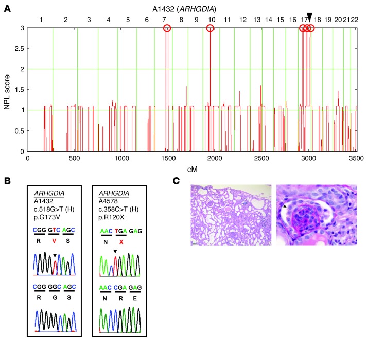
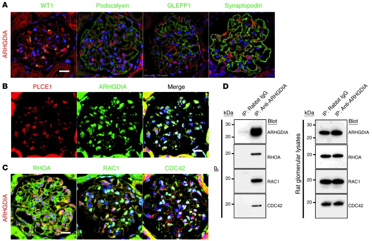
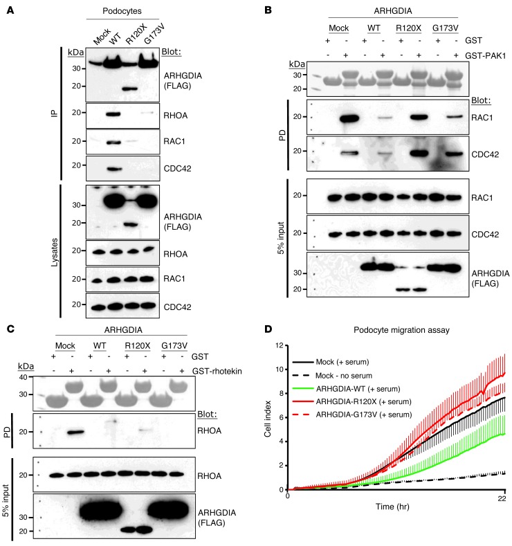
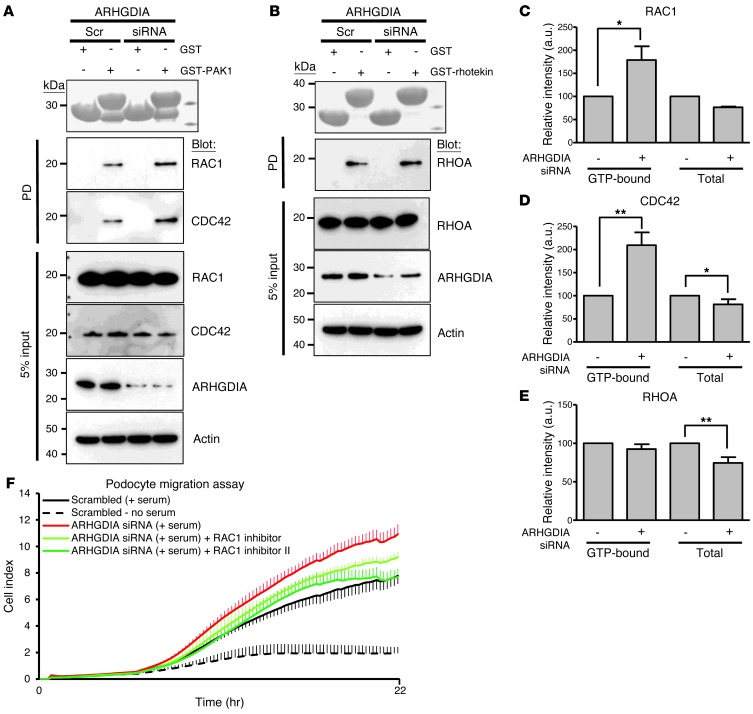
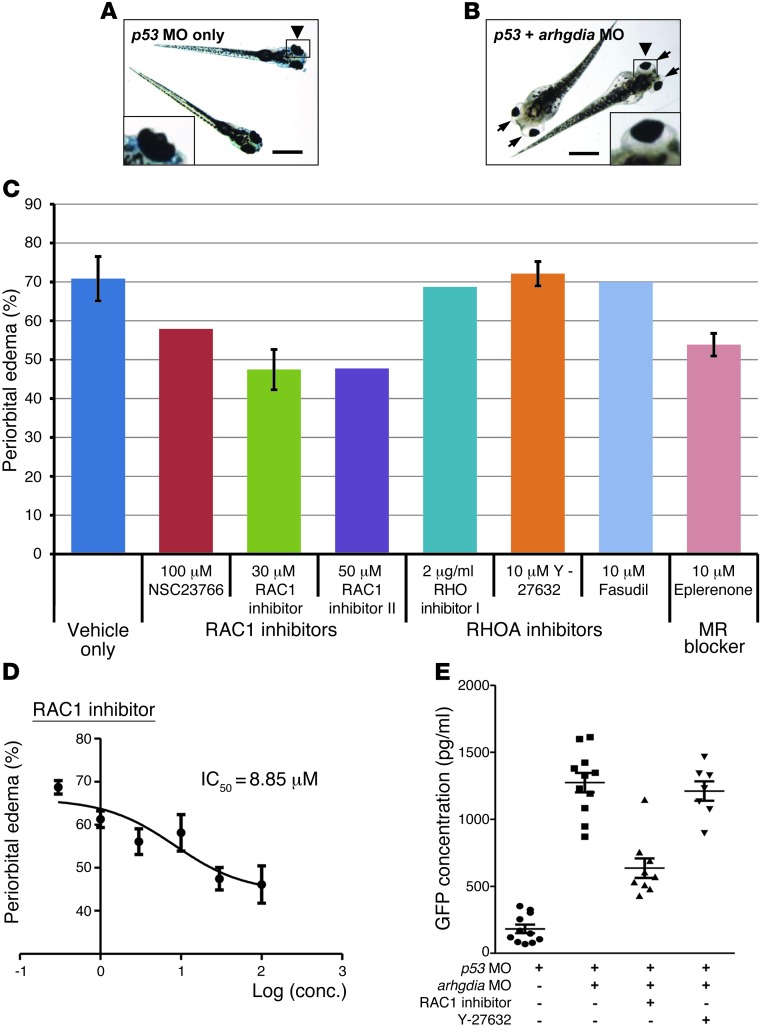
Nephrotic syndrome (NS) is divided into steroid-sensitive (SSNS) and -resistant (SRNS) variants. SRNS causes end-stage kidney disease, which cannot be cured. While the disease mechanisms of NS are not well understood, genetic mapping studies suggest a multitude of unknown single-gene causes. We combined homozygosity mapping with whole-exome resequencing and identified an ARHGDIA mutation that causes SRNS. We demonstrated that ARHGDIA is in a complex with RHO GTPases and is prominently expressed in podocytes of rat glomeruli. ARHGDIA mutations (R120X and G173V) from individuals with SRNS abrogated interaction with RHO GTPases and increased active GTP-bound RAC1 and CDC42, but not RHOA, indicating that RAC1 and CDC42 are more relevant to the pathogenesis of this SRNS variant than RHOA. Moreover, the mutations enhanced migration of cultured human podocytes; however, enhanced migration was reversed by treatment with RAC1 inhibitors. The nephrotic phenotype was recapitulated in arhgdia-deficient zebrafish. RAC1 inhibitors were partially effective in ameliorating arhgdia-associated defects. These findings identify a single-gene cause of NS and reveal that RHO GTPase signaling is a pathogenic mediator of SRNS.


Figures

Figure 1
Figure 1. Mapping and WER reveal a novel single-gene cause of NS (ARHGDIA).
(A) Homozygosity mapping in family A1432 consisting of 2 siblings with SRNS. NPL scores are plotted across the human genome. The x-axis shows SNP marker positions on human chromosomes concatenated from p-ter (left) to q-ter (right). Genetic distance is given in centimorgans. Maximum NPL peaks indicate the candidate regions of homozygosity by descent (red circles). The gene ARHGDIA is positioned (arrowhead) within one of the mapped candidate regions. (B) Chromatograms of ARHGDIA mutations in SRNS families A1432 and A4578. Gene symbols (underlined), family numbers, mutations, and predicted translational changes are given (see also Table 1). Sequence traces are shown for mutations above normal controls. Mutated nucleotides are indicated by arrowheads. (C) Renal histology of individual A4578-21 with DMS and an ARHGDIA mutation. Right kidney explanted at 2 months of age reveals on H&E staining advanced tubular dilation, atrophy, and casts (left). Original magnification, ×50. PAS stain (right) reveals the characteristic pattern of DMS featuring a small, sclerosed, simplified glomerulus with a corona of vacuolized podocytes (arrowhead). Original magnification, ×630.
Figure 2
Figure 2. ARHGDIA, RAC1, CDC42, and RHOA colocalize and interact in rat glomeruli.
(A) Coimmunofluorescence of ARHGDIA with podocyte marker proteins in rat glomeruli. ARHGDIA is highly expressed in podocytes, as identified by the expression of nuclear WT1. ARHGDIA partially colocalized with synaptopodin, but not with podocalyxin or GLEPP1. (B) ARHGDIA partially colocalized with the SRNS protein PLCε1 in proximal cell bodies and primary processes of podocytes. Direct fluorescent labeling of anti-PLCε1. (C) Coimmunofluorescence of ARHGDIA with the RHO GTPases RAC1, CDC42, and RHOA in adult rat glomeruli. ARHGDIA partially colocalized with RHOA, RAC1, and CDC42 in proximal cell bodies and primary processes, whereas RHOA, RAC1, and CDC42 exhibited a broad glomerular staining pattern in podocyte cell bodies and processes. Scale bars: 10 μm. (D) Coimmunoprecipitation of ARHGDIA in rat renal glomerular lysates. The protein complex precipitated by an anti-ARHDGIA antibody includes the RHO small GTPases RHOA, RAC1, and CDC42. The immunoprecipitation (IP) experiment is representative of more than 3 experiments.
Figure 3
Figure 3. Effects of disease-causing ARHGDIA mutations on protein-protein interaction, RHO GTPase activity, and podocyte migration.
(A) Interaction of wild-type ARHGDIA and 2 mutants (p.R120X and p.G173V) with RHO GTPases. FLAG-tagged ARHGDIA constructs were transfected into podocytes and were coimmunoprecipitated with endogenous RHO GTPases. Note that the R120X and G173V mutants abrogated interaction with RHOA, RAC1, and CDC42. (B) Active GTP-bound forms of RAC1 and CDC42 precipitated from podocytes expressing FLAG-ARHGDIA (wild-type and mutants) using a GST-PAK1 (CRIB, CDC42, and RAC interactive binding domain) pulldown assay. Five percent input represents the controls for equal loading. Note that, compared with mock cells, podocytes expressing ARHGDIA-WT exhibited a substantial decrease in active RAC1 and CDC42. This decrease was abrogated in the null mutant R120X and is diminished in the G173V mutant. (C) Active GTP-bound RHOA precipitated from podocytes expressing FLAG-ARHGDIA (wild-type and mutants) using a GST-rhotekin (RHO-binding domain [RBD]) pulldown assay. Overexpression of either wild-type or mutant ARHGDIA resulted in a substantial decrease in relative RHOA activity compared with mock cells. PD, pulldown. All IPs and PDs are representative of more than 3 experiments. (D) Effect on podocyte migration of wild-type ARHGDIA and 2 mutants found in patients with SRNS. Migration assay was performed using the xCELLigence system (described in Methods). Overexpression of wild-type ARHGDIA in podocytes inhibited serum-induced migration (green). However, the mutants G173V and R120X failed to inhibit migration (red). Error bars are shown in one direction only for clarity and indicate SDs for more than 3 independent experiments (see also Supplemental Figure 5).
Figure 4
Figure 4. Effects of ARHGDIA knockdown on RHO GTPase activity and podocyte migration in cultured human podocytes.
(A) Active GTP-bound RAC1 and CDC42 precipitated from podocytes transfected with scrambled (Scr) or ARHGDIA siRNA using a GST-PAK1 (CRIB) pulldown assay. Ponceau red staining at the top shows the GST proteins used. Compared with control podoctyes, podocytes transfected with ARHGDIA siRNA exhibited a significant increase in relative RAC1 and CDC42 (168% and 185%, respectively). The efficiency of knockdown by siRNA was confirmed by immunoblotting with an anti-ARHGDIA antibody (second to lowest panel). (B) Active GTP-bound RHOA precipitated from podocytes transfected with scrambled or ARHGDIA siRNA using a GST-rhotekin (RBD) pulldown assay. Cells transfected with scrambled control siRNA versus ARHGDIA siRNA exhibited no significant difference in relative RHOA activity. A and B represent 3 experiments each. (CE) Quantification of RAC1 (C), CDC42 (D), and RHOA (E) in ARHGDIA-depleted cells compared with control cells. Error bars indicate the SEM for greater than 4 independent experiments. *P < 0.05; **P < 0.01; difference from proteins in control podocytes transfected with scrambled siRNA. (F) Effect of ARHGDIA knockdown on podocyte migration. Podocytes transfected with ARHGDIA siRNA exhibited more active migration compared with those transfected with scrambled siRNA. Increase in podocyte migration by ARHGDIA knockdown (red) in serum-induced podocytes was reduced by 2 different RAC1 inhibitors (green). Error bars are shown in only one direction for clarity and indicate SDs for more than 4 independent experiments.
Figure 5
Figure 5. Functional analysis of arhgdia knockdown in zebrafish.
(A) Control zebrafish injected with p53 MO (0.2 mM). p53 MO did not produce any phenotype until 168 hpf (n >100). Scale bars: 1 mm (×3-fold for insets) (A and B). (B) Zebrafish coinjected with an arhgdia MO (0.2 mM) targeting the translation initiation site of zebrafish arhgdia and a p53 MO. At 120 hpf, the arhgdia morphants displayed the nephrosis phenotype of periorbital edema (“bug-eye”; arrows) and total body edema in 70% of embryos (255 of 360). (C) Effects of various RAC1 and RHOA inhibitors on the zebrafish nephrosis phenotype. Drugs were applied at 48 hpf in fish water and replenished every day. The number of arhgdia morphants that showed periorbital edema was counted at 144 hpf and is represented as a percentage. Two RAC1 inhibitors and eplerenone were partially effective in reducing edema, whereas RHOA inhibitors were not. Error bars indicate SDs for greater than 3 independent experiments in C and D. (D) Dose-response curve for the RAC1 inhibitor, which shows the greatest efficacy in C. The IC50 value of the RAC1 inhibitor for reducing periorbital edema was 8.85 μM. conc., concentration. (E) Proteinuria assay by ELISA against a fusion protein of vitamin D–binding protein and GFP in /-fabp::VDBP-GFP transgenic zebrafish. Note that knockdown of arhgdia caused significant proteinuria compared with the control. The RAC1 inhibitor was partially effective in reducing proteinuria in arhgdia morphants, whereas the RHO inhibitor Y-27632 was not. Error bars indicate the SEM for more than 3 independent experiments.

References

    1. Smith JM, Stablein DM, Munoz R, Hebert D, McDonald RA. Contributions of the Transplant Registry: The 2006 Annual Report of the North American Pediatric Renal Trials and Collaborative Studies (NAPRTCS). Pediatr Transplant. 2007;11(4):366–373. doi: 10.1111/j.1399-3046.2007.00704.x. - DOI - PubMed
    1. Hildebrandt F, Heeringa SF. Specific podocin mutations determine age of onset of nephrotic syndrome all the way into adult life. Kidney Int. 2009;75(7):669–671. doi: 10.1038/ki.2008.693. - DOI - PubMed
    1. Somlo S, Mundel P. Getting a foothold in nephrotic syndrome. Nat Genet. 2000;24(4):333–335. doi: 10.1038/74139. - DOI - PubMed
    1. Tryggvason K, Patrakka J, Wartiovaara J. Hereditary proteinuria syndromes and mechanisms of proteinuria. N Engl J Med. 2006;354(13):1387–1401. doi: 10.1056/NEJMra052131. - DOI - PubMed
    1. Kestila M, et al. Positionally cloned gene for a novel glomerular protein — nephrin — is mutated in congenital nephrotic syndrome. Mol Cell. 1998;1(4):575–582. doi: 10.1016/S1097-2765(00)80057-X. - DOI - PubMed

Publication types

MeSH terms

Substances