. 2013 Aug;123(8):3472-87.
doi: 10.1172/JCI60561. Epub 2013 Jul 8.

Macrophages regulate corpus luteum development during embryo implantation in mice

Affiliations

Macrophages regulate corpus luteum development during embryo implantation in mice

Alison S Care et al. J Clin Invest. 2013 Aug.

Abstract

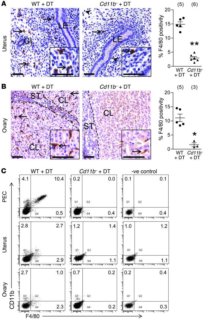
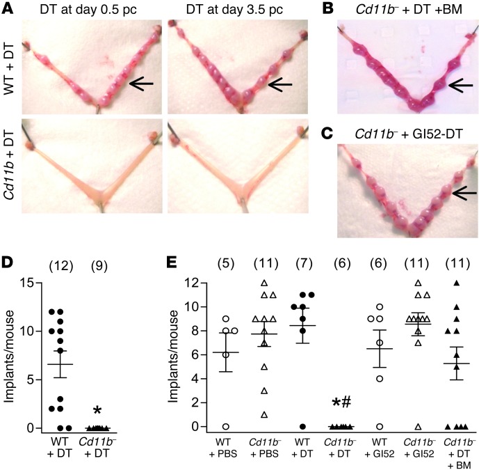
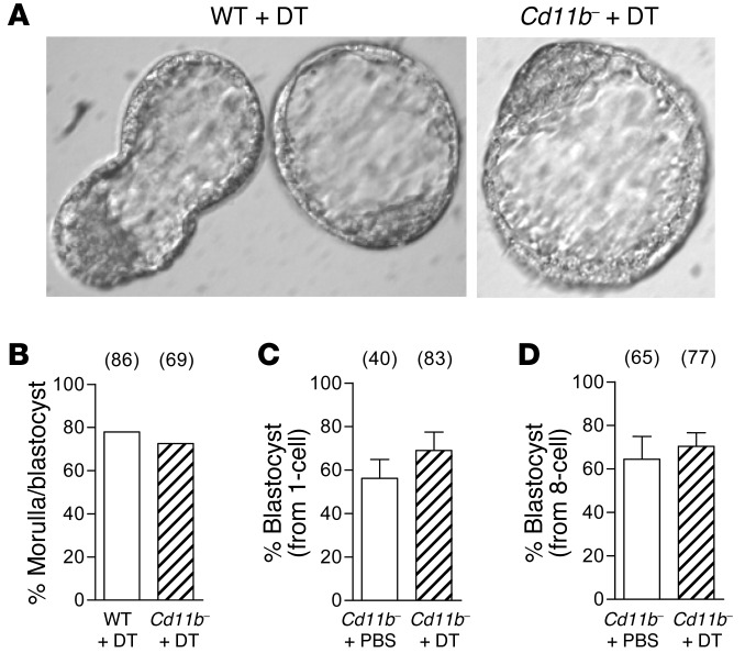
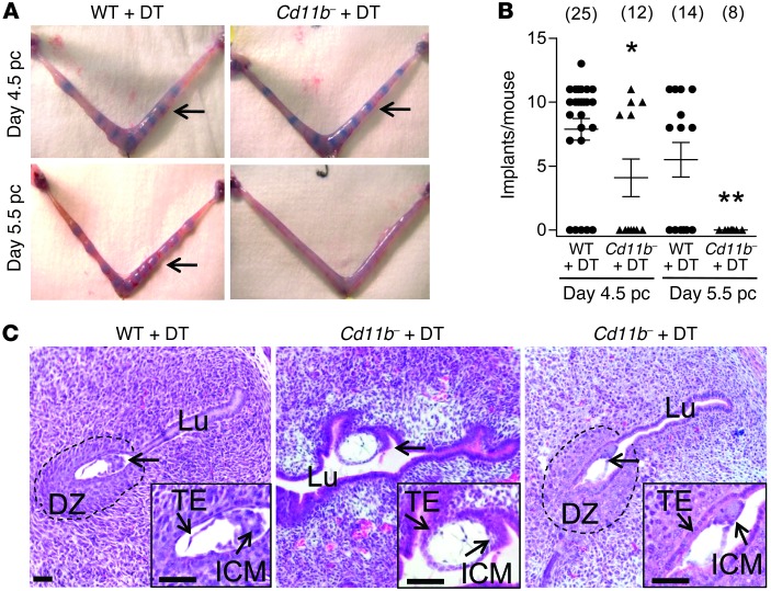
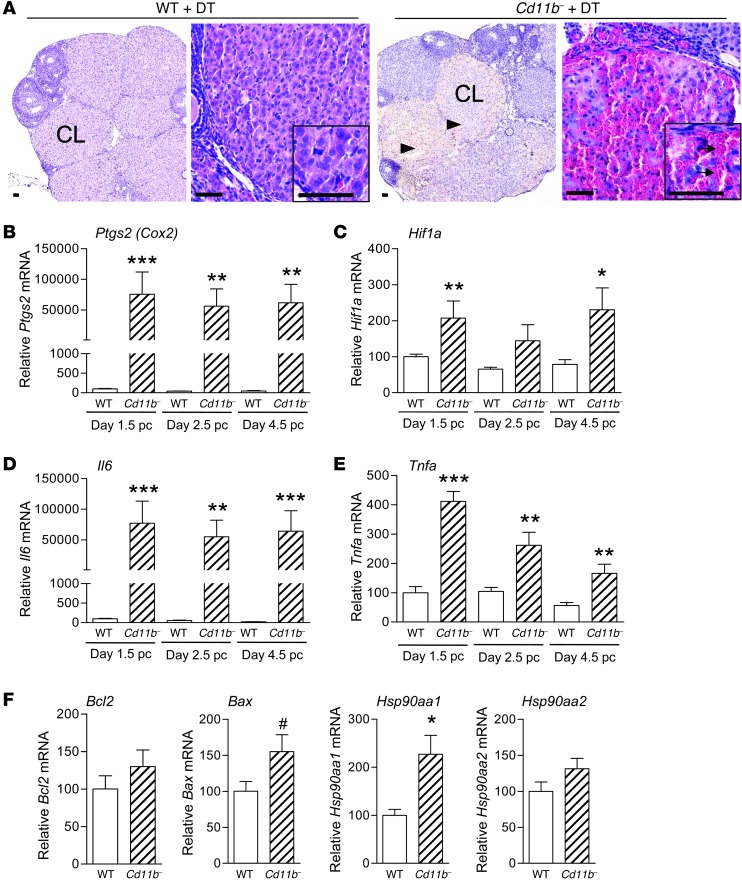
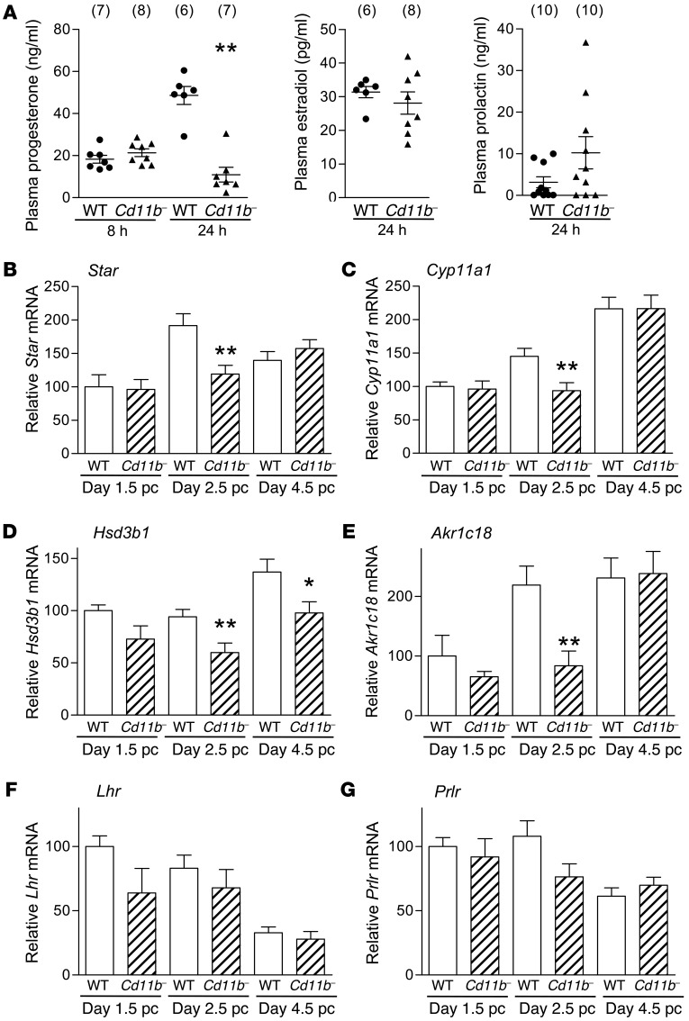
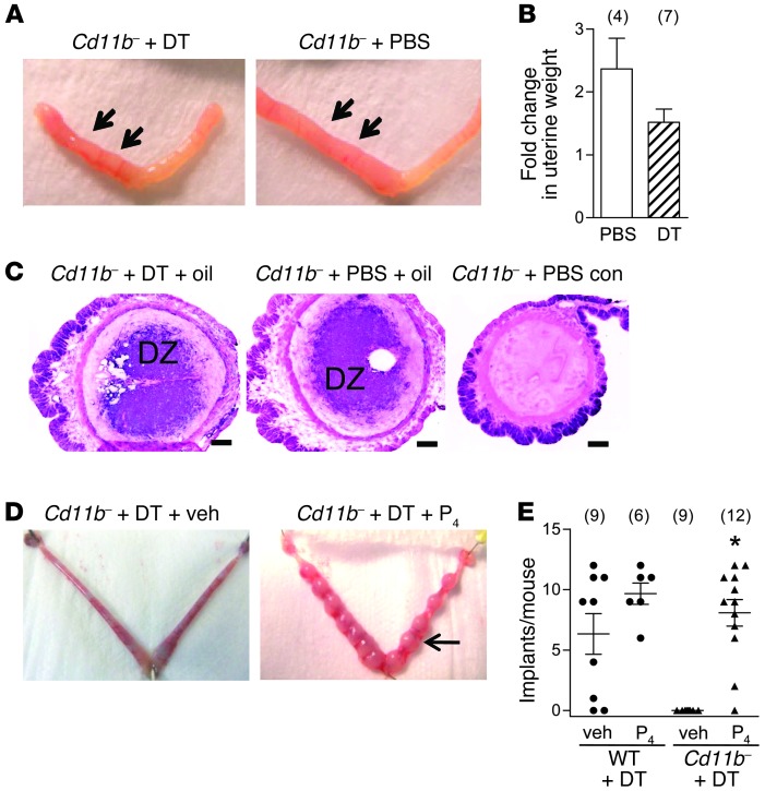
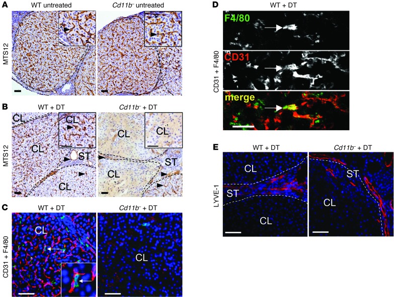
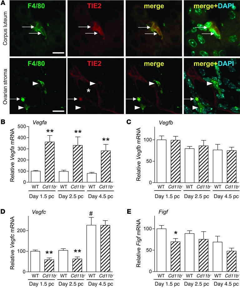
Macrophages are prominent in the uterus and ovary at conception. Here we utilize the Cd11b-Dtr mouse model of acute macrophage depletion to define the essential role of macrophages in early pregnancy. Macrophage depletion after conception caused embryo implantation arrest associated with diminished plasma progesterone and poor uterine receptivity. Implantation failure was alleviated by administration of bone marrow-derived CD11b+F4/80+ monocytes/macrophages. In the ovaries of macrophage-depleted mice, corpora lutea were profoundly abnormal, with elevated Ptgs2, Hif1a, and other inflammation and apoptosis genes and with diminished expression of steroidogenesis genes Star, Cyp11a1, and Hsd3b1. Infertility was rescued by exogenous progesterone, which confirmed that uterine refractoriness was fully attributable to the underlying luteal defect. In normally developing corpora lutea, macrophages were intimately juxtaposed with endothelial cells and expressed the proangiogenic marker TIE2. After macrophage depletion, substantial disruption of the luteal microvascular network occurred and was associated with altered ovarian expression of genes that encode vascular endothelial growth factors. These data indicate a critical role for macrophages in supporting the extensive vascular network required for corpus luteum integrity and production of progesterone essential for establishing pregnancy. Our findings raise the prospect that disruption of macrophage-endothelial cell interactions underpinning corpus luteum development contributes to infertility in women in whom luteal insufficiency is implicated.


Figures

Figure 1
Figure 1. DT administration to Cd11b-Dtr mice elicits macrophage depletion in the uterus and ovary.
Tissues were recovered from wild-type control or Cd11b-Dtr mice on day 4.5 pc, 24 hours after i.p. injection of DT (25 ng/g). (A and B) Sections of uterus (A) and ovary (B) labeled with anti-F4/80 indicated few macrophages (arrows) remaining in the uterus and ovary of Cd11b-Dtr mice (right panels; insets are high power) compared with wild-type mice (left panels). Some uterine F4/80+ cells were eosinophils (arrowheads), and these were retained after DT treatment (see Supplemental Figure 1). The percent positivity for F4/80+ cells is shown for wild-type mice (WT +DT) and macrophage-depleted Cd11b-Dtr mice (Cd11b- +DT) administered DT. Ten individual fields per uterus were analyzed, and the number of mice per group is indicated in parentheses. LE, luminal epithelium; Gl, uterine gland; CL, corpus luteum; ST, stroma. Scale bars: 50 μm. (C) Single-cell suspensions recovered from the peritoneal cavity (PEC) or prepared by enzymatic digestion of uterus and ovary were analyzed using FACS with anti-CD11b and anti-F4/80 antibodies, and show substantially diminished CD11b+F4/80+ macrophages in Cd11b-Dtr mice compared with wild-type controls. FACS plots are representative of 4–6 mice per group. Quantitative FACS data are given in Supplemental Table 1. *P < 0.05; **P < 0.01.
Figure 2
Figure 2. Macrophage depletion during the pre-implantation period causes complete infertility in Cd11b-Dtr mice.
(A) At autopsy on day 7.5 pc, macrophage-depleted Cd11b-Dtr mice had no visible implantation sites (lower panels) after injection with DT (25 ng/g) on day 0.5 pc or day 3.5 pc, compared with normal implantation sites (arrows) in wild-type mice given DT (upper panels). (B) Normal implantation sites (arrows) were seen when Cd11b-Dtr mice were reconstituted with wild-type bone marrow–derived CD11b+F4/80+ monocytes (Supplemental Figure 2) prior to DT injection on day 3.5 pc (Cd11b- +DT +BM). (C) Normal implantation sites (arrows) were also seen when Cd11b-Dtr mice were injected with biologically inactive [Glu52]-DT (Gl52-DT). (D) The number of implantation sites per mouse is shown for wild-type mice (WT +DT) and macrophage-depleted Cd11b-Dtr mice (Cd11b- +DT) administered DT on day 0.5 pc. (E) The number of implantation sites per mouse is shown for wild-type mice and Cd11b-Dtr mice administered PBS, DT or [Glu52]-DT on day 3.5 pc, or DT on day 3.5 pc after reconstitution with wild-type bone marrow-derived CD11b+F4/80+ monocytes (Cd11b- +DT +BM). Data are number of implantations per mouse, with mean ± SEM superimposed. The number of mice in each group is shown in parentheses. *P < 0.01, Cd11b- +DT versus WT +DT; #P < 0.01, Cd11b- +DT +BM versus Cd11b- +DT.
Figure 3
Figure 3. Implantation failure in macrophage-depleted Cd11b-Dtr mice is not due to adverse effects on pre-implantation embryo development.
(A) Embryos flushed on the morning of day 3.5 pc from macrophage-depleted Cd11b-Dtr mice, 24 hours following i.p. injection of DT (25 ng/g), were generally developed to blastocyst stage and often hatched from the zona pellucida, comparable to those flushed from wild-type mice treated with DT. (B) A comparable proportion of embryos flushed from the uterus on day 3.5 pc were developed to morula or blastocyst stage in wild-type or Cd11b-Dtr mice administered DT on day 2.5 pc. (C) Embryos that were flushed from the oviduct on day 0.5 pc (1-cell embryos) of gonadatropin-primed Cd11b-Dtr mice and cultured in the presence of DT (25 ng/ml) for 48 hours, and then without DT for a further 48 hours, developed to blastocyst stage at the same rate as embryos cultured without DT. (D) Embryos flushed from the uterus on day 2.5 pc (8-cell embryos) and cultured with DT (25 ng/ml) for 72 hours developed to blastocyst stage at the same rate as embryos cultured without DT. (BD) The number of embryos in each group is shown in parentheses. Data are mean ± SEM.
Figure 4
Figure 4. Infertility in macrophage-depleted Cd11b-Dtr mice results from implantation failure.
(A) Trypan blue clearly delineates bands of increased vascular permeability in the uterus, showing embryo implantation sites in some, but not all, macrophage-depleted Cd11b-Dtr mice (arrows) on day 4.5 pc (upper right), 24 hours after i.p. injection with DT (25 ng/g) on day 3.5 pc, compared with the majority of wild-type mice given DT (upper left). By day 5.5 pc, 48 hours after DT injection, no evidence of implantation was observed in any Cd11b-Dtr mice compared with normal implantation sites in wild-type mice. (B) The numbers of implantation sites per mouse at day 4.5 pc and day 5.5 pc are shown for wild-type mice (WT +DT) and macrophage-depleted Cd11b-Dtr mice (Cd11b- +DT), after injection of DT on day 3.5 pc. Data are number of implantations per mouse, with mean ± SEM superimposed. The number of mice per group is shown in parentheses. *P < 0.05, ** P < 0.01, Cd11b- +DT versus WT +DT. (C) Sections of uterus (H&E) from Cd11b-Dtr mice on day 4.5 pc (24 hours following DT injection) show that blastocyst-stage embryos (arrows) were attached laterally or in the middle of an open lumen with no decidual zone (10/12 embryos; middle panel), or less frequently were attached with a decidual zone, but incomplete uterine closure (2/12 embryos; right panel). This compared with wild-type mice, in which typical implantation sites in a narrowed endometrial lumen, with surrounding decidual zone, were consistently evident (left panel). Images are representative of 10–12 embryos in 4 WT and 5 Cd11b-Dtr mice. Insets are high-power. Lu, lumen; ICM, inner cell mass; TE, trophectoderm; DZ, decidual zone. Scale bars: 50 μm.
Figure 5
Figure 5. Infertility in macrophage-depleted Cd11b-Dtr mice is associated with structural demise of corpora lutea.
(A) Sections of ovary (low power, stained with hematoxylin) from macrophage-depleted Cd11b-Dtr mice collected on day 4.5 pc, 24 hours following i.p. injection of DT (25 ng/g), showed abnormal interstitial spaces within most corpora lutea (arrowheads). Sections of corpora lutea (inserts are high power, stained with H&E) were filled with red blood cells (arrows) and show atypical structure compared with wild-type control mice given DT. Scale bars: 50 μm. (BE) Expression of genes involved in luteolysis, hypoxia, and inflammation, including (B) Ptgs2 (Cox2), (C) Hif1a, (D) Il6, and (E) Tnfa, was upregulated in ovaries of Cd11b-Dtr mice on days 1.5, 2.5, and 4.5 pc, 24 hours after DT injection, compared with wild-type mice given DT. (F) Expression of genes regulating apoptosis, including Hsp90aa1 and Bax, was upregulated on day 4.5 pc 24 hours after DT injection, while expression of Bcl2 and Hsp90aa2 was unchanged. All genes were quantified by RT-PCR and normalized to Actb expression (n = 7–8 mice per group). Data are mean ± SEM relative to wild-type expression at day 1.5 (BE) or day 4.5 (F). #P = 0.06, *P < 0.05, **P < 0.01, ***P < 0.001, Cd11b- +DT versus WT +DT.
Figure 6
Figure 6. Luteal defects in macrophage-depleted Cd11b-Dtr mice are associated with diminished plasma progesterone and steroidogenic function.
(A) Plasma progesterone in macrophage-depleted Cd11b-Dtr mice was unchanged at 8 hours following i.p. injection of DT (25 ng/g) on day 3.5 pc, but by 24 hours was substantially lower than in wild-type mice given DT, while plasma estradiol and PRL were unchanged at 24 hours. Plasma PRL remained unchanged at 48 hours (Supplemental Figure 4). (BG) Expression of genes regulating steroidogenesis, including (B) Star, (C) Cyp11a, (D) Hsd3b1, and (E) Akr1c18, was downregulated in ovaries of Cd11b-Dtr mice on day 2.5 pc and day 4.5 pc, 24 hours after DT injection, compared with wild-type mice given DT. Expression of genes encoding (F) Lhr and (G) Prlr was unchanged on days 1.5, 2.5 and 4.5 pc following DT injection. All genes were quantified by RT-PCR and normalized to Actb expression (n = 7–8 mice per group). Data are mean ± SEM, relative to wild-type expression at day 1.5. *P < 0.05, **P < 0.01, Cd11b- +DT versus WT +DT.
Figure 7
Figure 7. Infertility in macrophage-depleted Cd11b-Dtr mice is rescued by administration of exogenous progesterone.
(A) Deciduoma formation was unchanged in Cd11b-Dtr mice given DT to deplete macrophages, compared with PBS-treated control Cd11b-Dtr mice. Deciduoma formation was measured after oil instillation into the left uterine horn (arrows), following ovariectomy and exogenous progesterone and estrogen replacement to mimic the physiological hormone environment of early pregnancy. (B) The fold-change in weight due to deciduoma (weight of oil-treated uterine horn / weight of control uterine horn) was comparable in Cd11b-Dtr mice given DT or PBS. (C) Alkaline phosphatase staining to detect decidual cells in sections of uterus from the oil-instilled left horn of the uterus showed a similar area of deciduoma in DT-treated and PBS-treated Cd11b-Dtr mice, with no deciduoma in the right, control horn. Scale bars: 200 μm. (D) At autopsy on day 7.5 pc, macrophage-depleted Cd11b-Dtr mice had normal implantation sites (arrow) when progesterone (P4) was administered following injection of DT (25 ng/g) on day 3.5 pc, compared with absence of implantation sites in macrophage-depleted Cd11b-Dtr mice given vehicle (veh; left panel). (E) The number of implantation sites per mouse is shown for wild-type mice (WT +DT) and macrophage-depleted Cd11b-Dtr mice (Cd11b- +DT) administered DT on day 3.5 pc, followed by progesterone or vehicle. Data represent the number of implantations per mouse, with mean ± SEM superimposed. The number of mice in each group is shown in parentheses. *P < 0.0001, Cd11b- +DT versus WT +DT.
Figure 8
Figure 8. The luteal defect in macrophage-depleted Cd11b-Dtr mice is accompanied by breakdown of the corpus luteum vasculature.
(AE) Ovaries were recovered from wild-type control or Cd11b-Dtr mice on day 3.5 pc before treatment (A) or on day 4.5 pc, 24 hours after i.p. injection of DT (25 ng/g) (BE). (A) Sections labeled with MTS-12 to detect blood vessel endothelial cells indicated a substantial network of intact vessels (arrows) in the corpus luteum of both Cd11b-Dtr and wild-type mice on day 3.5 pc. (B) Sections labeled with MTS-12 to detect blood vessel endothelial cells indicated few intact vessels in the corpus luteum of most Cd11b-Dtr compared with wild-type mice, while vessels in the ovarian stroma (arrowheads) and some corpora lutea (arrows) remained intact on day 4.5 pc, following DT injection 24 hours earlier. (C) Sections labeled with antibodies to both CD31 (red) to detect endothelial cells and F4/80 (green) to detect macrophages showed absence of blood vessels in the corpus luteum of Cd11b-Dtr compared with wild-type mice, highlighting the close spatial association between endothelial cells and macrophages (inset is high power). (D) In sections labeled with both CD31 and F4/80, cells co-expressing both markers (arrows) were evident. (E) Sections labeled with LYVE-1 (red) to detect lymphatic endothelial cells showed lymphatic vessels at the margins of a corpus luteum of Cd11b-Dtr mice, similar to wild-type mice. Photomicrographs are representative of 6–7 mice per group. Scale bars: 50 μm.
Figure 9
Figure 9. Macrophages in the corpus luteum have a proangiogenic phenotype, and macrophage depletion in Cd11b-Dtr mice is accompanied by altered expression of VEGF genes.
(A) Sections of ovary from wild-type mice on day 2.5 pc labeled with antibodies to TIE2 and F4/80 showed that macrophages in the corpus luteum were TIE2+, indicative of a proangiogenic phenotype. Both TIE2+ and TIE2 macrophages were seen adjacent to TIE2+ blood vessels in the ovarian stroma (arrows, F4/80+TIE2+ macrophages; short arrows, F4/80+TIE2 macrophages; *vessel lumen). Scale bars: 10 μm. (BE) Expression of angiogenesis genes was altered in ovaries in association with macrophage depletion from Cd11b-Dtr mice. (B) Vegfa was upregulated in ovaries of Cd11b-Dtr mice on days 1.5 pc, 2.5 pc, and 4.5 pc, 24 hours following DT injection, compared with wild-type mice given DT. (C) Vegfb was not changed at any time point following DT injection. (D) Vegfc was downregulated in ovaries of Cd11b-Dtr mice on days 1.5 and 2.5 pc following DT injection. (E) Figf was downregulated in ovaries of Cd11b-Dtr mice on day 1.5 pc following DT injection. All genes were quantified using RT-PCR and normalized to Actb expression (n = 6–10 mice per group). Data are mean ± SEM relative to wild-type expression at day 1.5. *P < 0.05, **P < 0.01, Cd11b- +DT versus WT +DT at same time point; #P < 0.01 WT +DT at day 4.5 pc versus day 1.5 pc.

References

    1. Gordon S. The macrophage: past, present and future. Eur J Immunol. 2007;37(suppl 1):S9–S17. doi: 10.1002/eji.200737638. - DOI - PubMed
    1. Sunderkotter C, Steinbrink K, Goebeler M, Bhardwaj R, Sorg C. Macrophages and angiogenesis. J Leukoc Biol. 1994;55(3):410–422. - PubMed
    1. Lamagna C, Aurrand-Lions M, Imhof BA. Dual role of macrophages in tumor growth and angiogenesis. J Leukoc Biol. 2006;80(4):705–713. doi: 10.1189/jlb.1105656. - DOI - PubMed
    1. Pollard JW. Trophic macrophages in development and disease. Nat Rev Immunol. 2009;9(4):259–270. doi: 10.1038/nri2528. - DOI - PMC - PubMed
    1. Hume DA, Ross IL, Himes SR, Sasmono RT, Wells CA, Ravasi T. The mononuclear phagocyte system revisited. J Leukoc Biol. 2002;72(4):621–627. - PubMed

Publication types

MeSH terms