

Concurrent TMS to the primary motor cortex augments slow motor learning
- PMID: 23867557
- PMCID: PMC4331120
- DOI: 10.1016/j.neuroimage.2013.07.024
Concurrent TMS to the primary motor cortex augments slow motor learning
Abstract
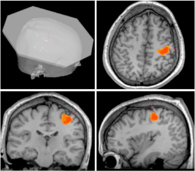
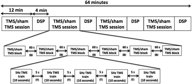
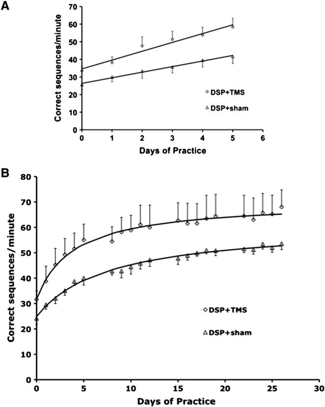
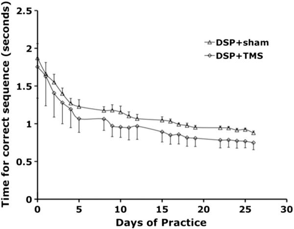
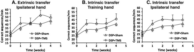
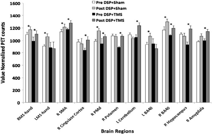
Transcranial magnetic stimulation (TMS) has shown promise as a treatment tool, with one FDA approved use. While TMS alone is able to up- (or down-) regulate a targeted neural system, we argue that TMS applied as an adjuvant is more effective for repetitive physical, behavioral and cognitive therapies, that is, therapies which are designed to alter the network properties of neural systems through Hebbian learning. We tested this hypothesis in the context of a slow motor learning paradigm. Healthy right-handed individuals were assigned to receive 5 Hz TMS (TMS group) or sham TMS (sham group) to the right primary motor cortex (M1) as they performed daily motor practice of a digit sequence task with their non-dominant hand for 4 weeks. Resting cerebral blood flow (CBF) was measured by H2(15)O PET at baseline and after 4 weeks of practice. Sequence performance was measured daily as the number of correct sequences performed, and modeled using a hyperbolic function. Sequence performance increased significantly at 4 weeks relative to baseline in both groups. The TMS group had a significant additional improvement in performance, specifically, in the rate of skill acquisition. In both groups, an improvement in sequence timing and transfer of skills to non-trained motor domains was also found. Compared to the sham group, the TMS group demonstrated increases in resting CBF specifically in regions known to mediate skill learning namely, the M1, cingulate cortex, putamen, hippocampus, and cerebellum. These results indicate that TMS applied concomitantly augments behavioral effects of motor practice, with corresponding neural plasticity in motor sequence learning network. These findings are the first demonstration of the behavioral and neural enhancing effects of TMS on slow motor practice and have direct application in neurorehabilitation where TMS could be applied in conjunction with physical therapy.
Keywords: Digit sequence practice; Hebbian learning; Hyperbolic function; Motor learning; Motor learning network; Motor system; Primary motor cortex; Skill transfer; TMS.
© 2013.
Figures

Similar articles
-
Neck muscle fatigue impacts plasticity and sensorimotor integration in cerebellum and motor cortex in response to novel motor skill acquisition.J Neurophysiol. 2020 Sep 1;124(3):844-855. doi: 10.1152/jn.00437.2020. Epub 2020 Aug 5. J Neurophysiol. 2020. PMID: 32755363 Free PMC article.
-
Motor cortex plasticity and visuomotor skill learning in upper and lower limbs of endurance-trained cyclists.Eur J Appl Physiol. 2022 Jan;122(1):169-184. doi: 10.1007/s00421-021-04825-y. Epub 2021 Oct 7. Eur J Appl Physiol. 2022. PMID: 34618222
-
Effect of slow repetitive TMS of the motor cortex on ipsilateral sequential simple finger movements and motor skill learning.Restor Neurol Neurosci. 2010;28(4):437-48. doi: 10.3233/RNN-2010-0562. Restor Neurol Neurosci. 2010. PMID: 20714068
-
Transcranial magnetic stimulation and the motor learning-associated cortical plasticity.Exp Brain Res. 2006 Aug;173(2):215-22. doi: 10.1007/s00221-006-0538-z. Epub 2006 May 30. Exp Brain Res. 2006. PMID: 16733699 Review.
-
Intensity-dependent regional cerebral blood flow during 1-Hz repetitive transcranial magnetic stimulation (rTMS) in healthy volunteers studied with H215O positron emission tomography: I. Effects of primary motor cortex rTMS.Biol Psychiatry. 2003 Oct 15;54(8):818-25. doi: 10.1016/s0006-3223(03)00002-7. Biol Psychiatry. 2003. PMID: 14550681 Review.
Cited by
-
Repetitive transcranial magnetic stimulation improves both hearing function and tinnitus perception in sudden sensorineural hearing loss patients.Sci Rep. 2015 Oct 14;5:14796. doi: 10.1038/srep14796. Sci Rep. 2015. PMID: 26463446 Free PMC article. Clinical Trial.
-
SeXX Matters in Multiple Sclerosis.Front Neurol. 2020 Jul 3;11:616. doi: 10.3389/fneur.2020.00616. eCollection 2020. Front Neurol. 2020. PMID: 32719651 Free PMC article. Review.
-
Low intensity repetitive transcranial magnetic stimulation modulates skilled motor learning in adult mice.Sci Rep. 2018 Mar 5;8(1):4016. doi: 10.1038/s41598-018-22385-8. Sci Rep. 2018. PMID: 29507375 Free PMC article.
-
Explaining Individual Differences in Motor Behavior by Intrinsic Functional Connectivity and Corticospinal Excitability.Front Neurosci. 2020 Feb 5;14:76. doi: 10.3389/fnins.2020.00076. eCollection 2020. Front Neurosci. 2020. PMID: 32116520 Free PMC article.
-
Online repetitive transcranial magnetic stimulation during working memory in younger and older adults: A randomized within-subject comparison.PLoS One. 2019 Mar 22;14(3):e0213707. doi: 10.1371/journal.pone.0213707. eCollection 2019. PLoS One. 2019. PMID: 30901345 Free PMC article. Clinical Trial.
References
-
- Adkins-Muir DL, Jones TA. Cortical electrical stimulation combined with rehabilitative training: enhanced functional recovery and dendritic plasticity following focal cortical ischemia in rats. Neurol. Res. 2003;25(8):780–788. - PubMed
-
- Agostino R, Iezzi E, Dinapoli L, Gilio F, Conte A, Mari F, Berardelli A. Effects of 5 Hz subthreshold magnetic stimulation of primary motor cortex on fast finger movements in normal subjects. Exp. Brain Res. 2007;180(1):105–111. - PubMed
-
- Albouy G, Sterpenich V, Balteau E, Vandewalle G, Desseilles M, ng-Vu T, et al. Both the hippocampus and striatum are involved in consolidation of motor sequence memory. Neuron. 2008;58:261–272. - PubMed
-
- Barker AT, Jalinous R, Freeston IL. Non-invasive magnetic stimulation of the human motor cortex. Lancet. 1985;1:1106–1107. - PubMed
-
- Boggio PS, Nunes A, Rigonatti SP, Nitsche MA, Pascual-Leone A, Fregni F. Repeated sessions of noninvasive brain DC stimulation is associated with motor function improvement in stroke patients. Restor. Neurol. Neurosci. 2007;25(2):123–129. - PubMed
Publication types
MeSH terms
Grants and funding
LinkOut - more resources
Full Text Sources
Other Literature Sources
