

High prevalence of contralateral ankle abnormalities in association with knee osteoarthritis and malalignment
- PMID: 23867580
- PMCID: PMC3795812
- DOI: 10.1016/j.joca.2013.07.008
High prevalence of contralateral ankle abnormalities in association with knee osteoarthritis and malalignment
Abstract
Objective: To evaluate ankle joint abnormalities in a knee osteoarthritis (OA) cohort.
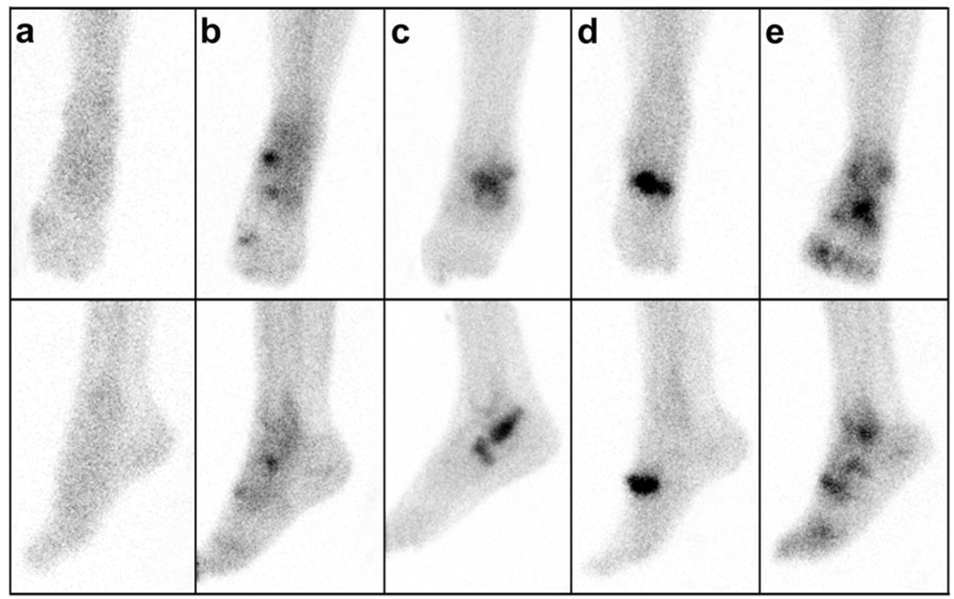
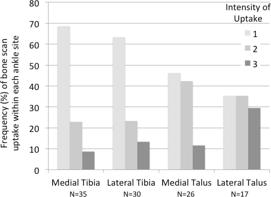
Methods: Participants (n = 159) with symptomatic and radiographic OA in at least one knee underwent technetium-99m methylene diphosphonate bone scan (scored 0-3) of the ankles and forefeet. Knee radiographs were graded for OA features of joint space narrowing (JSN) and osteophyte (OST). Ankle symptoms and history of ankle injury were assessed by self-report. Knee alignment was measured from a long-limb radiograph. Ankle radiographs were obtained on those who returned for follow-up (n = 138) and were graded for ankle tibiotalar JSN and OST.
Design: Ankle scintigraphic abnormalities were frequent (31% of individuals, one-third bilateral). Ankle symptoms were reported by 23% of individuals and history of ankle injury by 24%. Controlling for gender, age, body mass index (BMI), and contralateral predictor, ankle scintigraphic abnormalities were associated with: ipsilateral ankle symptoms (P = 0.005); contralateral knee JSN (P = 0.001), knee OST (P = 0.006) and knee malalignment (P = 0.08); and history of ankle injury or surgery of either ankle (P < 0.0001). At follow-up, scintigraphic abnormalities of the ankle were strongly associated with presence of tibiotalar radiographic OA (P < 0.0001).
Conclusions: Although considered rare, we observed a high prevalence of radiographic features of ankle OA in this knee OA cohort. History of overt ankle injury did not appear to account for the majority of ankle abnormalities. These results are consistent with a probable kinematic association of knee OA pathology and contralateral ankle abnormalities and suggest that interventions targeting mechanical factors may be needed to prevent ankle OA in the setting of knee OA.
Keywords: Alignment; Ankle; Knee; Osteoarthritis; Scintigraphy.
Copyright © 2013 Osteoarthritis Research Society International. Published by Elsevier Ltd. All rights reserved.
Figures
References
-
- Aurich M, Squires GR, Reiner A, Mollenhauer JA, Kuettner KE, Poole AR, et al. Differential matrix degradation and turnover in early cartilage lesions of human knee and ankle joints. Arthritis Rheum. 2005;52:112–119. - PubMed
-
- Huch K, Kuettner KE, Dieppe P. Osteoarthritis in ankle and knee joints. Semin Arthritis Rheum. 1997;26:667–674. - PubMed
-
- Kang Y, Koepp H, Cole AA, Kuettner KE, Homandberg GA. Cultured human ankle and knee cartilage differ in susceptibility to damage mediated by fibronectin fragments. J Orthop Res. 1998;16:551–556. - PubMed
-
- Huch K. Knee and ankle: human joints with different susceptibility to osteoarthritis reveal different cartilage cellularity and matrix synthesis in vitro. Arch Orthop Trauma Surg. 2001;121:301–306. - PubMed
-
- Eger W, Schumacher BL, Mollenhauer J, Kuettner KE, Cole AA. Human knee and ankle cartilage explants: catabolic differences. J Orthop Res. 2002;20:526–534. - PubMed
Publication types
MeSH terms
Grants and funding
LinkOut - more resources
Full Text Sources
Other Literature Sources
