Day-night differences in neural activation in histaminergic and serotonergic areas with putative projections to the cerebrospinal fluid in a diurnal brain
- PMID: 23867764
- PMCID: PMC3845805
- DOI: 10.1016/j.neuroscience.2013.07.007
Day-night differences in neural activation in histaminergic and serotonergic areas with putative projections to the cerebrospinal fluid in a diurnal brain
Abstract
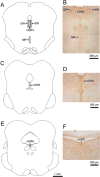
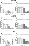
In nocturnal rodents, brain areas that promote wakefulness have a circadian pattern of neural activation that mirrors the sleep/wake cycle, with more neural activation during the active phase than during the rest phase. To investigate whether differences in temporal patterns of neural activity in wake-promoting regions contribute to differences in daily patterns of wakefulness between nocturnal and diurnal species, we assessed Fos expression patterns in the tuberomammillary (TMM), supramammillary (SUM), and raphe nuclei of male grass rats maintained in a 12:12 h light-dark cycle. Day-night profiles of Fos expression were observed in the ventral and dorsal TMM, in the SUM, and in specific subpopulations of the raphe, including serotonergic cells, with higher Fos expression during the day than during the night. Next, to explore whether the cerebrospinal fluid is an avenue used by the TMM and raphe in the regulation of target areas, we injected the retrograde tracer cholera toxin subunit beta (CTB) into the ventricular system of male grass rats. While CTB labeling was scarce in the TMM and other hypothalamic areas including the suprachiasmatic nucleus, which contains the main circadian pacemaker, a dense cluster of CTB-positive neurons was evident in the caudal dorsal raphe, and the majority of these neurons appeared to be serotonergic. Since these findings are in agreement with reports for nocturnal rodents, our results suggest that the evolution of diurnality did not involve a change in the overall distribution of neuronal connections between systems that support wakefulness and their target areas, but produced a complete temporal reversal in the functioning of those systems.
Keywords: 3V; 4V; 5HT; CP; CSF; CTB; DR; DTM; Fos; HA; ICC; LSd; LV; ME; MR; NDS; NGS; PB; PBS; PLP; PVN; SCN; SUM; TBS; TMM; TX; VTM; ZT; cerebrospinal fluid; cholera toxin subunit beta; choroid plexus; circadian; dorsal raphe; dorsal tuberomammillary nucleus; fourth ventricle; grass rat; histamine; immunocytochemistry; lDR; lateral dorsal raphe; lateral septal nucleus, dorsal part; lateral ventricle; mDR3–5; medial dorsal raphe levels 3 through 5; median eminence; median raphe; normal donkey serum; normal goat serum; paraformaldehyde–lysine–sodium periodate; paraventricular nucleus of the hypothalamus; phosphate buffer; phosphate-buffered saline; serotonin; suprachiasmatic nucleus; supramammillary nucleus; third ventricle; tris-buffered saline; triton-X; tuberomammillary nucleus; vSPZ; ventral subparaventricular zone; ventral tuberomammillary nucleus; zeitgeber time.
Copyright © 2013 IBRO. Published by Elsevier Ltd. All rights reserved.
Conflict of interest statement
The authors state no conflict of interest.
Figures


References
-
- Aghajanian GK, Gallager DW. Raphe origin of serotonergic nerves terminating in the cerebral ventricles. Brain Res. 1975;88:221–231. - PubMed
-
- Barassin S, Raison S, Saboureau M, Bienvenu C, Maitre M, Malan A, Pevet P. Circadian tryptophan hydroxylase levels and serotonin release in the suprachiasmatic nucleus of the rat. Eur J Neurosci. 2002;15:833–840. - PubMed
Publication types
MeSH terms
Substances
Grants and funding
LinkOut - more resources
Full Text Sources
Other Literature Sources
Research Materials
Miscellaneous

