. 2013 Jul 18;4(7):e728.
doi: 10.1038/cddis.2013.259.

Reolysin is a novel reovirus-based agent that induces endoplasmic reticular stress-mediated apoptosis in pancreatic cancer

Affiliations

Reolysin is a novel reovirus-based agent that induces endoplasmic reticular stress-mediated apoptosis in pancreatic cancer

J S Carew et al. Cell Death Dis. .

Abstract

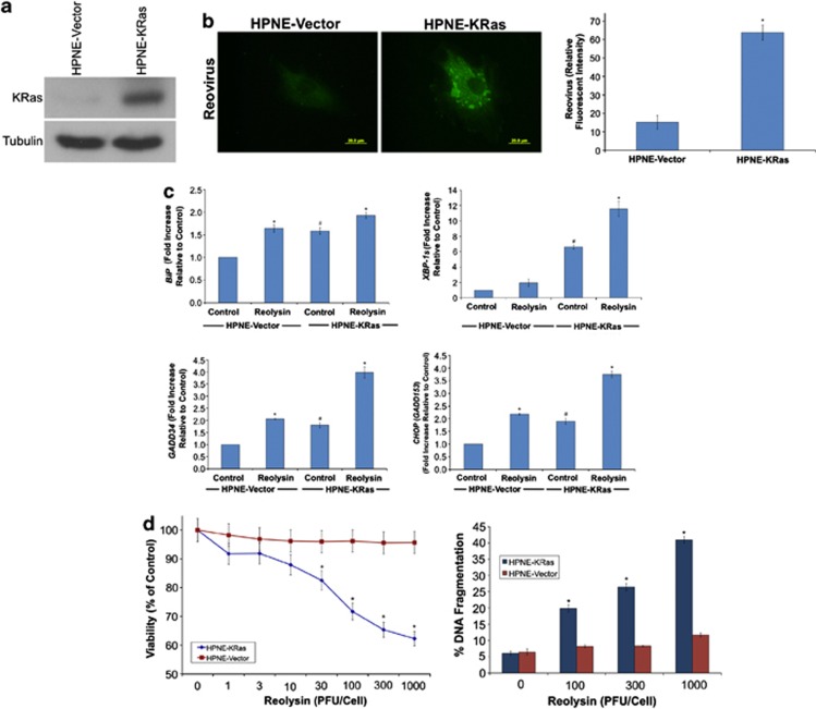
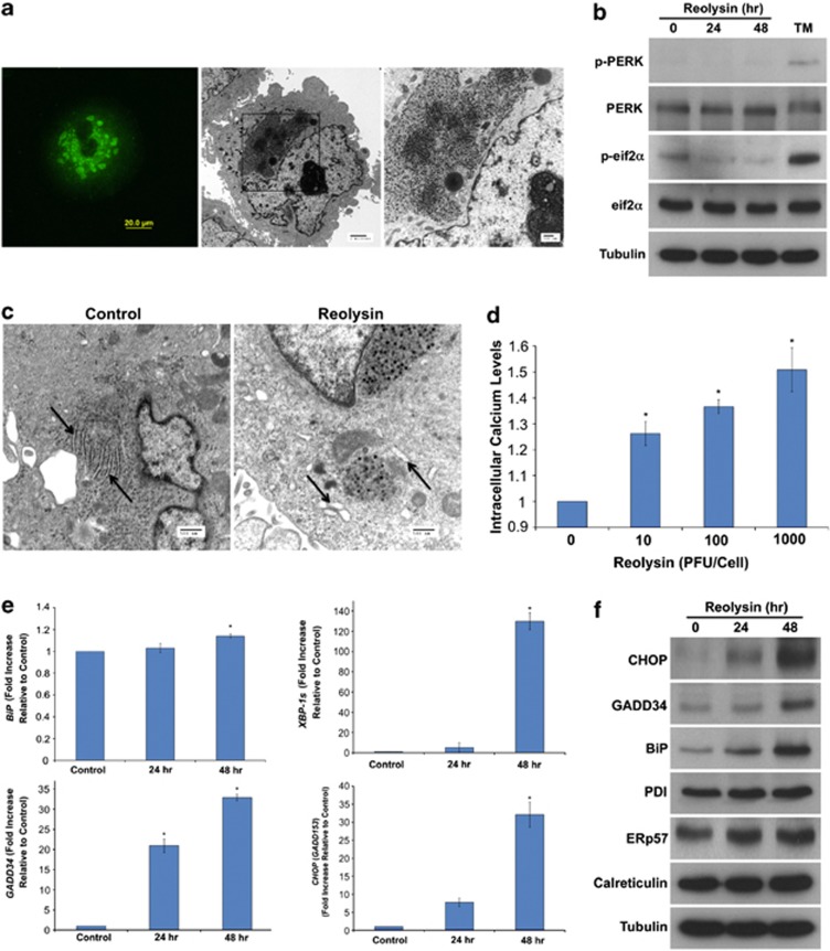
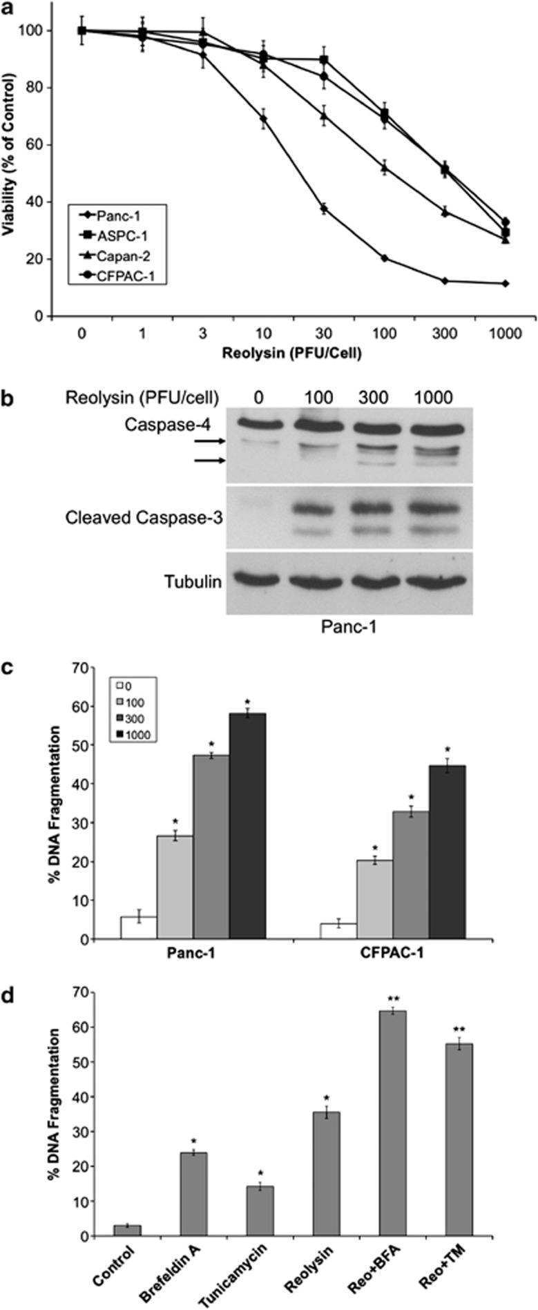
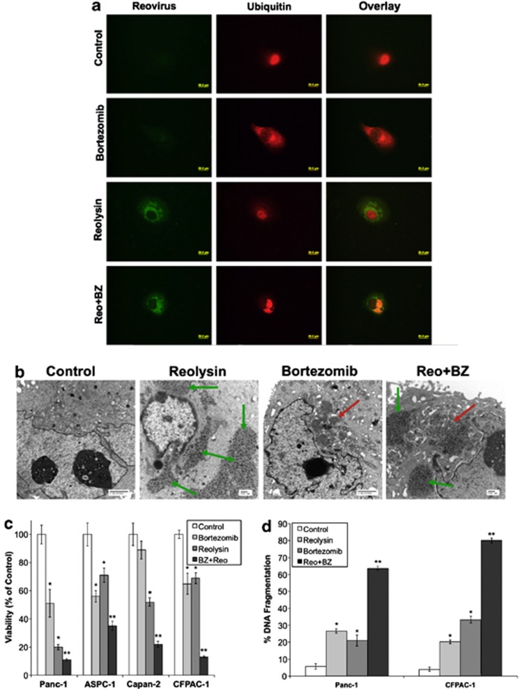
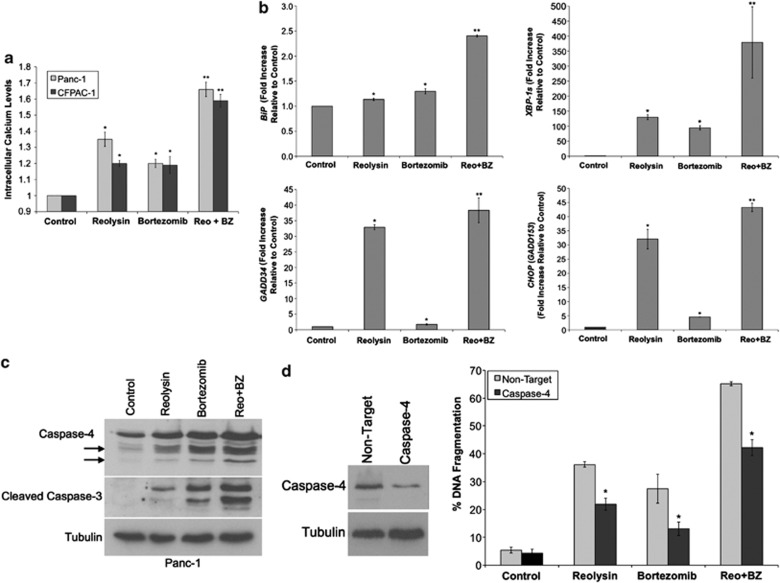
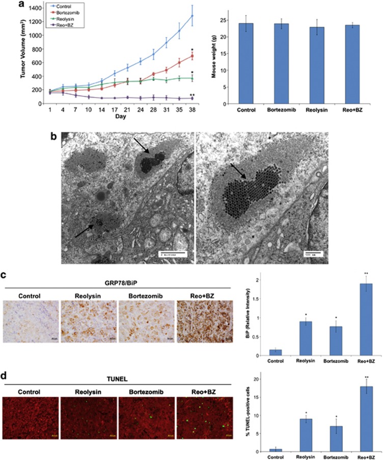
Activating mutation of KRas is a genetic alteration that occurs in the majority of pancreatic tumors and is therefore an ideal therapeutic target. The ability of reoviruses to preferentially replicate and induce cell death in transformed cells that express activated Ras prompted the development of a reovirus-based formulation for cancer therapy called Reolysin. We hypothesized that Reolysin exposure would trigger heavy production of viral products leading to endoplasmic reticular (ER) stress-mediated apoptosis. Here, we report that Reolysin treatment stimulated selective reovirus replication and decreased cell viability in KRas-transformed immortalized human pancreatic duct epithelial cells and pancreatic cancer cell lines. These effects were associated with increased expression of ER stress-related genes, ER swelling, cleavage of caspase-4, and splicing of XBP-1. Treatment with ER stress stimuli including tunicamycin, brefeldin A, and bortezomib (BZ) augmented the anticancer activity of Reolysin. Cotreatment with BZ and Reolysin induced the simultaneous accumulation of ubiquitinated and viral proteins, resulting in enhanced levels of ER stress and apoptosis in both in vitro and in vivo models of pancreatic cancer. Our collective results demonstrate that the abnormal protein accumulation induced by the combination of Reolysin and BZ promotes heightened ER stress and apoptosis in pancreatic cancer cells and provides the rationale for a phase I clinical trial further investigating the safety and efficacy of this novel strategy.


Figures

Figure 1
Figure 1
Reovirus preferentially replicates in KRas-transfected immortalized normal human pancreatic epithelial cells. (a) KRas transfected HPNE cells. Immunoblotting demonstrates KRas levels in HPNE cells. (b) Reovirus replicates preferentially in HPNE-KRas cells. Cells were treated with Reolysin for 48 h and stained with an anti-reovirus antibody. Immunocytochemistry reveals reovirus replication in KRas-transfected cells. Fluorescent intensity was quantified in HPNE and HPNE-KRas cells using Image-Pro Plus software version 6.2.1. Mean±S.D., n=5. *Indicates a significant difference compared with HPNE-vector cells. (c) KRas-transfected cells display higher levels of ER stress-related gene expression that can be further induced with reovirus exposure. HPNE-vector and HPNE-KRas cells were treated with 100 plaque-forming units (PFU)/cell Reolysin for 48 h. Gene expression was determined by qRT-PCR. Mean±S.D., n=3. #Represents a significant difference compared with vector control cells. *Indicates a significant difference compared with corresponding controls. (d) HPNE-KRas cells are sensitive to Reolysin-mediated cell death. Cells were treated for 72 h with the indicated concentrations of Reolysin, and cell viability was determined by MTT assay (left panel). Cells were treated for 48 h with Reolysin, and apoptosis was measured by PI-FACS analysis (right panel). Mean±S.D., n=3. *Indicates a significant difference compared with HPNE-vector cells treated with the same concentration of Reolysin P<0.05
Figure 2
Figure 2
Reolysin induces ER stress. (a) Reovirus replication in Panc-1 cells. Cells were treated with 100 PFU/cell Reolysin for 48 h. Reovirus replication was detected by immunocytochemistry and electron microscopy. (b) Reolysin does not promote PERK or eif2α phosphorylation. Panc-1 cells were treated with 100 PFU/cell Reolysin for 24 and 48 h or with 5 μg/ml tunicamycin (24 h) as a positive control. Proteins were detected by immunoblotting. (c) Reolysin promotes ER swelling. Panc-1 cells were treated with 100 PFU/cell Reolysin for 48 h, and ER morphology was visualized by electron microscopy. Arrows denote endoplasmic reticulum. (d) Reolysin treatment increases intracellular calcium levels. Panc-1 cells were treated with the indicated amounts of Reolysin for 16 h, and intracellular calcium levels were detected by calcium green-1 staining and flow cytometry. Mean±S.D., n=3. *Represents a significant difference compared with controls. (e) qRT-PCR analysis of BiP, GADD34, CHOP, and XBP-1s expression in Panc-1 cells. Cells were treated with 100 PFU/cell Reolysin for 24 and 48 h and then harvested for analysis. Levels of mRNAs were standardized to the expression of GAPDH. Mean±S.D., n=3. *Indicates a significant difference from the control. P<0.05. (f) Immunoblotting analysis of CHOP, GADD34, BiP, PDI, ERp57, and calreticulin. Panc-1 cells were treated with 100 PFU/cell Reolysin for 24 or 48 h. ER-related protein expression was measured by immunoblotting
Figure 3
Figure 3
Reolysin promotes caspase-4 processing and apoptosis and sensitizes cells to ER stress-mediated apoptosis. (a) Reolysin decreases cell viability in a panel of pancreatic cancer cell lines. Cells were treated with the indicated concentrations of Reolysin for 72 h. Cell viability was measured by MTT assay. Mean±S.D., n=3. (b) Reolysin promotes cleavage of caspase-4 and caspase-3. Panc-1 cells were treated with the indicated concentrations of Reolysin for 48 h, and caspase cleavage was measured by immunoblotting. Arrows denote cleaved caspase-4 fragments. (c) Reolysin induces apoptosis. Panc-1 and CFPAC-1 cells were treated with Reolysin for 48 h. Apoptosis was measured by PI-FACS analysis. Mean±S.D., n=3. *Indicates a significant difference compared with controls. (d) Reolysin augments ER stress-mediated apoptosis. Panc-1 cells were treated for 48 h with 300 PFU/cell Reolysin, 5 μg/ml tunicamycin, 5 μM brefeldin A, and combinations. Mean±S.D., n=3. *Indicates a significant difference compared with controls; **indicates a significant difference compared with either single-agent treatment P<0.05
Figure 4
Figure 4
Reolysin augments the anticancer activity of BZ. (a and b) Reolysin and BZ stimulate reoviral and ubiquitinated protein accumulation. Cells were treated with 100 PFU/cell Reolysin and 10 nM BZ for 48 h. Protein accumulation was visualized by (a) immunocytochemistry and (b) electron microscopy. Red arrows indicate ubiquitinated protein aggregates, and green arrows depict reovirus replication. (c) Reolysin and BZ reduce cell viability. Pancreatic cancer cells were treated with 100 PFU/cell Reolysin and 10 nM BZ for 72 h. Cell viability was measured by MTT assay. *Indicates a significant difference from controls and **denotes a significant difference compared to either single agent treatment group (P<0.05). (d) Reolysin and BZ induce apoptosis. Cells were treated with 100 PFU/cell Reolysin and 10 nM BZ for 48 h. Apoptosis was determined by PI-FACS analysis. Mean±S.D., n=3. *Represents a significant difference from controls. **Indicates a significant difference compared with single-agent treatment groups P<0.05
Figure 5
Figure 5
Reolysin enhances BZ-mediated ER stress and apoptosis. (a) The Reolysin and BZ combination increases intracellular calcium levels. Cells were treated with 100 PFU/cell Reolysin and 10 nM BZ for 16 h, and intracellular calcium levels were detected by calcium green-1 staining and flow cytometry. Mean±S.D., n=3. *Represents a significant difference compared with controls, and **indicates a significant difference compared with either single-agent treatment. (b) Reolysin augments BZ-induced increases in ER stress-related gene expression. Panc-1 cells were treated with 100 PFU/cell Reolysin and 10 nM BZ for 48 h and then harvested for analysis. Levels of mRNA were standardized to the expression of GAPDH. Mean±S.D., n=3. *Indicates a significant difference from the control. **Indicates a significant difference compared with single-agent treatment groups. (c) Reolysin enhances BZ-mediated cleavage of caspase-4 and caspase-3. Cells were treated with 100 PFU/cell Reolysin and 10 nM BZ for 48 h. Caspase cleavage was measured by immunoblotting. Arrows denote caspase-4 cleavage fragments. (d) Knockdown of caspase-4 reduces Reolysin- and BZ-induced apoptosis. siRNA-mediated knockdown of caspase-4 was determined by immunoblotting at 72 h post transfection. Cells were treated with Reolysin and BZ 24 h post transfection for 48 h. Apoptosis was measured by PI-FACS analysis. Mean±S.D., n=3. *Indicates a significant difference compared with non-target-transfected cells treated under the same conditions P<0.05
Figure 6
Figure 6
The combination of Reolysin and BZ strongly reduces tumor burden in the Panc-1 xenograft model. (a) Panc-1 cells (1 × 10⁷ per mouse) were injected into the flanks of nude mice. When tumors reached approximately 150 mm3 in size, mice were randomized into groups and treated with 0.5 mg BZ per kg Q3D, 5 × 10⁸ TCID50 Reolysin Q7D, or both agents for 5 weeks. Tumors were measured twice weekly. Mean±S.E.M., n=8. *Indicates a significant difference compared with vehicle, or **indicates a significant difference compared with either single-agent treatment. P<0.05. Reolysin and BZ are well tolerated in vivo. Animal body weight was determined at the end of the study (day 38) to quantify drug-induced weight loss. Mean±S.D., n=8. (b) Reovirus replicates in tumors in vivo. Electron microscopy was performed on tumors collected from Reolysin-treated animals and revealed the presence of reovirus. Images shown were taken from an animal treated with the Reolysin+BZ combination. Arrows denote the presence of reovirus. Similar results were observed in mice treated with Reolysin alone. (c) Reolysin and BZ increase BiP expression. BiP expression was measured by IHC, and staining intensity was quantified using ImageJ software. *Indicates a significant difference compared with controls, and **denotes a significant difference compared with either single-agent treatment group (P<0.05). (d) Apoptosis was measured by TUNEL staining. Quantification was conducted by manually counting TUNEL-positive cells. Mean±S.D., n=5. *Indicates a significant difference compared with controls, and **represents a significant difference compared with single-agent treatments P<0.05

References

    1. Bos JL. Ras oncogenes in human cancer: a review. Cancer Res. 1989;49:4682–4689. - PubMed
    1. Fearon ER. K-ras gene mutation as a pathogenetic and diagnostic marker in human cancer. J Natl Cancer Inst. 1993;85:1978–1980. - PubMed
    1. Malumbres M, Barbacid M. RAS oncogenes: the first 30 years. Nat Rev Cancer. 2003;3:459–465. - PubMed
    1. Kelly K, Nawrocki S, Mita A, Coffey M, Giles FJ, Mita M. Reovirus-based therapy for cancer. Expert Opin Biol Ther. 2009;9:817–830. - PubMed
    1. Maitra R, Ghalib MH, Goel S. Reovirus: a targeted therapeutic –progress and potential. Mol Cancer Res. 2012;10:1514–1525. - PMC - PubMed

Publication types

MeSH terms

LinkOut - more resources