. 2013 Jul 18;4(7):e729.
doi: 10.1038/cddis.2013.262.

Pax8 has a critical role in epithelial cell survival and proliferation

Affiliations

Pax8 has a critical role in epithelial cell survival and proliferation

T Di Palma et al. Cell Death Dis. .

Abstract

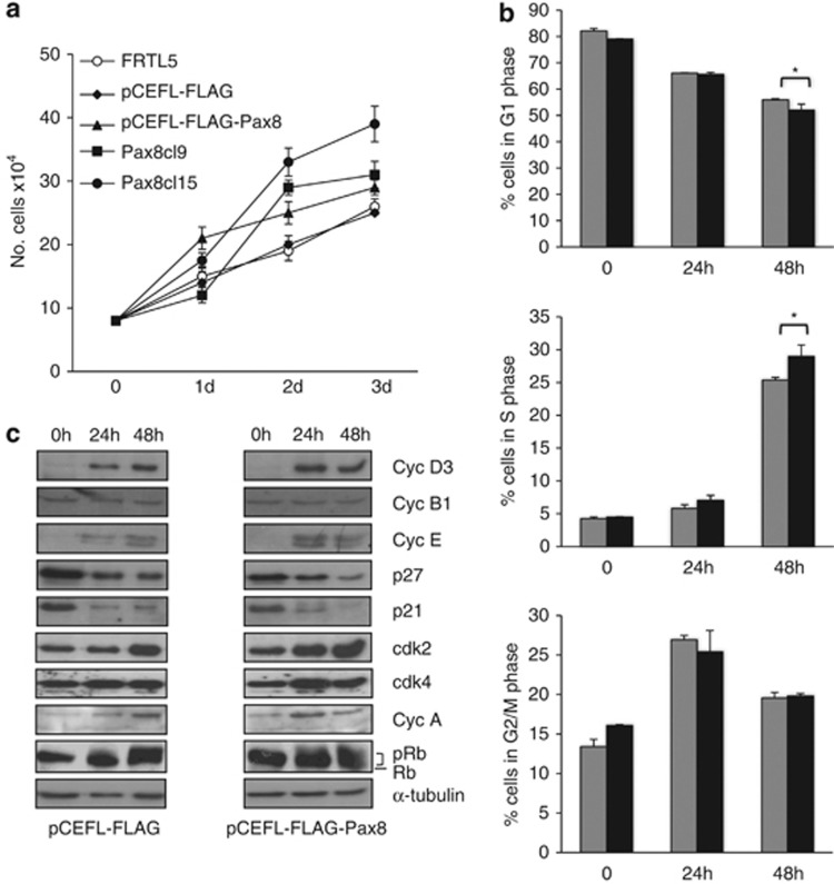
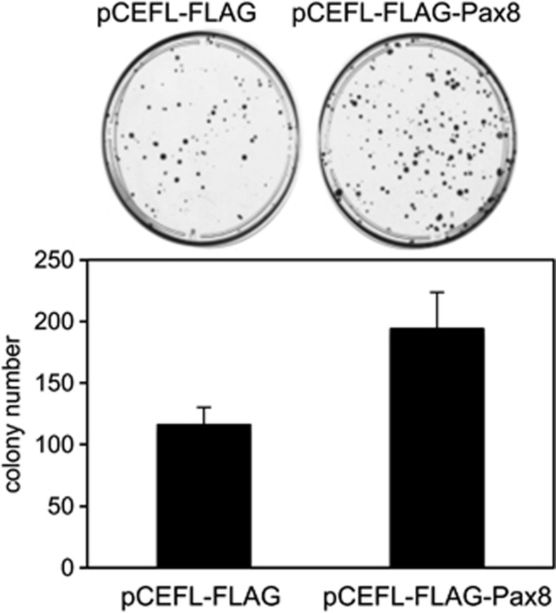
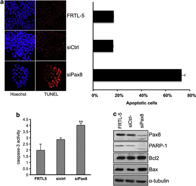
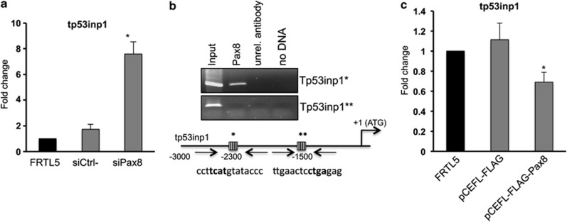
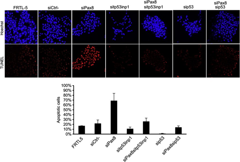
The transcription factor Pax8, a member of the Paired-box gene family, is a critical regulator required for proper development and differentiation of thyroid follicular cells. Despite being Pax8 well characterized with respect to its role in regulating genes responsible for thyroid differentiation, its involvement in cell survival and proliferation has been hypothesized but remains unclear. Here, we show that Pax8 overexpression significantly increases proliferation and colony-forming efficiency of Fischer rat thyroid line 5 epithelial cells, although it is not sufficient to overcome their hormone dependence. More interestingly, we show that Pax8-specific silencing induces apoptosis through a p53-dependent pathway that involves caspase-3 activation and cleavage of poly(ADP)ribose polymerase. Our data indicate that tumor protein 53 induced nuclear protein 1 (tp53inp1), a positive regulator of p53-dependent cell cycle arrest and apoptosis, is a transcriptional target of Pax8 and is upregulated by Pax8 knockdown. Remarkably, tp53inp1 silencing significantly abolishes Pax8-induced apoptosis thus suggesting that tp53inp1 may be the mediator of the observed effects. In conclusion, our data highlight that Pax8 is required for the survival of differentiated epithelial cells and its expression levels are able to modulate the proliferation rate of such cells.


Figures

Figure 1
Figure 1
Pax8 overexpression affects the cell growth and the cell cycle distribution of FRTL-5 cells. (a) Growth curves of wild-type FRTL-5 cells, individual Pax8 FRTL-5 stable clones and pools of clones expressing Pax8 or transfected with the empty vector. The results are the mean of three independent experiments. (b) Flow cytometry analysis of proliferating pCEFL-FLAG-Pax8 and pCEFL-FLAG cells starved for 3 days in 0.2% serum (t=0) and treated with complete medium at the indicated times. Gray and black bars correspond to pCEFL-FLAG and pCEFL-FLAG-Pax8 cells, respectively. Statistical analysis uses t-test (P=0.08). (c) Protein extracts of pCEFL-FLAG-Pax8 and pCEFL-FLAG cells starved for 3 days in 0.2% serum (t=0) and treated with complete medium at the indicated times were analyzed by western blot for the expression of cell cycle regulatory proteins. Filter was reblotted with α-tubulin to ensure protein loading and integrity
Figure 2
Figure 2
Colony-forming assays were performed with FRTL-5 cells transfected with a vector expressing Pax8 cDNA (pCEFL-FLAG-Pax8) or with the empty vector (pCEFL-FLAG). The results are the mean of two independent experiments. Cells transfected with Pax8 generated a higher number of colonies than that of cells transfected with the backbone vector
Figure 3
Figure 3
Pax8 knockdown promotes apoptosis of FRTL-5 cells. (a) Fluorescence microscopy representative images of TUNEL staining of FRTL-5 cells 48 h after Pax8 silencing. On the right side, the quantitative analysis of TUNEL-positive cells is shown. More than 500 cells were counted for each measurement, and experiments were repeated three times. Averages of TUNEL-positive cells (%) with standard error are shown in the graph. Statistical analysis uses t-test (P<0.01). (b) Pax8 silencing increases the caspase-3 activity. Cells were transfected with siRNA scramble (siCtrl-) and siPax8 and after 24 h the whole-cell lysates were subjected to caspase-3 activity assay. The results are the mean of three independent experiments (P<0.05). (c) Analysis by western blot of the expression levels of some genes involved in the control of apoptosis, upon Pax8 silencing. PARP1 was detected as a doublet indicative of its activation. The hybridization with α-tubulin is shown to ensure protein loading and integrity
Figure 4
Figure 4
Pax8 silencing directly modulates tp53inp1 gene expression. (a) Total RNA was isolated from FRTL-5 cells 72 h after transfection and subjected to qPCR analysis. The values are means±S.D. of three independent experiments in duplicate, normalized by the expression of β-actin and expressed as fold change with respect to FRTL-5 wild-type cells whose value was set at 1.0. Statistical analysis uses t-test (P=0.01). (b) Schematic representation of the upstream region of the rat tp53inp1 gene. Pax8-binding sites are represented as striped boxes and are marked by asterisks. Pax8 binding sequences are indicated below the boxes and the core sequences are in bold. ChIP analysis was performed using FRTL-5 cells. Crosslinked chromatin was immunoprecipitated with the antibody for Pax8 or with an unrelated antibody (α-tubulin). Immunoprecipitates from each sample were analyzed by PCR using primers specific for the tp53inp1 promoter (indicated by arrows). (c) RNA samples from wild-type FRTL-5 cells, pCEFL-FLAG and pCEFL-FLAG-Pax8 pools were used to perform qPCR to measure the expression of tp53inp1. The data shown are normalized mean values±S.D. of three independent experiments in duplicate. Statistical analyses were performed using t-test (P<0.05)
Figure 5
Figure 5
tp53inp1 depletion blocks Pax8-mediated apoptosis by a p53-dependent mechanism. TUNEL analysis of FRTL5 cells 48 h after silencing of Pax8 alone or in combination with tp53inp1 and p53. Averages of TUNEL-positive cells (%) with standard error are shown in the graph. Statistical analysis uses t-test (P<0.05)

References

    1. Dahl E, Koseki H, Balling R. Pax genes and organogenesis. Bioessays. 1997;19:755–765. - PubMed
    1. Walther C, Guenet JL, Simon D, Deutsch U, Jostes B, Goulding MD, et al. Pax: a murine multigene family of paired box-containing genes. Genomics. 1991;11:424–434. - PubMed
    1. Lang D, Powell SK, Plummer RS, Young KP, Ruggeri BA. PAX genes: roles in development, pathophysiology, and cancer. Biochem Pharmacol. 2007;73:1–14. - PubMed
    1. Muratovska A, Zhou C, He S, Goodyer P, Eccles MR. Paired-Box genes are frequently expressed in cancer and often required for cancer cell survival. Oncogene. 2003;22:7989–7997. - PubMed
    1. Plachov D, Chowdhury K, Walther C, Simon D, Guenet JL, Gruss P. Pax8, a murine paired box gene expressed in the developing excretory system and thyroid gland. Development. 1990;110:643–651. - PubMed

Publication types