

Abnormal functional network connectivity among resting-state networks in children with frontal lobe epilepsy
- PMID: 23868148
- PMCID: PMC7965212
- DOI: 10.3174/ajnr.A3608
Abnormal functional network connectivity among resting-state networks in children with frontal lobe epilepsy
Abstract
Background and purpose: Epilepsy is considered a disorder of neural networks. The aims of this study were to assess functional connectivity within resting-state networks and functional network connectivity across resting-state networks by use of resting-state fMRI in children with frontal lobe epilepsy and to relate changes in resting-state networks with neuropsychological function.
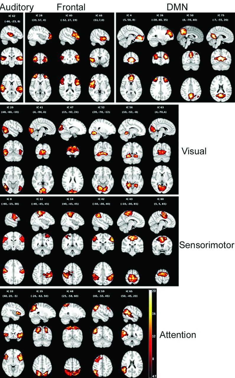
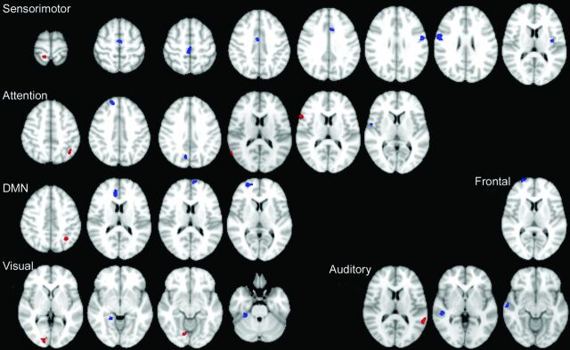
Materials and methods: Fifteen patients with frontal lobe epilepsy and normal MR imaging and 14 healthy control subjects were recruited. Spatial independent component analysis was used to identify the resting-state networks, including frontal, attention, default mode network, sensorimotor, visual, and auditory networks. The Z-maps of resting-state networks were compared between patients and control subjects. The relation between abnormal connectivity and neuropsychological function was assessed. Correlations from all pair-wise combinations of independent components were performed for each group and compared between groups.
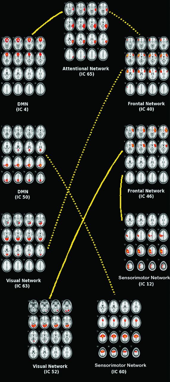
Results: The frontal network was the only network that showed reduced connectivity in patients relative to control subjects. The remaining 5 networks demonstrated both reduced and increased functional connectivity within resting-state networks in patients. There was a weak association between connectivity in frontal network and executive function (P = .029) and a significant association between sensorimotor network and fine motor function (P = .004). Control subjects had 79 pair-wise independent components that showed significant temporal coherence across all resting-state networks except for default mode network-auditory network. Patients had 66 pairs of independent components that showed significant temporal coherence across all resting-state networks. Group comparison showed reduced functional network connectivity between default mode network-attention, frontal-sensorimotor, and frontal-visual networks and increased functional network connectivity between frontal-attention, default mode network-sensorimotor, and frontal-visual networks in patients relative to control subjects.
Conclusions: We found abnormal functional connectivity within and across resting-state networks in children with frontal lobe epilepsy. Impairment in functional connectivity was associated with impaired neuropsychological function.
Figures
Similar articles
-
Salience, central executive, and sensorimotor network functional connectivity alterations in failed back surgery syndrome.Scand J Pain. 2017 Jul;16:10-14. doi: 10.1016/j.sjpain.2017.01.008. Epub 2017 Feb 20. Scand J Pain. 2017. PMID: 28850382
-
Betel quid chewing alters functional connectivity in frontal and default networks: A resting-state fMRI study.J Magn Reson Imaging. 2017 Jan;45(1):157-166. doi: 10.1002/jmri.25322. Epub 2016 May 26. J Magn Reson Imaging. 2017. PMID: 27227967 Clinical Trial.
-
Functional connectivity networks for preoperative brain mapping in neurosurgery.J Neurosurg. 2017 Jun;126(6):1941-1950. doi: 10.3171/2016.6.JNS1662. Epub 2016 Aug 26. J Neurosurg. 2017. PMID: 27564466
-
Resting state networks in temporal lobe epilepsy.Epilepsia. 2013 Dec;54(12):2048-59. doi: 10.1111/epi.12400. Epub 2013 Oct 10. Epilepsia. 2013. PMID: 24117098 Free PMC article. Review.
-
Functional Connectivity Changes on Resting-State fMRI after Mild Traumatic Brain Injury: A Systematic Review.AJNR Am J Neuroradiol. 2024 Jun 7;45(6):795-801. doi: 10.3174/ajnr.A8204. AJNR Am J Neuroradiol. 2024. PMID: 38637022 Free PMC article.
Cited by
-
Multimodal neurocognitive markers of frontal lobe epilepsy: Insights from ecological text processing.Neuroimage. 2021 Jul 15;235:117998. doi: 10.1016/j.neuroimage.2021.117998. Epub 2021 Mar 28. Neuroimage. 2021. PMID: 33789131 Free PMC article.
-
Impaired development of intrinsic connectivity networks in children with medically intractable localization-related epilepsy.Hum Brain Mapp. 2014 Nov;35(11):5686-700. doi: 10.1002/hbm.22580. Epub 2014 Jun 30. Hum Brain Mapp. 2014. PMID: 24976288 Free PMC article.
-
Chronnectome fingerprinting: Identifying individuals and predicting higher cognitive functions using dynamic brain connectivity patterns.Hum Brain Mapp. 2018 Feb;39(2):902-915. doi: 10.1002/hbm.23890. Epub 2017 Nov 15. Hum Brain Mapp. 2018. PMID: 29143409 Free PMC article.
-
Causal Cortical and Thalamic Connections in the Human Brain.Res Sq [Preprint]. 2024 May 30:rs.3.rs-4366486. doi: 10.21203/rs.3.rs-4366486/v1. Res Sq. 2024. Update in: Nat Neurosci. 2025 Aug;28(8):1797-1809. doi: 10.1038/s41593-025-02009-x. PMID: 38853954 Free PMC article. Updated. Preprint.
-
Dissociated multimodal hubs and seizures in temporal lobe epilepsy.Ann Clin Transl Neurol. 2015 Apr;2(4):338-52. doi: 10.1002/acn3.173. Epub 2015 Feb 25. Ann Clin Transl Neurol. 2015. PMID: 25909080 Free PMC article.
References
-
- Fox MD, Raichle ME. Spontaneous fluctuations in brain activity observed with functional magnetic resonance imaging. Nat Rev Neurosci 2007;8:700–11 - PubMed
Publication types
MeSH terms
LinkOut - more resources
Full Text Sources
Other Literature Sources
Medical