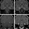
MRI findings in autoimmune voltage-gated potassium channel complex encephalitis with seizures: one potential etiology for mesial temporal sclerosis
- PMID: 23868165
- PMCID: PMC7966496
- DOI: 10.3174/ajnr.A3633
MRI findings in autoimmune voltage-gated potassium channel complex encephalitis with seizures: one potential etiology for mesial temporal sclerosis
Abstract
Background and purpose: Autoimmune voltage-gated potassium channel complex encephalitis is a common form of autoimmune encephalitis. Patients with seizures due to this form of encephalitis commonly have medically intractable epilepsy and may require immunotherapy to control seizures. It is important that radiologists recognize imaging characteristics of this type of autoimmune encephalitis and suggest it in the differential diagnosis because this seizure etiology is likely under-recognized. Our purpose was to characterize MR imaging findings in this patient population.
Materials and methods: MR imaging in 42 retrospectively identified patients (22 males; median age, 56 years; age range, 8-79 years) with seizures and voltage-gated potassium channel complex autoantibody seropositivity was evaluated for mesial and extratemporal swelling and/or atrophy, T2 hyperintensity, restricted diffusion, and enhancement. Statistical analysis was performed.
Results: Thirty-three of 42 patients (78.6%) demonstrated enlargement and T2 hyperintensity of mesial temporal lobe structures at some time point. Mesial temporal sclerosis was commonly identified (16/33, 48.5%) at follow-up imaging. Six of 9 patients (66.7%, P = .11) initially demonstrating hippocampal enhancement and 8/13 (61.5%, P = .013) showing hippocampal restricted diffusion progressed to mesial temporal sclerosis. Conversely, in 6 of 33 patients, abnormal imaging findings resolved.
Conclusions: Autoimmune voltage-gated potassium channel complex encephalitis is frequently manifested as enlargement, T2 hyperintensity, enhancement, and restricted diffusion of the mesial temporal lobe structures in the acute phase. Recognition of these typical imaging findings may help prompt serologic diagnosis, preventing unnecessary invasive procedures and facilitating early institution of immunotherapy. Serial MR imaging may demonstrate resolution or progression of radiologic changes, including development of changes involving the contralateral side and frequent development of mesial temporal sclerosis.
Figures


References
-
- Gultekin SH, Rosenfeld MR, Voltz R, et al. . Paraneoplastic limbic encephalitis: neurological symptoms, immunological findings and tumour association in 50 patients. Brain 2000;123:1481–94 - PubMed
-
- Lucchinetti CF, Kimmel DW, Lennon VA. Paraneoplastic and oncologic profiles of patients seropositive for type 1 antineuronal nuclear autoantibodies. Neurology 1998;50:652–57 - PubMed
Publication types
MeSH terms
Substances
Supplementary concepts
Grants and funding
LinkOut - more resources
Full Text Sources
Other Literature Sources
Medical
