

Prevalence of subdural collections in children with macrocrania
- PMID: 23868166
- PMCID: PMC7965194
- DOI: 10.3174/ajnr.A3588
Prevalence of subdural collections in children with macrocrania
Abstract
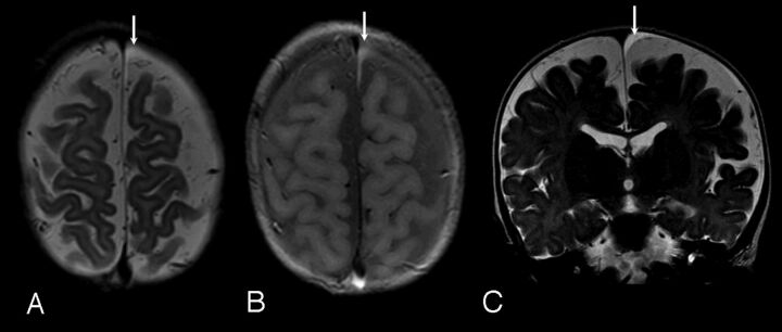
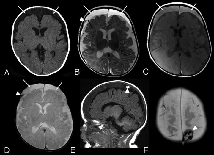
Background and purpose: The relationship between enlarged subarachnoid spaces and subdural collections is poorly understood and creates challenges for clinicians investigating the etiology of subdural collections. The purpose of this study was to determine the prevalence of subdural collections on cross sectional imaging in children with macrocephaly correlating with subarachnoid space enlargement.
Materials and methods: The radiology information system of a large pediatric medical center was reviewed for "macrocrania" and "macrocephaly" on reports of cranial MRI/CT examinations in children <24 months of age, over a 24-month period. Head circumference was obtained from the clinical record. Studies were reviewed blindly for subdural collection presence and subarachnoid space size. Children with prior cranial surgery, parenchymal abnormalities, hydrocephalus, or conditions predisposing to parenchymal volume loss were excluded. Chart review was performed on those with subdural collections.
Results: Imaging from 177 children with enlarged head circumference was reviewed. Nine were excluded, for a final cohort of 168 subjects (108 with enlarged subarachnoid space). Subdural collections were identified in 6 (3.6%), all with enlarged subarachnoid space (6/108, 5.6%). In 4, subdural collections were small, homogeneous, and nonhemorrhagic. In 2, the collections were complex (septations or hemorrhage). Two children were reported as victims of child abuse (both with complex collections). No definitive etiology was established in the other cases.
Conclusions: The prevalence of subdural collections in imaged children with macrocrania was 3.6%, all occurring in children with enlarged subarachnoid space. Our results suggest that enlarged subarachnoid space can be associated with some subdural collections in this cohort. Despite this, we believe that unexpected subdural collections in children should receive close clinical evaluation for underlying causes, including abusive head trauma.
Figures
References
-
- Ment LR, Duncan CC, Geehr R. Benign enlargement of the subarachnoid spaces in the infant. J Neurosurg 1981;54:504–08 - PubMed
-
- Raybaud AB, Barkovich AJ. Hydrocephalus. In: Barkovich AJ, ed. Pediatric Neuroimaging. 5th ed. Philadelphia: Lippincott Williams & Wilkins; 2012
-
- Babcock DS, Han BK, Dine MS. Sonographic findings in infants with macrocrania. AJR Am J Roentgenol 1988;150:1359–65 - PubMed
-
- Carolan PL, McLaurin RL, Towbin RB, et al. Benign extra-axial collections of infancy. Pediatr Neurosci 1985;12:140–44 - PubMed
MeSH terms
LinkOut - more resources
Full Text Sources
Other Literature Sources
Medical