. 2013 Jul 18;499(7458):306-11.
doi: 10.1038/nature12345.

LRG1 promotes angiogenesis by modulating endothelial TGF-β signalling

Affiliations

LRG1 promotes angiogenesis by modulating endothelial TGF-β signalling

Xiaomeng Wang et al. Nature. .

Abstract

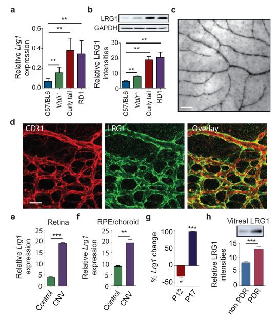
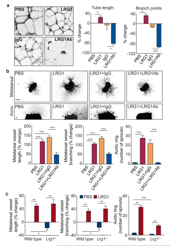
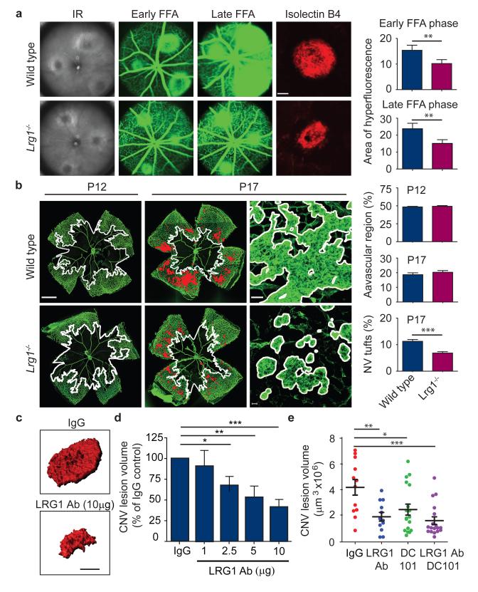
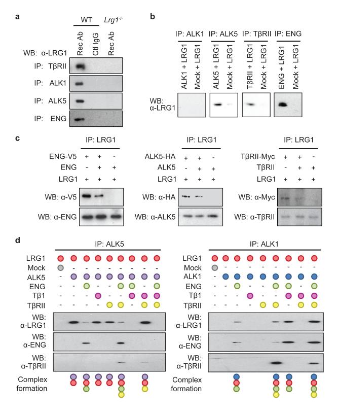
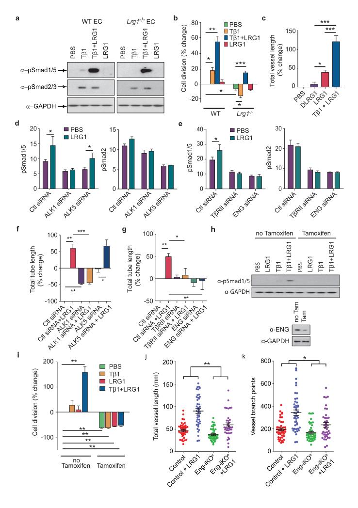
Aberrant neovascularization contributes to diseases such as cancer, blindness and atherosclerosis, and is the consequence of inappropriate angiogenic signalling. Although many regulators of pathogenic angiogenesis have been identified, our understanding of this process is incomplete. Here we explore the transcriptome of retinal microvessels isolated from mouse models of retinal disease that exhibit vascular pathology, and uncover an upregulated gene, leucine-rich alpha-2-glycoprotein 1 (Lrg1), of previously unknown function. We show that in the presence of transforming growth factor-β1 (TGF-β1), LRG1 is mitogenic to endothelial cells and promotes angiogenesis. Mice lacking Lrg1 develop a mild retinal vascular phenotype but exhibit a significant reduction in pathological ocular angiogenesis. LRG1 binds directly to the TGF-β accessory receptor endoglin, which, in the presence of TGF-β1, results in promotion of the pro-angiogenic Smad1/5/8 signalling pathway. LRG1 antibody blockade inhibits this switch and attenuates angiogenesis. These studies reveal a new regulator of angiogenesis that mediates its effect by modulating TGF-β signalling.


Figures

Figure 1
Figure 1. LRG1 is over-expressed in pathogenic retinal vasculature
a, Quantification of Lrg1 mRNA and b, LRG1 protein expression showing up-regulation in the retina of mice exhibiting retinal vascular changes. c, Lrg1 in situ hybridisation at P21. Scale bar = 50μm. d, Immunohistochemical detection of CD31 (red) and LRG1 (green) at P10 showing LRG1 expression in the retinal vasculature. e, Up-regulation of Lrg1 mRNA in the retina and f, RPE/choroid in CNV mice. g, Reduced Lrg1 transcript levels in OIR at P12 and increased levels at P17. h, Elevation of LRG1 protein in the vitreous of patients with PDR. All images shown are representative and values are expressed as means ± s.e.m of ≥ 3 independent experimental groups. *P<0.05, **P<0.01, ***P<0.001.
Figure 2
Figure 2. LRG1 promotes angiogenesis
a, Increased HUVEC tube and branch formation following addition of LRG1 and inhibition by a LRG1 neutralizing antibody. Scale bar 160μm. b, Vessel outgrowth in the metatarsal (top row) and aortic ring (bottom row) assay is enhanced by LRG1 and attenuated by a LRG1 neutralizing antibody. Scale bar = 1,500μm. c, Comparison of vessel growth from metatarsals and aortic rings isolated from WT and Lrg1−/− mice shows reduced angiogenesis in the latter that could be rescued by the addition of LRG1. All images shown are representative and values are expressed as means ± s.e.m of n ≥ 3 independent experimental groups. *P<0.05, **P<0.01, ***P<0.001.
Figure 3
Figure 3. Lrg1 contributes to pathogenic neovascularisation
a, Representative images of WT and Lrg1−/− mouse laser-burn lesions by infra red (IR) fundus imaging. At 7 days post laser, early and late-phase fundus fluorescein angiography (FFA) revealed a reduction in CNV lesion size and a decrease in fluorescein leakage respectively in Lrg1−/− mice. Representative images of isolectin B4 stained (red) CNV in choroidal/RPE flat-mount 7 days after induction confirming decreased lesion size in Lrg1−/− mice. Scale bar = 100μm. b, In OIR Lrg1 deletion does not affect the size of the avascular region at P12 (delineated by white boundary line) or the organised normal revascularisation at P17 (scale bar = 1000μm) but does decrease the formation of pathological neovascular (NV) tufts (highlighted in red and delineated in higher power by white boundary line). Scale bar = 50μm. c, Volume-rendered examples of PECAM-1 stained CNV lesions in WT mice following intravitreal injection of irrelevant IgG or LRG1 neutralising antibody (scale bar = 100μm). d, Dose-dependent anti-LRG1 antibody reduction of CNV lesion volume. e, Combination of anti-LRG1 and DC101 (anti-VEGFR2) in CNV in WT mice resulted in enhanced reduction of lesion volume compared to single treatments. All values represent means ± s.e.m of n ≥ 10 for each group. *P<0.05; **P<0.01; ***P<0.001.
Figure 4
Figure 4. LRG1 modifies the TGFβ receptor complex
a, Immunoprecipitation of TβRII, ALK1, ALK5 and ENG with anti-receptor antibodies (Rec Ab) from WT mouse brain EC lysates co-precipitates LRG1. Control IgG in WT ECs or Rec Ab in Lrg1−/− ECs did not co-precipitate LRG1. b, Immunoprecipitation of peptide-tagged extracellular domains of ALK5 (HA-tagged), TβRII (Myc-tagged) or ENG (V5-tagged) added individually to His-tagged LRG1 resulted in co-precipitation of LRG1 indicating direct interactions with these receptors. Immunoprecipitation of ALK1 (HA-tagged) in the presence of LRG1 did not co-precipitate the latter. c, Addition of appropriate soluble non-tagged extracellular domains of ENG, ALK5 and TβRII competed off peptide-tagged receptor binding to LRG1. d, LRG1 was incubated in vitro with different combinations of TGFβ receptor extracellular domains and TGFβ1. In the presence of ENG binding between LRG1 and ALK5 is diminished and with the further addition of TGFβ1 is completely lost. Conversely, ENG facilitates the association between LRG1 and ALK1, which is enhanced in the presence of TGFβ1. Although TβRII has no impact on LRG1/ALK1 or LRG1/ALK5 interactions, it is recruited to the complex in the presence of ENG. All data are representative western blots of n ≥ 3 for each experiment.
Figure 5
Figure 5. LRG1 promotes angiogenesis via a switch in TGFβ signalling
a, In WT brain ECs, TGFβ1 (Tβ1) stimulates Smad 2/3 phosphorylation and low levels of Smad 1/5 phosphorylation but in Lrg1−/− brain ECs only Smad 2/3 is phosphorylated. Addition of LRG1 has no effect on Smad phosphorylation in WT or Lrg1 null cells but co-treatment with TGFβ1 and LRG1 enhances Smad 1/5 phosphorylation without affecting Smad2/3 phosphorylation (n ≥ 3). b, Proliferation of brain ECs isolated from WT control and Lrg1−/− mice after exogenous TGFβ1 and/or LRG1 treatment normalized to control (n ≥ 3 ). Non-treated Lrg1−/− ECs are less proliferative than WT ECs. Addition of TGFβ1 to WT ECs results in enhanced proliferation but reduces proliferation in Lrg1−/− ECs whereas TGFβ1 and LRG1 co-treatment results in enhanced proliferation in WT and Lrg1−/− ECs. c, Addition of exogenous TGFβ1 and LRG1, compared to LRG1 alone or denatured (D)LRG1, enhances microvessel formation in the mouse metatarsal angiogenesis assay (n=3 independent experiments, n ≥ 30 metatarsals per treatment). d, siRNA knockdown of ALK1 or ALK5 in HUVEC results in reduced Smad1/5 and Smad2 phosphorylation respectively. ALK1, but not ALK5, knockdown results in prevention of LRG1-induced Smad1/5 phosphorylation. Histograms show semi-quantification of Smad phosphorylation relative to GAPDH (n ≥ 3). e, siRNA knockdown of TβRII or ENG inhibits LRG1-induced Smad1/5 phosphorylation. Histograms show semi-quantification of Smad phosphorylation relative to GAPDH (n ≥ 3). f and g, Knockdown of ALK1, TβRII or ENG, but not ALK5, reduces LRG1-mediated HUVEC Matrigel tube formation. (n=3 independent groups for each assay). h, Treatment of lung ECs isolated from Rosa26-CreERT:Engfl/fl mice (MLEC;Engfl/fl) with a combination of TGFβ1 and LRG1 results in Smad 1/5 phosphorylation, whereas following pre-treatment with 4OH-tamoxifen to delete ENG (MLEC;Eng−/−) the response is lost. i, Treatment of MLEC;Engfl/fl with a combination of TGFβ1 and LRG1 stimulates cell division. In MLEC;Eng−/− cell division is reduced and refractive to treatment with TGFβ1 ± LRG1 (n=3 independent experiments). j, 4OH-tamoxifen treatment of metatarsals isolated from Engfl/fl (control) and Cdh5(PAC)-CreERT2;Engfl/fl (Eng-iKOe) mice results in loss of ENG expression in the latter (Supplementary Fig 30) and decreases LRG1 induced metatarsal vessel length and k, vessel branching (metatarsals from 5 independent litters). All values represent means ± s.e.m. * P<0.05; ** P<0.01; *** P<0.001.

References

    1. Leung DW, Cachianes G, Kuang WJ, Goeddel DV, Ferrara N. Vascular endothelial growth factor is a secreted angiogenic mitogen. Science. 1989;246:1306–1309. - PubMed
    1. Carmeliet P, et al. Abnormal blood vessel development and lethality in embryos lacking a single VEGF allele. Nature. 1996;380:435–439. - PubMed
    1. Ferrara N, et al. Heterozygous embryonic lethality induced by targeted inactivation of the VEGF gene. Nature. 1996;380:439–442. - PubMed
    1. Holderfield MT, Hughes CC. Crosstalk between vascular endothelial growth factor, notch, and transforming growth factor-beta in vascular morphogenesis. Circ Res. 2008;102:637–652. - PubMed
    1. Chung AS, Ferrara N. Developmental and pathological angiogenesis. Annu Rev Cell Dev Biol. 2010 - PubMed

Publication types

MeSH terms

Substances