. 2013 Jul-Aug;41(4):213-23.
doi: 10.1002/bmb.20706.

Protein structure in context: the molecular landscape of angiogenesis

Affiliations

Protein structure in context: the molecular landscape of angiogenesis

Elise A Span et al. Biochem Mol Biol Educ. 2013 Jul-Aug.

Abstract

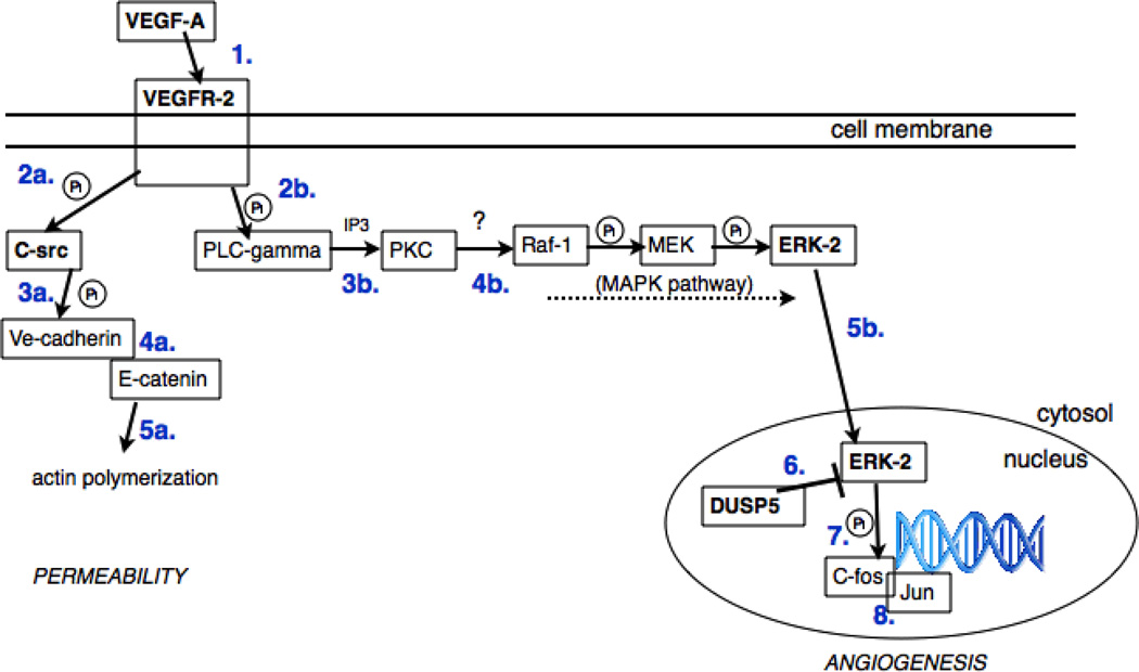
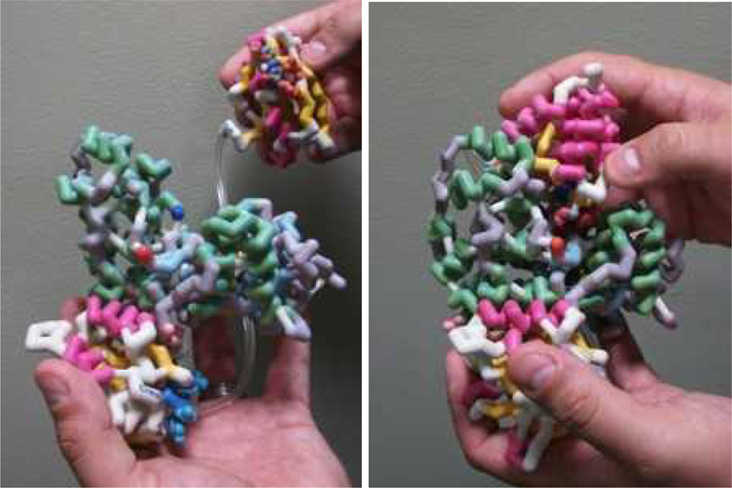
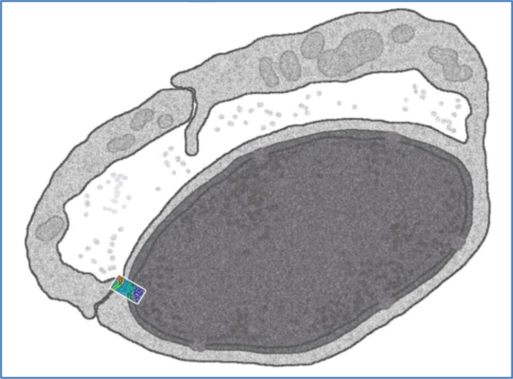
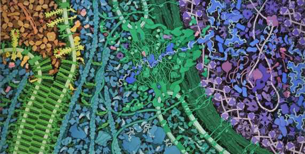
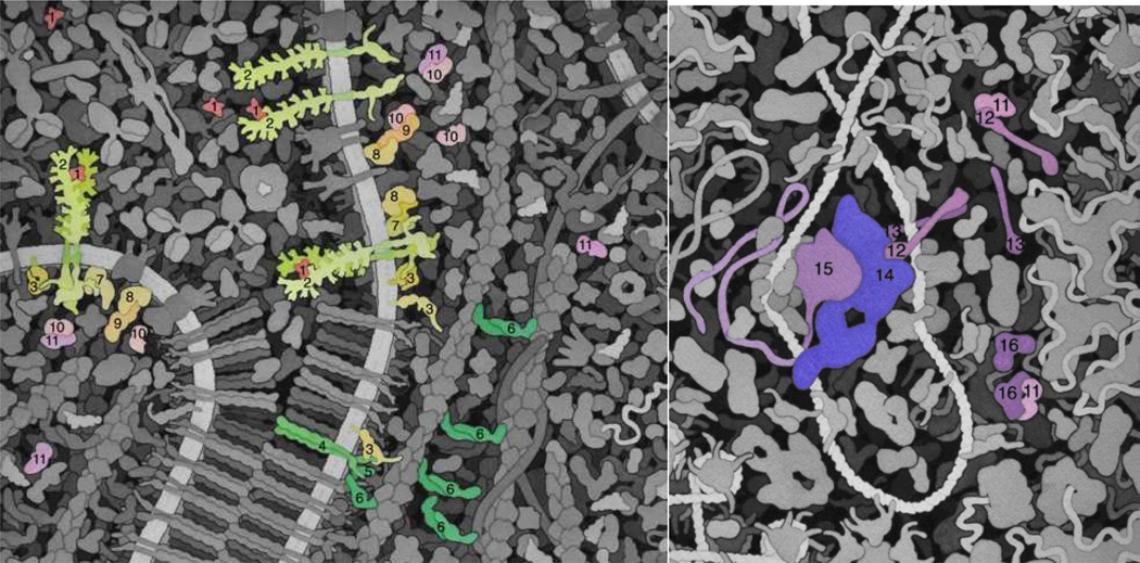
A team of students, educators, and researchers has developed new materials to teach cell signaling within its cellular context. Two nontraditional modalities are employed: physical models, to explore the atomic details of several of the proteins in the angiogenesis signaling cascade, and illustrations of the proteins in their cellular environment, to give an intuitive understanding of the cellular context of the pathway. The experiences of the team underscore the use of these types of materials as an effective mode for fostering students' understanding of the molecular world and the scientific method used to define it.

Keywords: VEGF signaling; angiogenesis; protein structure; visual learning.


Figures

Figure 1
Figure 1
Schematic of the VEGF the pathways to be depicted in the landscape. The DUSP5/ERK-2 interaction that occurs in the nucleus (bottom right), is represented using physical models in Fig. 2.
Figure 2
Figure 2
Physical models of DUSP-5 and ERK-2, demonstrating the binding interaction known to occur between these proteins. DUSP-5 in comprised of two domains, an ERK binding domain and a phosphatase domain, connected by an unstructured linker region (plastic tubing in the model).
Figure 3
Figure 3
Summary of the proteins in the VEGF-mediated angiogenesis signaling cascade (Fig. 1), including relevant structural information needed for drawing the molecular landscape – such as relative size and quaternary structure.
Figure 4
Figure 4
(a) An initial sketch for the molecular landscape painting depicting angiogenesis signaling, and (b) a sketch after three rounds of revisions, with markings suggesting further alterations. Relative protein sizes and shapes are based on the data compiled from Fig. 3.
Figure 5
Figure 5
Simulated electron micrograph showing two cells from the vascular endothelium. The size and location of the portion of the vascular endothelium that is to be depicted in the molecular landscape painting is shown in color. This is the cellular context within which the angiogenesis signaling cascade is to be presented.
Figure 6
Figure 6
The final molecular landscape depicting VEGF signaling. Blood serum is shown in tan at the upper left. The adherens junction between two cells is in green at left, with surface VEGF receptors shown in yellow. The cytoplasmic proteins are in turquoise, and the nuclear pore is at the center in green. The nucleus is at the right, with proteins shown in blues and purples.
Figure 7
Figure 7
Expanded and color-coded views of the molecular landscape from Fig. 6. (a) VEGF signaling, across the cell membrane into the cytosol. VEGF-A (1) in blood serum binds to VEGFR (2) and causes it to dimerize. This activates the VEGFR tyrosine kinase domains inside the cell. Two downstream pathways are shown. In one, C-src (3) is phosphorylated, causing it to open up and phosphorylate cadherins (4) in the adherens junctions, releasing alpha-catenin (6), which dimerizes and bundles actin (beta-catenin (5), which is involved in the adherens junction structure, is also shown). In the other pathway, the receptor initiates a cascade of phosphorylation reactions through PLC-gamma (7), PKC (8), Raf-1 (9), MEK (10), and ultimately ERK-2 (11). Phosphorylated ERK-2 (pERK-2) is transported through the nuclear pore (the large structure at the center of the full painting, not depicted here). (b) Continuing the signaling cascade into the nucleus. pERK-2 (11) phosphorylates C-fos (12), causing it to form a heterodimer with Jun (13) and becoming active as a transcription factor effecting transcription of proteins needed for blood vessel growth. It is shown here in an enhancer binding to the transcription mediator (14) and RNA polymerase (15). Finally, DUSP5 (16) terminates the process by dephosphorylating pERK-2. This final interaction is depicted with the physical models in Fig. 2.

References

    1. Harris MA, et al. A Combination of Hand-held Models and Computer Imaging Programs Helps Students Answer Oral Questions about Molecular Structure and Function: A Controlled Investigation of Student Learning. CBE—Life Sciences Ed. 2009;8:29–43. - PMC - PubMed
    1. http://www.rcsb.org/pdb/
    1. http://www.bmrb.wisc.edu/
    1. Nye LS. The Mind's Eye—Biomedical Visualization: The Most Powerful Tool in Science. Biochem. Mol. Biol. Ed. 2004;32:123–131. - PubMed
    1. Herman T, et al. Tactile Teaching—Exploring Protein Structure/Function Using Physical Models. Biochem. Mol. Biol. Ed. 2006;34:247–254. - PubMed

Publication types

LinkOut - more resources