

doi: 10.1093/database/bat049.
Print 2013.
CREDO: a structural interactomics database for drug discovery
Affiliations
- PMID: 23868908
- PMCID: PMC3715132
- DOI: 10.1093/database/bat049
Item in Clipboard
CREDO: a structural interactomics database for drug discovery
Database (Oxford).
.
Abstract
CREDO is a unique relational database storing all pairwise atomic interactions of inter- as well as intra-molecular contacts between small molecules and macromolecules found in experimentally determined structures from the Protein Data Bank. These interactions are integrated with further chemical and biological data. The database implements useful data structures and algorithms such as cheminformatics routines to create a comprehensive analysis platform for drug discovery. The database can be accessed through a web-based interface, downloads of data sets and web services at http://www-cryst.bioc.cam.ac.uk/credo. Database URL: http://www-cryst.bioc.cam.ac.uk/credo.
Figures
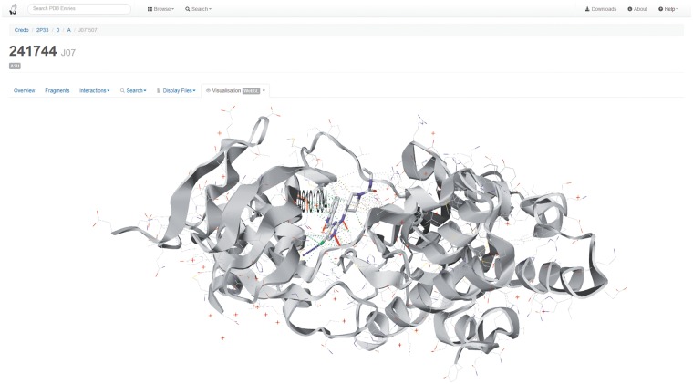
Screenshot of the CREDO website displaying the WebGL-based visualization of the protein–ligand complex [aminopyrimidine inhibitor of c-Jun N-Terminal Kinase (JNK)] found in PDB entry 2P33. The rendering also includes the interactions for ligand J07 as well as a highlighting (residue side chain carbons in pale red) of a polymorphism from dbSNP that can be linked to a binding site-lining residue.
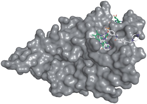
Complex of Imatinib with human NQO2 at 1.75 Å resolution (PDB: 3FW1). Imatinib is displayed with white, FAD with green carbon atoms. Only PDB chain A is shown for clarity.
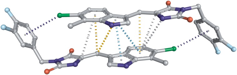
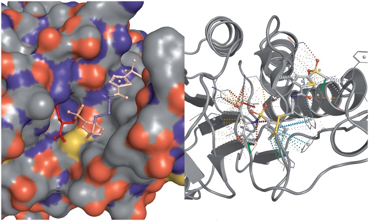
Interactions between aromatic rings as well as atoms and aromatic rings in the dimeric small molecule complex in the p53 pocket of MDM2 (PDB: 3VBG). The inhibitor complex leads to the dimerization of MDM2 and MDMX proteins, eventually causing cell cycle arrest and apoptosis. Aromatic–ring interactions are displayed using a cyan dashed line, halogen-pi interactions are shown as dark blue dashed line, carbon-pi in orange and undefined atom-ring interactions simply in grey. Only interactions within 4.0 Å are shown.
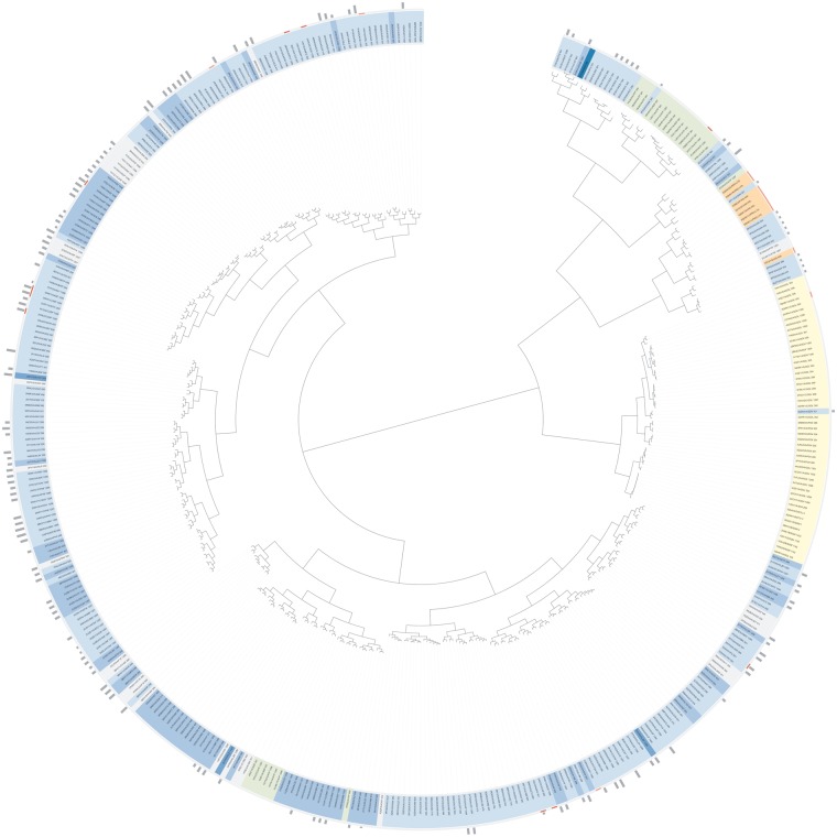
SIFt clustering for all ligands binding to UniProt entry P24941, Cyclin-dependent kinase 2.
FCD visualized in the crystal structure of cysteine aspartyl protease-3 (caspase-3) in complex with a non-peptidic inhibitor. The image was rendered as grid in PyMOL, atomic positions are identical on both sides. The FCD (from blue to red) is shown for the set of smallest fragments resulting from the fragmentation of this ligand, chemical component 160.
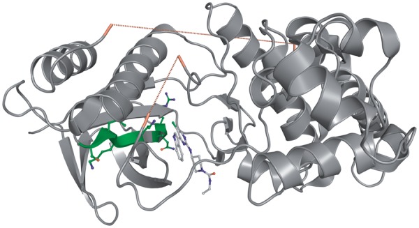
Visualization of missing regions and a secondary structure fragment in PDB entry 2P33. The two regions of missing amino acids inside are displayed with a red dashed line that connects the Cα atoms of the flanking residues. N- and C-terminal missing regions are not shown. The secondary structure fragment 2P33/0/A/PF:13, an extended beta strand, is visualized with amino acid side chains and carbon atoms in green.
References
-
- Ondetti MA, Cushman DW. Enzymes of the renin-angiotensin system and their inhibitors. Ann. Rev. Biochem. 1982;51:283–308. - PubMed
-
- Szelke M, Leckie B, Hallett A, et al. Potent new inhibitors of human renin. Nature. 1982;299:555–557. - PubMed
-
- Blundell T, Sibanda BL, Pearl L. Three-dimensional structure, specificity and catalytic mechanism of renin. Nature. 1983;304:273–275. - PubMed
-
- Miller M, Schneider J, Sathyanarayana BK, et al. Structure of complex of synthetic HIV-1 protease with a substrate-based inhibitor at 2.3 A resolution. Science. 1989;246:1149–1152. - PubMed
-
- Lapatto R, Blundell T, Hemmings A, et al. X-ray analysis of HIV-1 proteinase at 2.7 A resolution confirms structural homology among retroviral enzymes. Nature. 1989;342:299–302. - PubMed
Publication types
MeSH terms
Substances
Grants and funding
LinkOut - more resources
Full Text Sources
Other Literature Sources
