. 2013 Oct 1;126(Pt 19):4349-57.
doi: 10.1242/jcs.125328. Epub 2013 Jul 18.

Src mediates the mechanical activation of myogenesis by activating TNFα-converting enzyme

Affiliations

Src mediates the mechanical activation of myogenesis by activating TNFα-converting enzyme

Airu Niu et al. J Cell Sci. .

Abstract

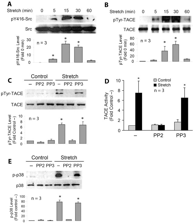
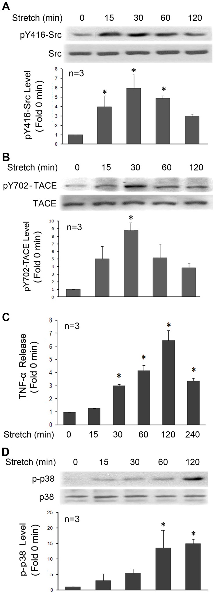
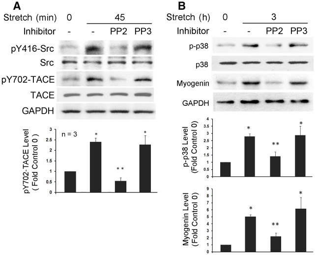
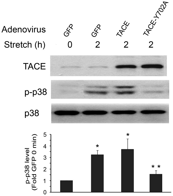
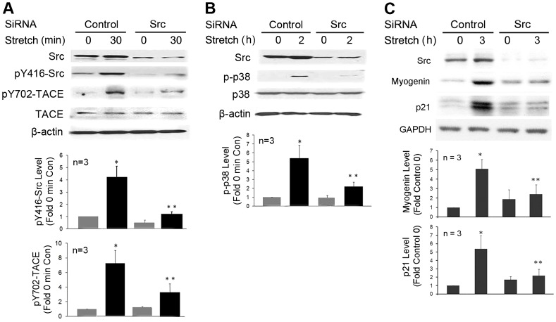
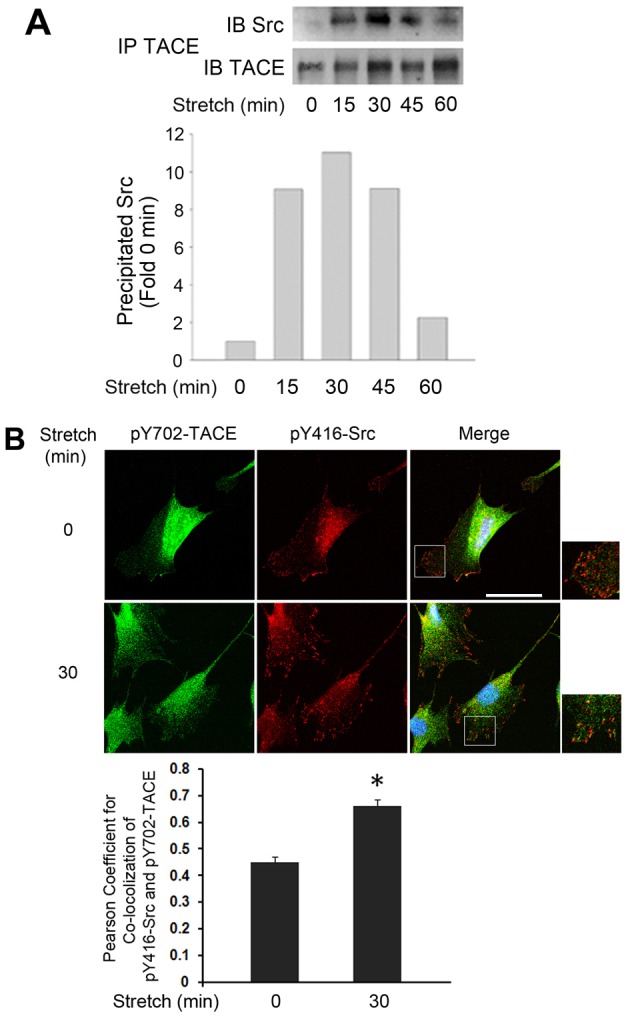
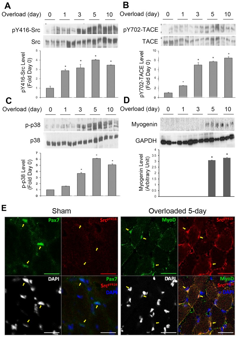
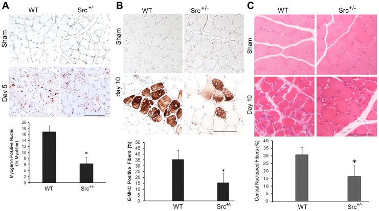
Mechanical stimulation affects many biological aspects in living cells through mechanotransduction. In myogenic precursor cells (MPCs), mechanical stimulation activates p38 mitogen-activated protein kinase (MAPK), a key regulator of myogenesis, via activating TNFα-converting enzyme (TACE, also known as ADAM17), to release autocrine TNFα. However, the signaling mechanism of mechanical activation of TACE is unknown. Because TACE possesses the structural features of substrates of the non-receptor tyrosine kinase Src, we tested the hypothesis that Src mediates mechanical activation of TACE in MPCs. We observed that mechanical stretch of C2C12 or primary rat myoblasts rapidly activates Src, which in turn interacts and colocalizes with TACE, resulting in tyrosine phosphorylation and activation of TACE. Particularly, Src activates TACE via the phosphorylation of amino acid residue Tyr702 in the intracellular tail of TACE, resulting in increased TNFα release and p38 activation. Src inhibition or deficiency blocks stretch activation of the TACE-p38-MAPK signaling, resulting in impaired myogenic gene expression. In response to functional overloading, Src and TACE are activated in mouse soleus muscle. Further, overloading-induced myogenesis and regeneration are impaired in the soleus of Src(+/-) mice. Therefore, Src mediates mechano-activation of TACE and myogenesis.

Keywords: ADAM17; Mechanotransduction; Muscle regeneration; Myogenic gene expression; Src; TACE; p38 MAPK.


Figures

Fig. 1.
Fig. 1.
Mechanical stretch activates TACE through Src-mediated phosphorylation in C2C12 myoblasts. C2C12 myoblasts were statically stretched for the indicated time period using the Flexcell® FX-5000™ Tension System. Cell lysate was prepared and subjected to western blotting or immunoprecipitation analysis. (A) Mechanical stretch activates Src. Antibodies specific for Src or Src that is phosphorylated at the Tyr416 residue (pY416) were used in western blot analysis to assess Src activity. (B) Mechanical stretch activates tyrosine phosphorylation of TACE. TACE was immunoprecipitated from cell lysate and analyzed for tyrosine phosphorylation by western blot analysis using antibodies against phosphorylated tyrosine (pTyr) or TACE. (C) The SFK inhibitor PP2 blocks mechanical activation of TACE phosphorylation. C2C12 myoblasts were stretched for 30 minutes with or without pretreatment with 10 µM PP2 or PP3. TACE was immunoprecipitated from cell lysate and analyzed by western blotting with antibodies against phosphorylated tyrosine or TACE. (D) PP2 blocks mechanical activation of TACE. C2C12 myoblasts were stretched for 30 minutes with or without pretreatment with 10 µM PP2 or PP3. TACE activity in cell lysates was analyzed by measuring the rate of cleavage of the peptide containing the TACE-specific cleavage site in pro-TNFα. (E) PP2 blocks mechanical activation of myogenic marker p38 MAPK. On the basis of the previously determined time course of stretch-activation of p38 (Zhan et al., 2007), C2C12 myoblasts were stretched for 120 minutes with or without 10 µM PP2 or PP3. Cell lysate was analyzed for p38 activation using western blotting. Western blots were quantified by densitometry. Data (means±s.e.) were analyzed by ANOVA (A,B,C and E) or Student's t-test (D); *P<0.05 compared with the non-stretched control.
Fig. 2.
Fig. 2.
Mechanical stretch activates Src, TACE phosphorylation at Tyr702, TNFα release and p38 MAPK activity in primary rat myoblasts. Primary myoblasts prepared from newborn rats were subjected to static stretch for the indicated time periods. Activation of Src (A), TACE phosphorylation at Tyr702 (B) and p38 MAPK (D) in cell lysate was analyzed by western blotting. TACE activity was measured by determining the concentration of TNFα released into cell culture medium using ELISA (C). Data were analyzed by ANOVA; *P<0.05 compared with the non-stretched control.
Fig. 3.
Fig. 3.
Mechanical stretch-activated myogenesis in primary rat myoblasts is dependent on Src activation. Primary rat myoblasts were pre-incubated with PP2 or PP3 and stretched for the indicated time periods. Western blotting was performed to analyze Src activation and TACE phosphorylation on Tyr702 (A), or p38 MAPK activation and myogenin expression (B). Optical density data was analyzed by ANOVA. *P<0.05 compared with the non-stretched myoblasts (0 minute); **P<0.05 compared with the stretched myoblasts that were not treated with PP2 or PP3.
Fig. 4.
Fig. 4.
Tyr702 phosphorylation of TACE is crucial to its activation by mechanical stretch. C2C12 myoblasts were transduced with adenovirus encoding GFP, TACE or a TACE mutant in which Tyr702 was replaced with alanine (TACE-Y702A). Myoblasts were stretched for 2 hours before assessment of p38 activation. Optical density data was analyzed by ANOVA, *P<0.05 compared with the non-stretched myoblasts (0 minutes) that were transduced with GFP-encoding adenovirus; **P<0.05 compared with the stretched myoblasts that were transduced with GFP-encoding adenovirus.
Fig. 5.
Fig. 5.
Src is required for mechanical activation of TACE phosphorylation at Tyr702. C2C12 myoblasts were transfected with Src-specific siRNA or scrambled siRNA as control, and stretched for 30 minutes to assess Src activity and TACE phosphorylation at Tyr702 (A), 2 hours to assess p38 activation (B) or 3 hours to assess myogenin and p21 expression (C) using western blotting. Optical density data were analyzed by ANOVA. *P<0.05 compared with the non-stretched myoblasts (0 minute) that were transfected with control siRNA, **P<0.05 compared with the stretched myoblasts that were transfected with control siRNA.
Fig. 6.
Fig. 6.
Mechanically activated Src and TACE interact and colocalize in clusters. (A) Mechanical stretch induces interaction between Src and TACE. C2C12 myoblasts were stretched for the indicated time period. Cell lysate was subjected to immunoprecipitation with an antibody against TACE and analyzed by western blotting for Src co-precipitation with TACE. (B) Mechanical stretch induces colocalization of Src and TACE into clusters. C2C12 myoblasts were stretched for 30 minutes and then subjected to immunofluorescence staining with antibodies against pY416-Src (red) and pY702-TACE (green). Scale bar: 20 µm. The Pearson coefficient for the colocalization in control and stretched cells was analyzed by Student's t-test, *P<0.01 compared with the non-stretched control.
Fig. 7.
Fig. 7.
Src and TACE are activated in overloaded mouse soleus. The gastrocnemius of adult male mice (C57BL/6) was severed bilaterally to create functional overloading of soleus and plantaris. Sham-operated mice were used as control (day 0). At the indicated number of days after the surgery, the soleus was collected for western blot analysis of activation of Src (A), TACE (B) and p38 (C), as well as expression of myogenin (D). Each sample shown represents one mouse. Bar graphs summarize optical density data of the activated forms of the proteins normalized against the total protein or GAPDH. Data were analyzed by ANOVA. *P<0.05 compared with control (day 0). For D, the bar graph used arbitrary units owing to the zero expression of myogenin on day 0. (E) Cross sections of soleus prepared from mice that had been sham-operated or overloaded for 5 days were examined by immunohistofluorescence staining to visualize the localization of active Src (Red). Scale bars: 20 µm.
Fig. 8.
Fig. 8.
Src is essential for overloading induced TACE activation, myogenic differentiation and regeneration in mouse soleus. Adult Src+/− mice and wild-type (WT) littermates were subjected to bilateral ablation of the gastrocnemius. Myogenin expression on day 5 (A) and embryonic MHC (eMHC) expression on day 10 (B) of overloading were compared by immunohistochemistry of cross sections of soleus. The percentage of myofibers that express myogenin or eMHC in overloaded soleus are quantified in the bar graph. Cross sections of soleus that had been overloaded for 10 days were stained by H&E (C). Centralized nuclei were counted as an indication of regeneration. Scale bars: 100 µm. Data were analyzed by Student's t-test, *P<0.05 compared with wild-type soleus.

Similar articles

Cited by

References

    1. Aikawa R., Nagai T., Kudoh S., Zou Y., Tanaka M., Tamura M., Akazawa H., Takano H., Nagai R., Komuro I. (2002). Integrins play a critical role in mechanical stress-induced p38 MAPK activation. Hypertension 39, 233–238 10.1161/hy0202.102699 - DOI - PubMed
    1. Alexandropoulos K., Baltimore D. (1996). Coordinate activation of c-Src by SH3- and SH2-binding sites on a novel p130Cas-related protein, Sin. Genes Dev. 10, 1341–1355 10.1101/gad.10.11.1341 - DOI - PubMed
    1. Bidwell J. P., Pavalko F. M. (2010). The load-bearing mechanosome revisited. Clin. Rev. Bone Miner Metab 8, 213–223 10.1007/s12018-010-9075-1 - DOI - PMC - PubMed
    1. Black R. A. (2002). Tumor necrosis factor-alpha converting enzyme. Int. J. Biochem. Cell Biol. 34, 1–5 10.1016/S1357-2725(01)00097-8 - DOI - PubMed
    1. Black R. A., Rauch C. T., Kozlosky C. J., Peschon J. J., Slack J. L., Wolfson M. F., Castner B. J., Stocking K. L., Reddy P., Srinivasan S. et al.(1997). A metalloproteinase disintegrin that releases tumour-necrosis factor-alpha from cells. Nature 385, 729–733 10.1038/385729a0 - DOI - PubMed

Publication types

MeSH terms