. 2013 Jun;46(2):93-100.
doi: 10.5115/acb.2013.46.2.93. Epub 2013 Jun 30.

Surgical anatomy of the upper eyelid relating to upper blepharoplasty or blepharoptosis surgery

Affiliations

Surgical anatomy of the upper eyelid relating to upper blepharoplasty or blepharoptosis surgery

Kun Hwang. Anat Cell Biol. 2013 Jun.

Abstract

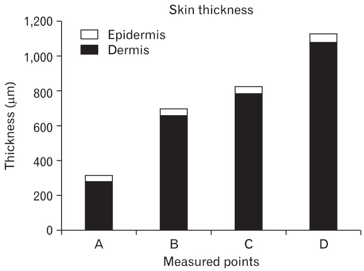
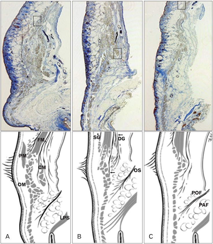
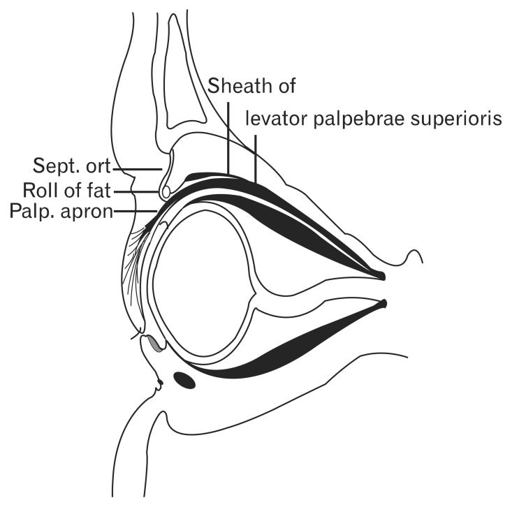
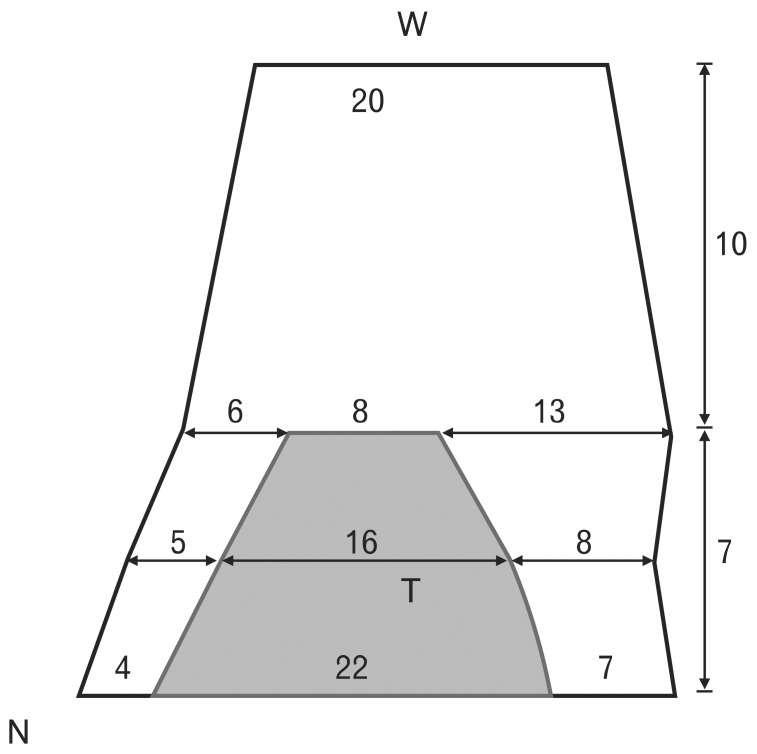
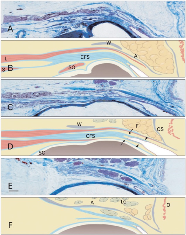
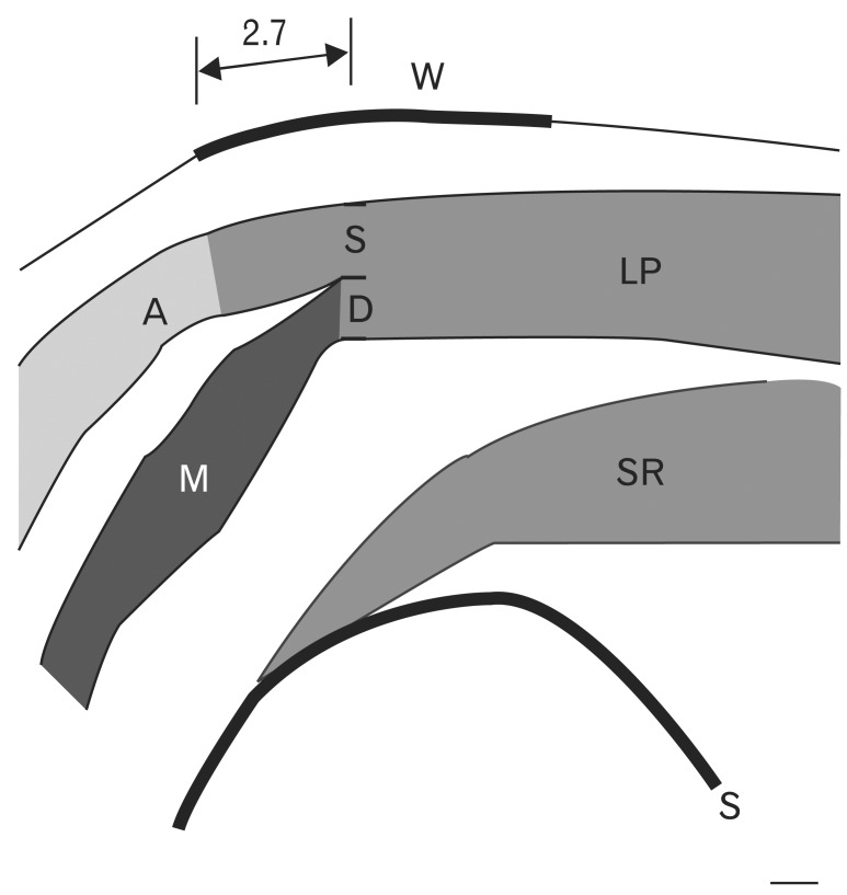
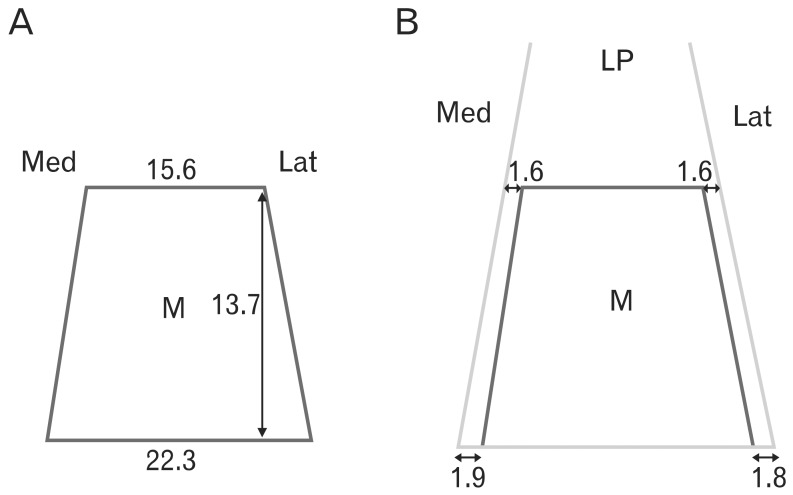
Eyelid anatomy, including thickness measurements, was examined in numerous age groups. The thickest part of the upper eyelid is just below the eyebrow (1.127±238 µm), and the thinnest near the ciliary margin (320±49 µm). The thickness of skin at 7 mm above the eyelashes was 860±305 µm. The results revealed no significant differences among the age groups. Fast fibers (87.8±3.7%) occupied a significantly larger portion of the orbicularis oculi muscle (OOM) than nonfast fibers (12.2±3.7%). The frontalis muscle passed through and was inserted into the bundles of the OOM on the superior border of the eyebrow at the middle and medial portions of the upper eyelid. Laterally, the frontalis muscle inserted about 0.5 cm below the superior border of the eyebrow. Fast fibers occupied a significantly larger portion of the OOM than did non-fast fibers. The oculomotor nerve ends that extend forward to the distal third of the levator muscle are exposed and vulnerable to local anesthetics and may be numbed during blepharoplasty. The orbital septum consists of 2 layers. The outer layer of loose connective tissue descends to interdigitate with the levator aponeurosis and disperses inferiorly. The inner layer follows the outer layer, then reflects and continues posteriorly with the levator sheath. Widths of the tarsal plate at its lower border, mid-height, and upper border were 21.8±1.8, 16.2±1.6, and 8.3±1.0 mm, respectively. The widths of the levator aponeurosis were 32.0±2.2, 29.2±3.5, and 27.2±3.9 mm, respectively. Below the levator, the "conjoint fascial sheath" (CFS) is attached to the conjunctival fornix. The CFS was 12.2±2.0 mm anteroposterior length and 1.1±0.1 mm thick. The shape was equilateral trapezoid with a longer base anteriorly. The superior palpebral muscle was trapezoidal. The lengths of its sides were 15.58±1.82 and 22.30±5.25 mm, and its height was 13.70±2.74 mm. The width of the levator aponeurosis was approximately 4 mm wider than the superior palpebral muscle.

Keywords: Anatomy and histology; Blepharoplasty; Blepharoptosis; Eyelids.


Figures

Fig. 1
Fig. 1
Histograms showing epidermis and dermis thickness as measured at various points. A, near the ciliary margin; B, midtarsus; C, upper tarsal border; D, lower border of the eyebrow.
Fig. 2
Fig. 2
Sagittal sections of the upper eyelid. The anterior surface of the frontalis muscle is enveloped by superficial galea (SG, arrowhead) and deep galea (DG, arrows) on its posterior surface. The frontalis muscle (FM) passes through and inserts in the bundles of the orbicularis oculi muscle (OM). (A) Medial section. The FM inserts in the superior border of the eyebrow. The most distal part of the FM is located deep to the procerus muscle (PM) and superficial to the corrugator muscle (CM). (B) Middle section. The FM inserts in the superior border of the eyebrow. (C) Lateral section (the FM inserts about 0.5 cm below the superior border of the eyebrow). OS, orbital septum; PAF, pre-aponeurotic fat; POF, pre-orbicularis fascia.
Fig. 3
Fig. 3
Schema of a sagittal section of the eyelid. Reproduced from Whitnall SE. J Anat Physiol 1911;45:131-9 [21], with permission from John Wiley & Sons Ltd. Sept. ort, septum orbitale; Palp. aporn, palpebral aponeurosis.
Fig. 4
Fig. 4
Schema of the levator aponeurosis (black trapezoid) and tarsal plate (T, gray trapezoid). Upper: Levator aponeurosis width was measured at the lower border, mid-region, and upper border of the tarsal plate to be 32.0±2.2 mm, 29.2±3.5 mm, 27.2±3.9 mm, respectively. Lower: The medial brim of the levator aponeurosis at the lower border, mid-region, and upper border of the tarsal plate was 3.6±1.1 mm, 5.1±1.0 mm, and 6.2±1.1 mm, respectively. The lateral brim of the levator aponeurosis at the lower border, mid-region, and upper border of the tarsal plate was 6.6±0.9 mm, 7.9±2.6 mm, and 12.7±3.7 mm, respectively. N, nasal side; W, superior transverse ligament of Whitnall.
Fig. 5
Fig. 5
Microscopic findings of the upper eyelid. (A, B) Through the medial limbus line, (C, D) midpupillary line, (E, F) through the lateral limbus line. The conjoint fascial sheath (CFS) was located in the intermuscular space between the anterior third of the superior rectus (S) and the levator (L). Posteriorly, it extended from the fascia of the levator and superior rectus. Anteriorly, superficial and deep extensions (arrows) of CFS continued approximately 2 mm to the superior conjunctival fornix and then 2-3 mm distally (arrowheads) along and beneath the palpebral and bulbar conjunctiva. A, levator aponeurosis; F, pre-aponeurotic fat; LG, lacrimal gland; O, orbicularis oculi muscle; OS, orbital septum; SO, superior oblique; W, Whitnall's superior transverse ligament. Scale bar in (E)=2 mm (A-F).
Fig. 6
Fig. 6
Schema of a parasagittal section through the midpupillary line. The levator palpebrae superior muscle divides into superficial (S) and deep (D) parts beneath the superior transverse ligament. The levator aponeurosis originates from this superficial portion. The superior palpebral muscle originates from the deep portion. A, levator aponeurosis; M, superior palpebral involuntary muscle (Müller's muscle); LP, levator palpebrae muscle; SR, superior rectus muscle.
Fig. 7
Fig. 7
Schema of the superior palpebral muscle (M) and levator aponeurosis. (A) The width of the superior palpebral muscle was 15.6 mm at the anterior border of the superior transverse ligament and 22.3 mm at the upper border of the tarsal plate. The height was 13.7 mm. (B) The width of the levator aponeurosis was wider than that of the superior palpebral muscle. The width of the levator aponeurosis was 19.3 mm at the anterior border of the superior transverse ligament and 26.8 mm at the upper border of the tarsal plate. Medially, the levator aponeurosis was 1.9 and 1.6 mm wider at the level of the superior transverse ligament and the upper tarsal border, respectively. Laterally, the levator aponeurosis was 1.8 and 1.6 mm wider at the level of the superior transverse ligament and at the upper tarsal border, respectively. LP, levator palpebrae muscle.

References

    1. Barker DE. Skin thickness in the human. Plast Reconstr Surg (1946) 1951;7:115–116. - PubMed
    1. Lee Y, Hwang K. Skin thickness of Korean adults. Surg Radiol Anat. 2002;24:183–189. - PubMed
    1. Hwang K, Kim DJ, Hwang SH. Thickness of Korean upper eyelid skin at different levels. J Craniofac Surg. 2006;17:54–56. - PubMed
    1. Gonzalez-Ulloa M, Flores ES. Senility of the face: basic study to understand its causes and effects. Plast Reconstr Surg. 1965;36:239–246. - PubMed
    1. Hykin PG, Bron AJ. Age-related morphological changes in lid margin and meibomian gland anatomy. Cornea. 1992;11:334–342. - PubMed

LinkOut - more resources