. 2013 Jun;46(2):113-21.
doi: 10.5115/acb.2013.46.2.113. Epub 2013 Jun 30.

Expression of surface markers and myogenic potential of rat bone marrow- and adipose-derived stem cells: a comparative study

Affiliations

Expression of surface markers and myogenic potential of rat bone marrow- and adipose-derived stem cells: a comparative study

Vahid Bayati et al. Anat Cell Biol. 2013 Jun.

Abstract

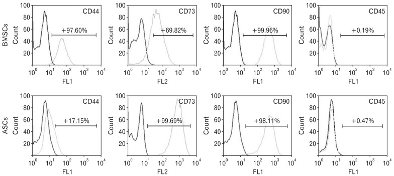
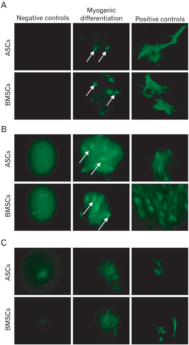
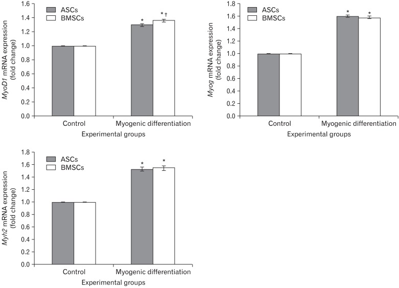
In recent years, examination and comparison of the biological characteristics of bone marrow- and adipose-derived mesenchymal stem cells (MSCs) from various perspectives have come into the focus of stem cell research, as these cells should be well characterized in order to utilize them in future cellular therapies. Therefore, in the present study, surface protein markers and the skeletal myogenic differentiation potential of rat bone marrow- and adipose-derived MSCs were examined. The expression of CD44, CD45, CD73, and CD90 on bone marrow- and adipose-derived MSCs was characterized using flow cytometry. Subsequently, the stem cells were differentiated into myogenic lineages, and the expression of the skeletal myogenic markers MyoD1, Myog, and Myh2 was studied in cells using real time polymerase chain reaction and immunofluorescence. Our results reveal that the pattern of CD marker expression differs between these 2 types of MSCs to some extent, whereas no significant difference was observed with respect to their myogenic differentiation potential. Therefore, we concluded that despite the differences observed in the biological features of these 2 types of MSCs, their myogenic potential appears to be similar, and that adipose-derived stem cells may be useful in skeletal muscle tissue engineering, due to their easy isolation and capacity for rapid expansion in a short time span.

Keywords: Adipose-derived stem cells; Bone marrow-derived mesenchymal stem cells; Myogenic potential; Rat; Surface antigens.


Figures

Fig. 1
Fig. 1
Mononuclear cells isolated from bone marrow after 24 hours in culture. (A) The majority of adherent cells were round (arrowheads) (inverted microscope, ×100) although (B) occasionally some of them had processes (arrows) (inverted microscope, ×250).
Fig. 2
Fig. 2
Bone marrow-derived mesenchymal stem cells (BMSCs) after 5 days in culture. (A) A colony of BMSCs on the fifth day (inverted microscope, ×100). (B) By observing with high magnification, BMSCs seemed to contain cells with 2 different morphologies: small cells with various appearances and large cells that were multiform and polygonal. Arrow points to a large cell (inverted microscope, ×250).
Fig. 3
Fig. 3
Stromal-vascular cell fraction (SVF) of the adipose tissue on the first day of culture. (A) A variety of cell populations with different sizes and morphologies appeared in the culture. (B) SVF cells after reaching confluence in less than 1 week (inverted microscope, ×250).
Fig. 4
Fig. 4
Flow cytometric results demonstrated that the bone marrow-derived mesenchymal stem cells (BMSCs) and adipose-derived stem cells (ASCs) differed in the expression of CD44 and CD73 on their surfaces.
Fig. 5
Fig. 5
Expression and the intracellular position of myogenic markers MyoD1, Myog, and myosin (fast skeletal) in bone marrow-derived mesenchymal stem cells (BMSCs) and adipose-derived stem cells (ASCs) in the absence or presence of chemical growth factors. MyoD1 (A) and Myog (B), as skeletal muscle transcription factors, were mostly concentrated in the nuclei (arrows). (C) Myosin (fast skeletal). Undifferentiated stem cells were taken as a negative control, and the cell line L6 was considered as positive control (A-C, ×630).
Fig. 6
Fig. 6
Expression levels of MyoD1, Myog, and Myh2 mRNA in bone marrow-derived mesenchymal stem cells (BMSCs) and adipose-derived stem cells (ASCs). *P<0.001 between the myogenic differentiation group and the control; P<0.05 between BMSCs and ASCs.

References

    1. Ahn SM, Simpson R, Lee B. Genomics and proteomics in stem cell research: the road ahead. Anat Cell Biol. 2010;43:1–14. - PMC - PubMed
    1. Wang S, Qu X, Zhao RC. Clinical applications of mesenchymal stem cells. J Hematol Oncol. 2012;5:19. - PMC - PubMed
    1. Chamberlain G, Fox J, Ashton B, Middleton J. Concise review: mesenchymal stem cells: their phenotype, differentiation capacity, immunological features, and potential for homing. Stem Cells. 2007;25:2739–2749. - PubMed
    1. Gimble JM, Katz AJ, Bunnell BA. Adipose-derived stem cells for regenerative medicine. Circ Res. 2007;100:1249–1260. - PMC - PubMed
    1. Bunnell BA, Flaat M, Gagliardi C, Patel B, Ripoll C. Adipose-derived stem cells: isolation, expansion and differentiation. Methods. 2008;45:115–120. - PMC - PubMed