Migration of a Hem-o-Lok Clip to the Ureter Following Laparoscopic Partial Nephrectomy Presenting With Lower Urinary Tract Symptoms
- PMID: 23869274
- PMCID: PMC3713248
- DOI: 10.5213/inj.2013.17.2.90
Migration of a Hem-o-Lok Clip to the Ureter Following Laparoscopic Partial Nephrectomy Presenting With Lower Urinary Tract Symptoms
Abstract
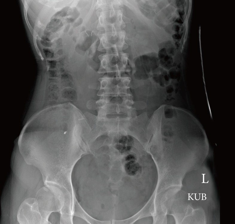
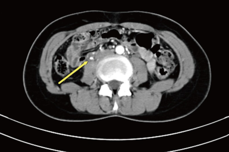
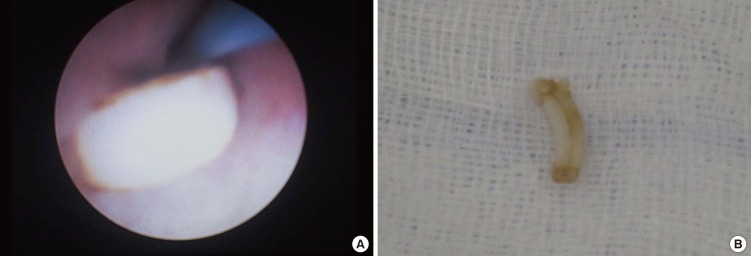
We report a case of ureteral migration of a surgical clip after partial nephrectomy in which the clip was misdiagnosed as a ureteral stone. A 37-year-old woman had undergone laparoscopic partial nephrectomy of right renal cell carcinoma at another hospital 2 years previously. Postoperatively, she had gradually acquired lower urinary tract symptoms. Then, she complained of sudden right flank pain for a week. A plain X-ray and enhanced abdominopelvic computed tomography scan were performed. A 0.5 cm×1.0 cm right upper ureteral opacity with borderline hydronephrosis was seen but could not be found on the X-ray. Ureteroscopy revealed a medium-sized Hem-o-Lok clip on the right upper ureter that was removed with a stone basket. We concluded that a Hem-o-Lok clip used for collecting system sealing had migrated to the ureter and had been misdiagnosed as a ureteral stone on a computed tomography scan.
Keywords: Nephrectomy; Surgical instruments; Ureteral calculi.
Conflict of interest statement
No potential conflict of interest relevant to this article was reported.
Figures
References
-
- Marszalek M, Meixl H, Polajnar M, Rauchenwald M, Jeschke K, Madersbacher S. Laparoscopic and open partial nephrectomy: a matched-pair comparison of 200 patients. Eur Urol. 2009;55:1171–1178. - PubMed
-
- Parsons JK, Palazzi K, Chang D, Stroup SP. Patient safety and the diffusion of surgical innovations: a national analysis of laparoscopic partial nephrectomy. Surg Endosc. 2013;27:1674–1680. - PubMed
-
- Msezane LP, Katz MH, Gofrit ON, Shalhav AL, Zorn KC. Hemostatic agents and instruments in laparoscopic renal surgery. J Endourol. 2008;22:403–408. - PubMed
-
- Miller M, Anderson JK, Pearle MS, Cadeddu JA. Resorbable clip migration in the collecting system after laparoscopic partial nephrectomy. Urology. 2006;67:845.e7–845.e8. - PubMed
LinkOut - more resources
Full Text Sources
Other Literature Sources