The effect of continuous epidural electrical stimulation on neuronal proliferation in cerebral ischemic rats
- PMID: 23869327
- PMCID: PMC3713286
- DOI: 10.5535/arm.2013.37.3.301
The effect of continuous epidural electrical stimulation on neuronal proliferation in cerebral ischemic rats
Abstract
Objective: To investigate the effect of electrical stimulation (ES) on the recovery of motor skill and neuronal cell proliferation.
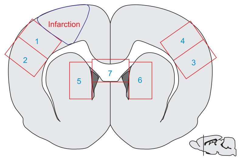
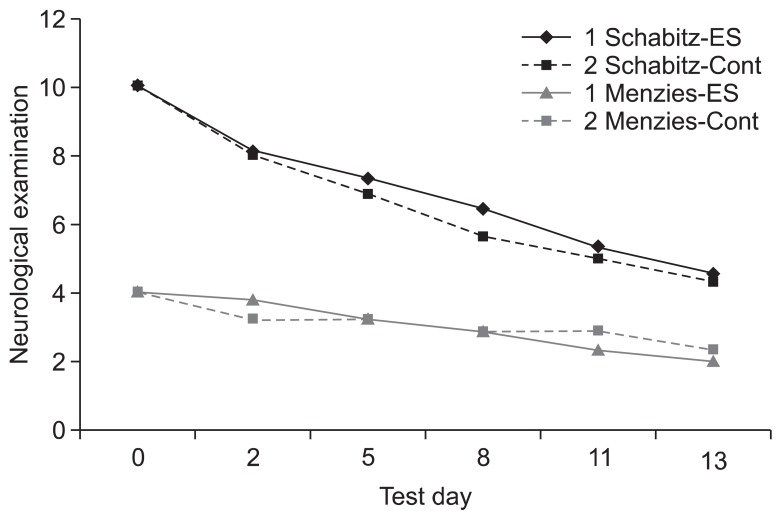
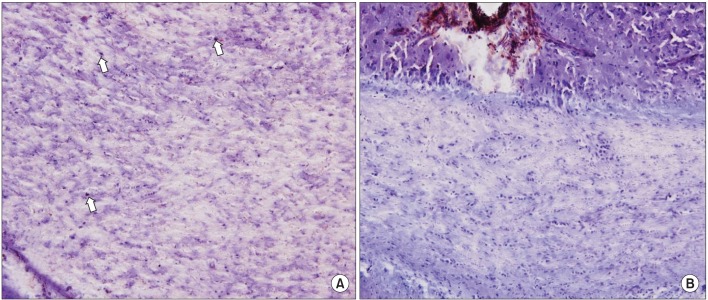
Methods: The male Sprague-Dawley rats were implanted with an epidural electrode over the peri-ischemic area after photothrombotic stroke in the dominant sensorimotor cortex. All rats were randomly assigned into the ES group and control group. The behavioral test of a single pellet reaching task (SPRT) and neurological examinations including the Schabitz's photothrombotic neurological score and the Menzies test were conducted for 2 weeks. After 14 days, coronal sections were obtained and immunostained for neuronal cell differentiation markers including bromodeoxyuridine (BrdU), neuron-specific nuclear protein (NeuN), and doublecortin (DCX).
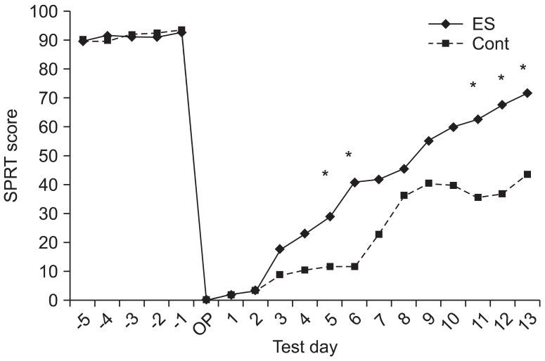
Results: On the SPRT, the motor function in paralytic forelimbs of the ES group was significantly improved. There were no significant differences in neurological examinations and neuronal cell differentiation markers except for the significantly increased number of DCX+ cells in the corpus callosum of the ES group (p<0.05). But in the ES group, the number of NeuN+ cells in the ischemic cortex and the number of NeuN+ cells and DCX+ cells in the ischemic striatum tended to increase. In the ES group, NeuN+ cells in the ischemic hemisphere and DCX+ cells and BrdU+ cells in the opposite hemisphere tended to increase compared to those in the contralateral.
Conclusion: The continuous epidural ES of the ischemic sensorimotor cortex induced a significant improvement in the motor function and tended to increase neural cell proliferation in the ischemic hemisphere and the neural regeneration in the opposite hemisphere.
Keywords: Cell proliferation; Cerebral ischemia; Electrical stimulation; Motor skills; Stroke.
Conflict of interest statement
No potential conflict of interest relevant to this article was reported.
Figures
Similar articles
-
Effects of extradural cortical stimulation on motor recovery in a rat model of subacute stroke.Restor Neurol Neurosci. 2015;33(5):589-96. doi: 10.3233/RNN-140445. Restor Neurol Neurosci. 2015. PMID: 25735240
-
Functional electrical stimulation increases neural stem/progenitor cell proliferation and neurogenesis in the subventricular zone of rats with stroke.Chin Med J (Engl). 2013 Jun;126(12):2361-7. Chin Med J (Engl). 2013. PMID: 23786955
-
Effect of Epidural Electrical Stimulation and Repetitive Transcranial Magnetic Stimulation in Rats With Diffuse Traumatic Brain Injury.Ann Rehabil Med. 2015 Jun;39(3):416-24. doi: 10.5535/arm.2015.39.3.416. Epub 2015 Jun 30. Ann Rehabil Med. 2015. PMID: 26161348 Free PMC article.
-
Effect of skilled reaching training and enriched environment on generation of oligodendrocytes in the adult sensorimotor cortex and corpus callosum.BMC Neurosci. 2017 Mar 9;18(1):31. doi: 10.1186/s12868-017-0347-2. BMC Neurosci. 2017. PMID: 28279169 Free PMC article.
-
Bioactive components of Chinese herbal medicine enhance endogenous neurogenesis in animal models of ischemic stroke: A systematic analysis.Medicine (Baltimore). 2016 Oct;95(40):e4904. doi: 10.1097/MD.0000000000004904. Medicine (Baltimore). 2016. PMID: 27749547 Free PMC article. Review.
Cited by
-
Progress in the Field of Micro-Electrocorticography.Micromachines (Basel). 2019 Jan 17;10(1):62. doi: 10.3390/mi10010062. Micromachines (Basel). 2019. PMID: 30658503 Free PMC article. Review.
-
Merging DBS with viral vector or stem cell implantation: "hybrid" stereotactic surgery as an evolution in the surgical treatment of Parkinson's disease.Mol Ther Methods Clin Dev. 2016 Jan 13;3:15051. doi: 10.1038/mtm.2015.51. eCollection 2016. Mol Ther Methods Clin Dev. 2016. PMID: 26817024 Free PMC article. Review.
-
Soluble Nogo receptor 1 fusion protein protects neural progenitor cells in rats with ischemic stroke.Neural Regen Res. 2019 Oct;14(10):1755-1764. doi: 10.4103/1673-5374.257531. Neural Regen Res. 2019. PMID: 31169193 Free PMC article.
-
Effects of Electric Cortical Stimulation (ECS) and Transcranial Direct Current Stimulation (tDCS) on Rats With a Traumatic Brain Injury.Ann Rehabil Med. 2018 Aug;42(4):502-513. doi: 10.5535/arm.2018.42.4.502. Epub 2018 Aug 31. Ann Rehabil Med. 2018. PMID: 30180518 Free PMC article.
-
Using dual polarities of transcranial direct current stimulation in global cerebral ischemia and its following reperfusion period attenuates neuronal injury.Metab Brain Dis. 2022 Jun;37(5):1503-1516. doi: 10.1007/s11011-022-00985-8. Epub 2022 May 2. Metab Brain Dis. 2022. PMID: 35499797
References
-
- Statistics Korea. Annual reports on the cause of death statistics 2010. Deajeon: Statistics Korea; 2011.
-
- Tsubokawa T, Katayama Y, Yamamoto T, Hirayama T, Koyama S. Chronic motor cortex stimulation for the treatment of central pain. Acta Neurochir Suppl (Wien) 1991;52:137–139. - PubMed
-
- Brown JA, Lutsep H, Cramer SC, Weinand M. Motor cortex stimulation for enhancement of recovery after stroke: case report. Neurol Res. 2003;25:815–818. - PubMed
-
- Brown JA, Lutsep HL, Weinand M, Cramer SC. Motor cortex stimulation for the enhancement of recovery from stroke: a prospective, multicenter safety study. Neurosurgery. 2006;58:464–473. - PubMed
-
- Franzini A, Ferroli P, Dones I, Marras C, Broggi G. Chronic motor cortex stimulation for movement disorders: a promising perspective. Neurol Res. 2003;25:123–126. - PubMed
LinkOut - more resources
Full Text Sources
Other Literature Sources