. 2013 Sep;89(6):1084-98.
doi: 10.1111/mmi.12335. Epub 2013 Aug 4.

Differentiated roles for MreB-actin isologues and autolytic enzymes in Bacillus subtilis morphogenesis

Affiliations
Free PMC article

Differentiated roles for MreB-actin isologues and autolytic enzymes in Bacillus subtilis morphogenesis

Patricia Domínguez-Cuevas et al. Mol Microbiol. 2013 Sep.
Free PMC article

Abstract

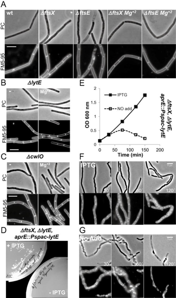
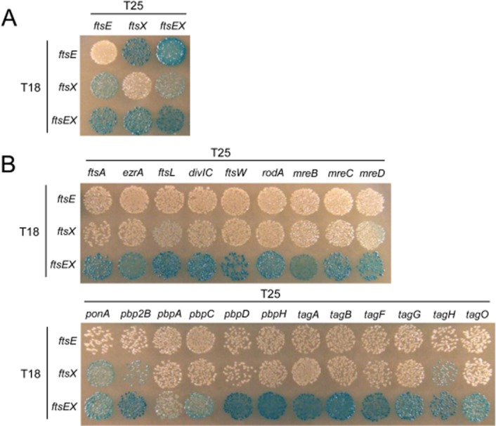
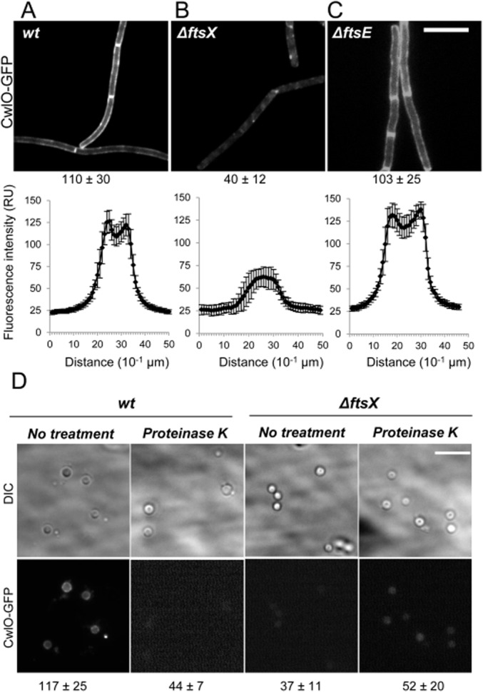
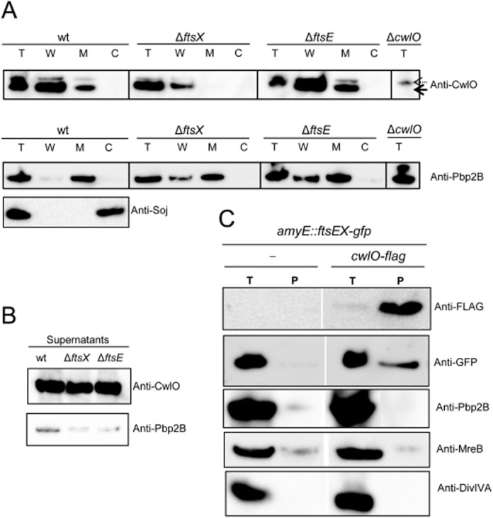
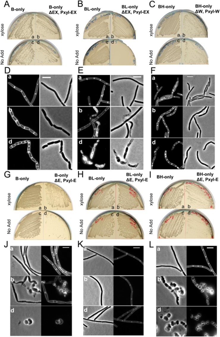
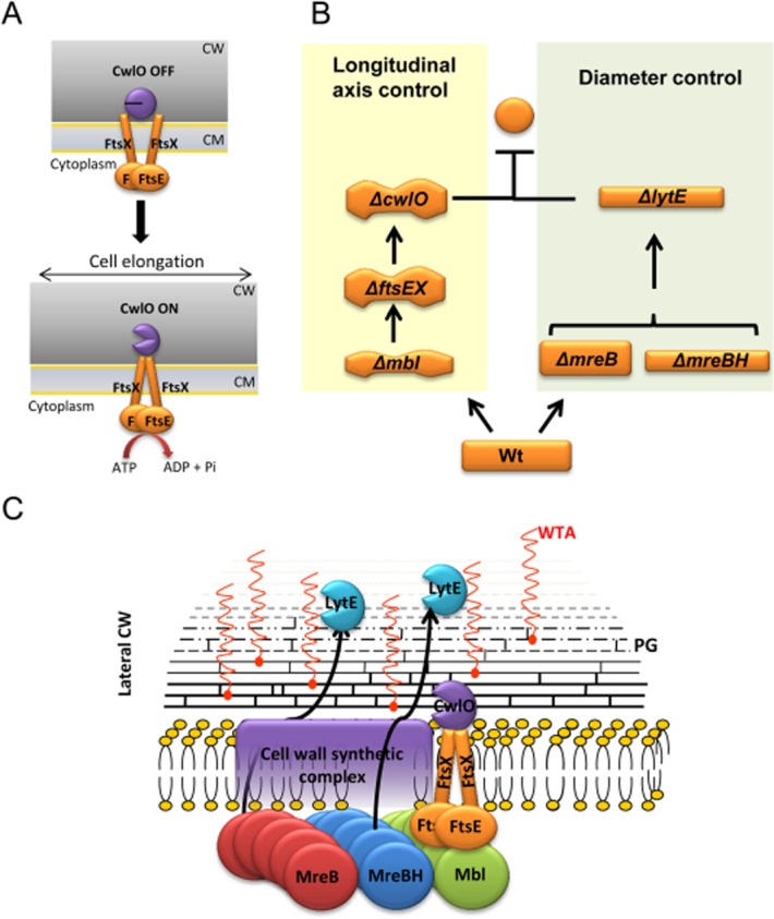
Cell morphogenesis in most bacteria is governed by spatiotemporal growth regulation of the peptidoglycan cell wall layer. Much is known about peptidoglycan synthesis but regulation of its turnover by hydrolytic enzymes is much less well understood. Bacillus subtilis has a multitude of such enzymes. Two of the best characterized are CwlO and LytE: cells lacking both enzymes have a lethal block in cell elongation. Here we show that activity of CwlO is regulated by an ABC transporter, FtsEX, which is required for cell elongation, unlike cell division as in Escherichia coli. Actin-like MreB proteins are thought to play a key role in orchestrating cell wall morphogenesis. B. subtilis has three MreB isologues with partially differentiated functions. We now show that the three MreB isologues have differential roles in regulation of the CwlO and LytE systems and that autolysins control different aspects of cell morphogenesis. The results add major autolytic activities to the growing list of functions controlled by MreB isologues in bacteria and provide new insights into the different specialized functions of essential cell wall autolysins.


Figures

Fig. 1
Fig. 1
FtsEX mutants are similar to ΔcwlO and synthetic lethal with ΔlytE. A. Cell morphologies of typical fields of wild-type B. subtilis strain 168, ΔftsX (4501) and ΔftsE (4503) mutant strains growing in a NA plates or in NA plates with supplement of 20 mM Mg2+, as indicated. Scale bar represents 5 μm. B and C. Cell morphologies of typical fields of strains PDC464 (ΔlytE::cat) and PDC463 (ΔcwlO::spec) cultured on NA plates in the presence or absence of Mg2+ as indicated. Scale bar represents 5 μm. D. Growth of strain PDC492 (ΔftsX::neo ΔlytE::cat aprE::Pspac-LytE) on NA plates with or without 0.5 mM IPTG. E. Growth of strain PDC492 on LB liquid medium in the presence or absence of IPTG. Growth curves (IPTG 0.5 mM, closed symbols; no addition, open symbols). F and G. Effect of LytE depletion on cell morphology. Phase-contrast micrographs and the corresponding membrane staining images were taken at the indicated times during the growth curves in (E). (F) 0.5 mM IPTG added; (G) no IPTG addition. Scale bar represents 5 μm.
Fig. 2
Fig. 2
Bacterial two-hybrid analysis of FtsEX protein interactions. A. Bacterial two-hybrid analysis of interaction between FtsE and FtsX. B. Bacterial two-hybrid analysis of interaction with FtsE and FtsX. Escherichia coli strain BTH101 was co-transformed with two-hybrid vector plasmids (pUT18 and pKT25) expressing C-terminal fusions of the cyaA T18 domain to ftsE, ftsX and ftsEX, and N-terminal fusions of cyaA T25 domain to various genes, as indicated. Transformants were spotted onto nutrient agar plates containing X-Gal and incubated at 30°C for 40 h. Blue colouration indicates a positive interaction.
Fig. 3
Fig. 3
CwlO localizes at the cell membrane in an FtsX-dependent manner. A–C. Epifluorescence microscopy of strains expressing the fluorescent fusion amyE::Pxyl-cwlO-gfpsf. The different panels correspond to (A) strain PDC528 (Bs168CA wprA::hyg, epr::tet amyE::Pxyl-cwlO-gfpsf) and isogenic strains (B) PDC560 (ΔftsX), (C) PDC594 (ΔftsE), as indicated. Scale bar represents 5 μm. Fluorescent images were taken with the same acquisition settings and exposure times, with the maximum averaged value of quantified fluorescence intensity over the lateral wall of the cells (see Experimental procedures) indicated below. Lower panels: Profiles of fluorescence intensity corresponding to strains in (A)–(C) respectively. Averaged fluorescence intensity quantified in segments of equal size across the cell's longitudinal axis. The y-axis represents fluorescence intensity (relative units, RU), while the x-axis represents distance (10−1 μm). Error bars represent standard deviation of fluorescent intensity measurements. D. Cells of strains PDC528 (wt, left panels) and PDC560 (ΔftsX::neo, right panels) were grown in CH media in the presence of 0.5% xylose and protoplasted (see Experimental procedures). Images show the relative fluorescence intensity from CwlO–GFPsf protoplasts treated with proteinase K or not, as indicated. Maximum averaged value of quantified fluorescence intensities across the cells are shown below together with calculated standard deviations. Scale bar: 5 μm.
Fig. 4
Fig. 4
CwlO and FtsX form part of a protein complex in the cell membrane. A. Reduction in CwlO membrane fraction levels in the absence of FtsX. Fractionation of wt, ΔftsE and ΔftsX mutants. Total fraction from ΔcwlO mutant strain was analysed to discard unspecific bands. Solid black arrow indicates CwlO band after Western blotting. Dashed arrow indicates unspecific band. Lower panels correspond to cell fractionation controls using polyclonal antibodies against Pbp2B and Soj membrane and cytosolic proteins respectively. B. CwlO is detected predominantly in the supernatant fraction. C. Pull-down of CwlO-FLAG complexes in membranes of exponentially growing cells treated with formaldehyde (described in Experimental procedures). Bands on the Western blots were detected with anti-FLAG, anti-GFP, anti-Pbp2B, anti-MreB and anti-DivIVA antibodies, as indicated. Left lanes correspond to the control strain PDC528 were CwlO remains untagged, while expresses an FtsX–GFP fusion. Right lanes correspond to the strain PDC612 that coexpresses CwlO–FLAG and FtsX–GFP fusion proteins. T, total-cell extract prior cross-linking; P, heated pull-down fraction; the experiment was performed three times with similar results.
Fig. 5
Fig. 5
CwlO activity depends on the Mbl actin homologue. A–C. Growth of strains B-only, BL-only and BH-only (a/c), respectively, and its derivatives PDC664, PDC643 and PDC659 (b/d) on NA 20 mM Mg2+, supplemented with appropriate concentration of IPTG in each case (Kawai et al., 2009a), in the presence (a–b) or absence of xylose (c–d), as indicated. D–F. Cell morphologies of typical fields of strains in (A)–(C) respectively. Phase-contrast and membrane-stained fluorescent images (FM5-95) of parental strains (a) and its derivatives in the presence (b) or absence (d) of xylose for 3 h at 37°C. G–I. Growth of strains B-only, BL-only and BH-only (a/c), respectively, and its derivatives (ΔlytE::spec, aprE::Pxyl-lytE) PDC688, PDC678 and PDC697 (b/d) on NA 20 mM Mg2+, supplemented with appropriate concentration of IPTG in each case (Kawai et al., 2009a), in the presence (a–b) or absence of xylose (c–d). J–L. Cell morphologies of typical fields of strains in (G)–(I) respectively. Phase-contrast and membrane-stained fluorescent images (FM5-95) of parental strains (a) and its derivatives in the presence (b) or absence (d) of xylose for 3 h at 37°C.
Fig. 6
Fig. 6
Model for actin cytoskeleton function in co-ordination of CW hydrolytic activities CwlO and LytE during cell elongation. A. Schematic representation of the FtsEX ATPase cycle during activation of the CwlO CW hydrolase activity in B. subtilis. FtsEX is shown in the model as a tetramer, although other stoichiometries are possible on the basis of the two-hybrid data. B. Two distinct pathways for CW hydrolytic activity at the lateral cell wall in B. subtilis. C. Co-ordination by the actin cytoskeleton to ensure the balance between cell wall synthesis and hydrolysis during cell elongation. See text for further details.

References

    1. Abhayawardhane Y, Stewart GC. Bacillus subtilis possesses a second determinant with extensive sequence similarity to the Escherichia coli mreB morphogene. J Bacteriol. 1995;177:765–773. - PMC - PubMed
    1. Anagnostopoulos C, Spizizen J. Requirements for transformation in Bacillus subtilis. J Bacteriol. 1961;81:741–746. - PMC - PubMed
    1. Bhavsar AP, Brown ED. Cell wall assembly in Bacillus subtilis: how spirals and spaces challenge paradigms. Mol Microbiol. 2006;60:1077–1090. - PubMed
    1. Bisicchia P, Noone D, Lioliou E, Howell A, Quigley S, Jensen T, et al. The essential YycFG two-component system controls cell wall metabolism in Bacillus subtilis. Mol Microbiol. 2007;65:180–200. - PubMed
    1. den Blaauwen T, de Pedro MA, Nguyen-Disteche M, Ayala JA. Morphogenesis of rod-shaped sacculi. FEMS Microbiol Rev. 2008;32:321–344. - PubMed

Publication types

MeSH terms