

Stacked endoplasmic reticulum sheets are connected by helicoidal membrane motifs
- PMID: 23870120
- PMCID: PMC3767119
- DOI: 10.1016/j.cell.2013.06.031
Stacked endoplasmic reticulum sheets are connected by helicoidal membrane motifs
Abstract
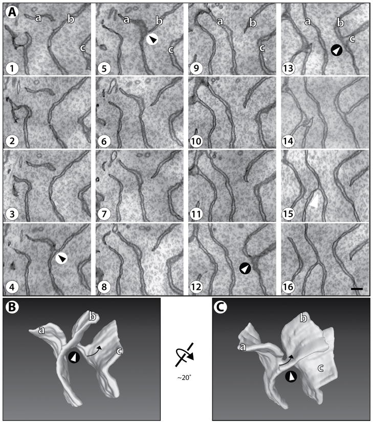
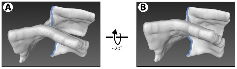
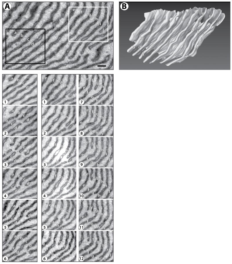
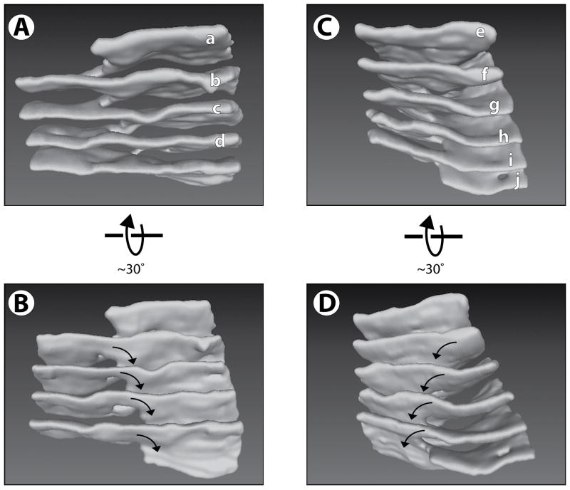
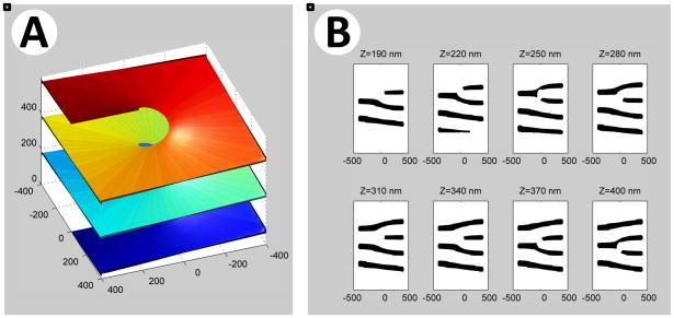
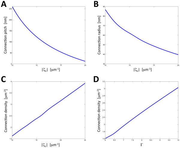
The endoplasmic reticulum (ER) often forms stacked membrane sheets, an arrangement that is likely required to accommodate a maximum of membrane-bound polysomes for secretory protein synthesis. How sheets are stacked is unknown. Here, we used improved staining and automated ultrathin sectioning electron microscopy methods to analyze stacked ER sheets in neuronal cells and secretory salivary gland cells of mice. Our results show that stacked ER sheets form a continuous membrane system in which the sheets are connected by twisted membrane surfaces with helical edges of left- or right-handedness. The three-dimensional structure of tightly stacked ER sheets resembles a parking garage, in which the different levels are connected by helicoidal ramps. A theoretical model explains the experimental observations and indicates that the structure corresponds to a minimum of elastic energy of sheet edges and surfaces. The structure allows the dense packing of ER sheets in the restricted space of a cell.
Copyright © 2013 Elsevier Inc. All rights reserved.
Figures

Comment in
-
Differential geometry meets the cell.Cell. 2013 Jul 18;154(2):265-6. doi: 10.1016/j.cell.2013.06.032. Cell. 2013. PMID: 23870115
References
-
- Baumann O, Walz B. Endoplasmic reticulum of animal cells and its organization into structural and functional domains. Int Rev Cytol. 2001;205:149–214. - PubMed
Publication types
MeSH terms
Substances
Grants and funding
LinkOut - more resources
Full Text Sources
Other Literature Sources
