. 2013 Jul 18;154(2):416-29.
doi: 10.1016/j.cell.2013.06.043.

Sulfur amino acids regulate translational capacity and metabolic homeostasis through modulation of tRNA thiolation

Affiliations

Sulfur amino acids regulate translational capacity and metabolic homeostasis through modulation of tRNA thiolation

Sunil Laxman et al. Cell. .

Abstract

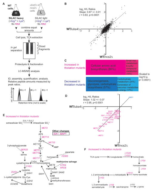
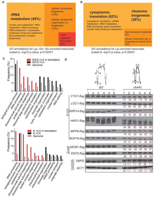
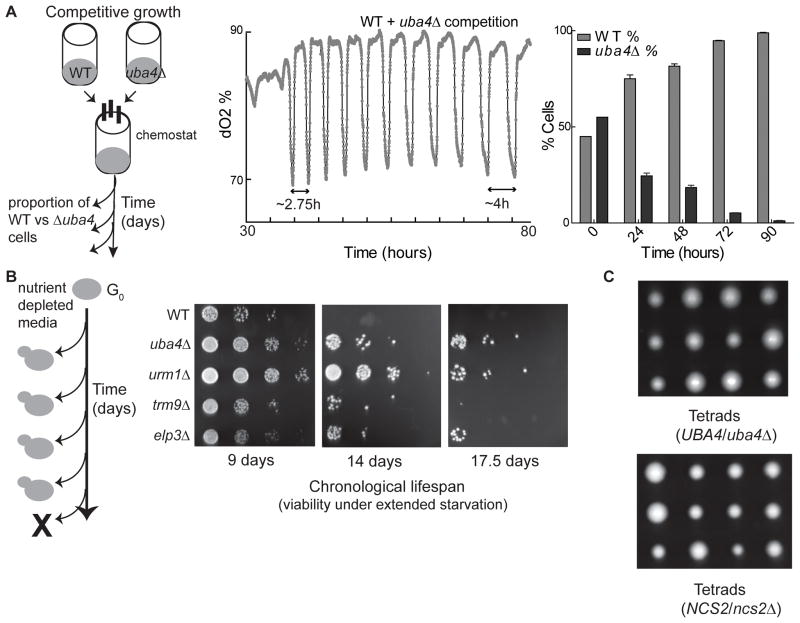
Protein translation is an energetically demanding process that must be regulated in response to changes in nutrient availability. Herein, we report that intracellular methionine and cysteine availability directly controls the thiolation status of wobble-uridine (U34) nucleotides present on lysine, glutamine, or glutamate tRNAs to regulate cellular translational capacity and metabolic homeostasis. tRNA thiolation is important for growth under nutritionally challenging environments and required for efficient translation of genes enriched in lysine, glutamine, and glutamate codons, which are enriched in proteins important for translation and growth-specific processes. tRNA thiolation is downregulated during sulfur starvation in order to decrease sulfur consumption and growth, and its absence leads to a compensatory increase in enzymes involved in methionine, cysteine, and lysine biosynthesis. Thus, tRNA thiolation enables cells to modulate translational capacity according to the availability of sulfur amino acids, establishing a functional significance for this conserved tRNA nucleotide modification in cell growth control.


Figures

Figure 1
Figure 1. tRNA uridine thiolation levels reflect sulfur amino acid availability
(A) tRNA structure, wobble uridine modifications, and structures of methoxycarbonylmethyl (mcm5) and methoxycarbonylmethyl + thiolation (mcm5s2) uridine modifications. (B) Experimental approach for measuring tRNA uridine modifications by LC-MS/MS. (C) tRNA uridine thiolation amounts decrease in minimal media. Relative abundance of thiolated (mcm5s2) or mcm5-modified tRNA uridines in cells grown in rich (YPD, YPL) and minimal media (SD, SL) were measured. (Mean±SD, n=4, with experimental duplicates. *** indicates p< 0.001, ** indicates p<0.01, * indicates p< 0.05). See also Figure S1. (D) tRNA uridine thiolation amounts are rescued by sulfur-containing amino acids. Cells were grown as described in (B), with or without supplementation of the indicated amino acids, ammonium sulfate (NH4)2SO4, or a non-sulfur amino acid mixture (non-S). Tyrosine was excluded due to poor solubility. (Mean±SD, n=3, with experimental duplicates). See also Figure S1. (E) Simplified schematic of sulfur amino acid metabolism in budding yeast. Cysteine and methionine are derived from homocysteine, and can be interconverted. (F) Intracellular sulfur amino acid levels decrease in SD minimal medium. Cells were grown in YPD, SD, or SD supplemented with methionine (150 μM) (see panel B), and sulfur metabolites were measured by LC-MS/MS. Note: relative decreases of both cysteine and methionine in SD mirror the decrease in uridine thiolation abundance. (Mean±SD, n=4, with experimental duplicates). See also Table S1 and Figure S1.
Figure 2
Figure 2. tRNA uridine thiolation is required for normal metabolic cycles
(A) Schematic describing the yeast metabolic cycle (YMC) and its three phases, OX, RB, and RC. Numbers indicate time points of typical sample collections for Figure S2. (B) Thiolation-deficient strains cannot undergo normal metabolic cycles. Metabolic cycles of WT, uba4Δ and urm1Δ (uridine thiolation-deficient), and elp3Δ and trm9Δ (uridine mcm5- deficient) cells are shown across a 20 h time period. See also Figure S2. (C) Schematic of the tRNA uridine thiolation/sulfur transfer pathway and urmylation pathway. The tRNA uridine thiolation pathway shares components with the protein urmylation pathway. Uba4p/Urm1p are required for both pathways, while Ncs2p/Ncs6p are only required for tRNA uridine thiolation. Ahp1p is the only known urmylated yeast protein. See also Figure S2. (D) Uba4p catalytic activity is required for normal metabolic cycles. The metabolic cycles of catalytically dead uba4 mutant (C225A and C397A) compared to WT are shown, along with the location of the catalytic cysteine residues within Uba4p. (E) Protein urmylation is not required for metabolic cycles. ahp1Δ strains show normal metabolic cycles. ncs2Δ and ncs6Δ strains are incapable of tRNA uridine thiolation, but are not involved in protein urmylation. Both ncs2Δ and ncs6Δ strains exhibit cycles comparable to uba4Δ and urm1Δ strains. See also Figure S2.
Figure 3
Figure 3. tRNA uridine thiolation regulates amino acid biosynthesis and sugar metabolism
(A) Schematic of the SILAC quantitative proteomic approach used to assess global changes in protein levels in thiolation-deficient cells (Supplemental Information). (B) tRNA uridine thiolation is responsible for most uba4Δ phenotypes. Correlation plot comparing normalized log2 H/L ratios from WT/ubaΔ (X-axis) and WT/ncs2Δ (Y-axis) samples, for all proteins detected in both samples (~1900 total). ncs2Δ and uba4Δ strains showed similar profiles (slope=0.87, r=0.83). See also Table S2 for complete data, Table S3 and S4 for GO analysis data. (C) tRNA uridine thiolation regulates sugar and carbohydrate metabolism as well as sulfur and lysine amino acid metabolism. Proteins increased (top panel) or decreased (bottom panel) by at least 1.4 fold in thiolation-deficient strains (compared to WT cells) were analyzed by GO term grouping, to find significantly enriched terms (significance threshold p<0.0001). Consolidated GO-term groups were visualized using the descriptive, scaled TreeMaps shown (Supplemental Information). (D) Proteins related to sulfur amino acid metabolism or lysine metabolism that increase or decrease in uba4Δ or ncs2Δ mutant cells were compared using log2 H/L ratio plots. WT/uba4Δ (X-axis) ratios for these proteins are compared against WT/ncs2Δ (Y-axis). Both samples showed a nearly 1:1 correlation (r=0.95, slope=1). (E) Sulfur amino acid metabolism-related proteins that increase in tRNA uridine thiolation-deficient mutants. Proteins involved in methionine or cysteine biosynthesis or salvage that specifically increased in both uba4Δ and ncs2Δ mutants are indicated in magenta. See also Figure S3. (F) Lysine metabolism-related proteins that increased in tRNA uridine thiolation-deficient mutants (uba4Δ, ncs2Δ) compared to WT are indicated in magenta.
Figure 4
Figure 4. Uba4p is negatively regulated during sulfur amino acid starvation
(A) npr2Δ and npr3Δ strains exhibit excessive tRNA thiolation during sulfur starvation. Relative tRNA thiolation amounts were measured in WT, npr2Δ and npr3Δ cells grown in YPL or SL medium. Thiolation increased in npr2Δ and npr3Δ strains compared to WT cells only in SL (Mean±SD, n=3, with experimental duplicates. ** indicates p<0.01). See also Figure S4. (B) Thiolation-deficient npr2Δ/uba4Δ strains exhibit slower growth compared to npr2Δ in SL medium. Growth curves of the indicated strains following switch from YPL to SL medium were measured over ~50 hours in batch cultures starting at an OD600 of ~0.1. (Mean±SD, n=3, *** indicates p<0.001). See also Figure S4 (C) npr2Δ cells have abnormally higher levels of sulfur-containing metabolites. Relative amounts of cysteine, methionine and SAM were measured in WT and npr2Δ cells growing in YPL or SL by LC-MS/MS (D) Uba4p abundance decreases upon switch to SL medium. Western blot shows amounts of Myc-UBA4 present over time in cells growing in YPL, SL or SL+0.5mM methionine (Mean±SD, n=3) (E) UBA4 and URM1 transcript levels remain relatively stable upon sulfur amino acid starvation. Relative amounts of UBA4 and URM1 transcripts in cells growing in the indicated conditions were measured by RT-qPCR (normalized to snR14). See also Figure S4.
Figure 5
Figure 5. Lys, Gln, Glu codon-enriched genes are overrepresented in translation and growth-related functions
(A) From a genome-wide analysis of codon usage frequency (Table S5), the genes with the highest frequencies of Lys, Gln and Glu (K, Q, E) (Z≥2) were selected and analyzed by Gene Ontology (GO) for highly significant GO terms (209 genes, p<0.00001), and visualized using scaled TreeMaps (See also Supplemental Information, Figure S5 and Table S6) (B) Genes with the highest frequencies of Lys (K) codons (Z≥2) were selected and analyzed by Gene Ontology (GO) for highly significant GO terms (204 genes, p<0.00001), and visualized using scaled TreeMaps. (See also Table S6 and Figure S5 for genes overrepresented for Gln and Glu codons) (C) Granular GO annotations for K, Q, E or K codon enriched genes to GO terms by biological processes, shown in comparison to genome-wide frequencies of the same biological process. Biological processes related to translation are shown in red (D) Decreased levels of K and Q-rich proteins in tRNA thiolation-deficient cells. Several genes enriched for K or Q codons were arbitrarily selected, flag epitope tagged at their C-termini, and the encoded proteins were detected by Western blot in WT and uba4Δ mutant cells grown under continuous glucose-limitation. Quantifications of relative protein amounts comparing uba4Δ with WT are shown in red. Note: the 6 independent time points from each strain cycle can be compared since cells were maintained simultaneously at exactly the same cell density and fed the same medium. See also Figure S5.
Figure 6
Figure 6. tRNA uridine modifications promote growth at the cost of survivability
(A) Competitive growth between WT and tRNA thiolation-deficient cells during nutrient limited growth in chemostats was measured as depicted on the left. The percentage of each strain within the population was measured for several days. Thiolation-deficient cells were out-competed by WT cells in less than 3 days. See also Figure S6 (B) Chronological lifespan of mutants with disrupted tRNA uridine modifications. WT or mutant cells were grown in batch cultures in SD medium as depicted on the left. Cell survivability was assessed after the indicated number of days. (C) Tetrads of sporulated diploid cells carrying a single copy uba4Δ or ncs2Δ deletion germinated on YPD medium. All the slower-growing spores were verified to be uba4Δ or ncs2Δ.
Figure 7
Figure 7. tRNA uridine thiolation gauges sulfur amino acid availability to regulate cell growth and translation
In sulfur amino acid-replete conditions (left), cells have high amounts of tRNA uridine thiolation, which increases translational capacity and cell growth. Sulfur amino acid starvation (right) results in decreased tRNA uridine thiolation, due to a decrease in Uba4p amounts as well as reduced sulfur equivalents. A reduction in tRNA thiolation functions to down-regulate translation and cell growth. This results in a feedback mechanism which serves to increase methionine, cysteine, and lysine synthesis and salvage. Decreased tRNA uridine thiolation may also limit translation of components of the translation machinery, which often harbor a large number of Lys, Gln and Glu codons. Thus, the abundance of thiolated tRNAs enables cells to integrate sulfur amino acid availability with supportable rates of translation and cell growth.

Similar articles

Cited by

References

    1. Abdullah U, Cullen PJ. The tRNA modification complex elongator regulates the Cdc42-dependent mitogen-activated protein kinase pathway that controls filamentous growth in yeast. Eukaryot Cell. 2009;8:1362–1372. - PMC - PubMed
    1. Begley U, Dyavaiah M, Patil A, Rooney JP, DiRenzo D, Young CM, Conklin DS, Zitomer RS, Begley TJ. Trm9-catalyzed tRNA modifications link translation to the DNA damage response. Mol Cell. 2007;28:860–870. - PMC - PubMed
    1. Blagosklonny MV, Hall MN. Growth and aging: a common molecular mechanism. Aging (Albany NY) 2009;1:357–362. - PMC - PubMed
    1. Brandman O, Stewart-Ornstein J, Wong D, Larson A, Williams CC, Li GW, Zhou S, King D, Shen PS, Weibezahn J, et al. A ribosome-bound quality control complex triggers degradation of nascent peptides and signals translation stress. Cell. 2012;151:1042–1054. - PMC - PubMed
    1. Brauer MJ, Huttenhower C, Airoldi EM, Rosenstein R, Matese JC, Gresham D, Boer VM, Troyanskaya OG, Botstein D. Coordination of growth rate, cell cycle, stress response, and metabolic activity in yeast. Mol Biol Cell. 2008;19:352–367. - PMC - PubMed

Publication types

MeSH terms