. 2013 Jul 25;51(2):156-73.
doi: 10.1016/j.molcel.2013.07.001.

Tandem stem-loops in roX RNAs act together to mediate X chromosome dosage compensation in Drosophila

Affiliations

Tandem stem-loops in roX RNAs act together to mediate X chromosome dosage compensation in Drosophila

Ibrahim Avsar Ilik et al. Mol Cell. .

Abstract

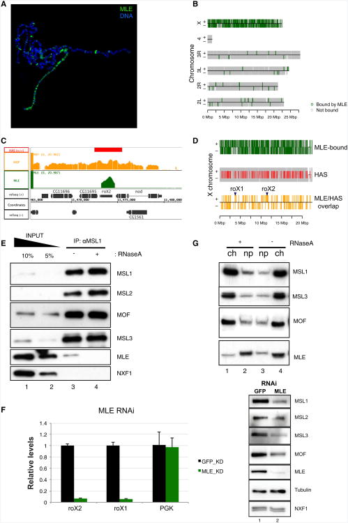
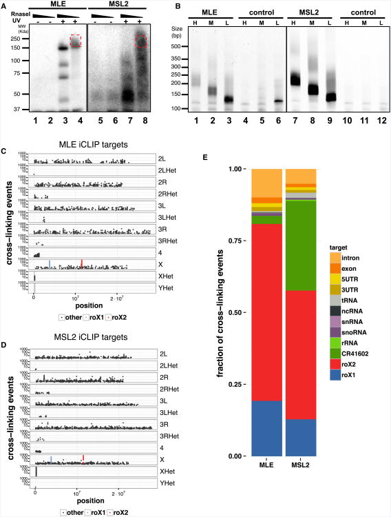
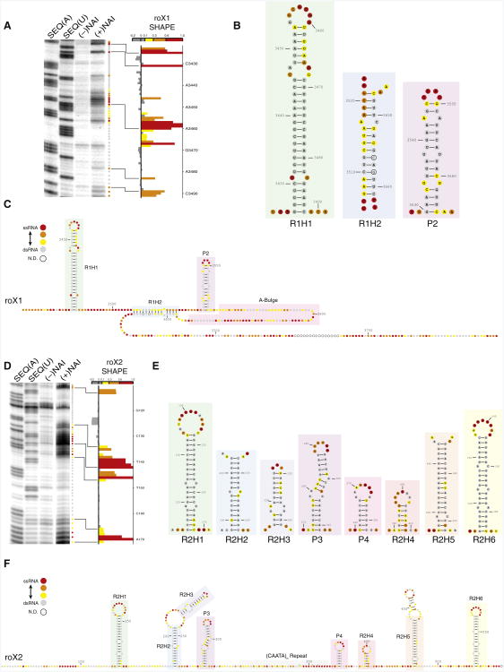
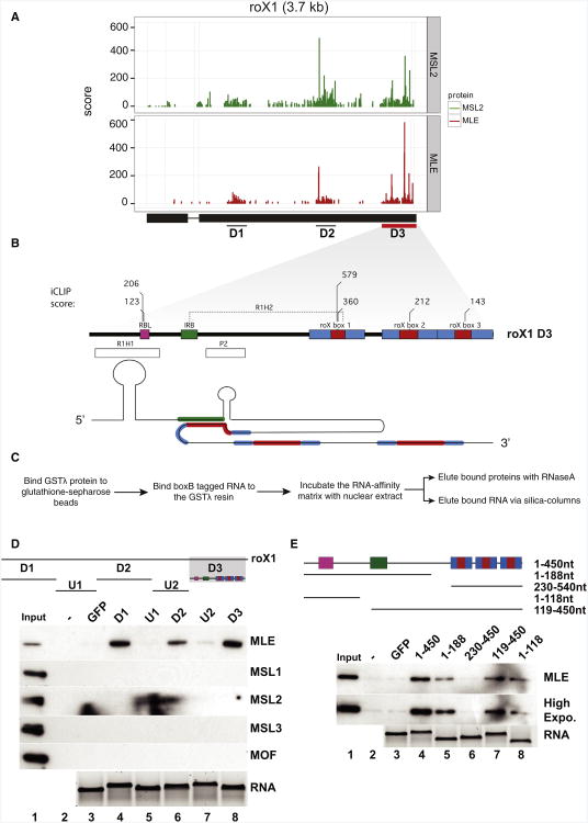
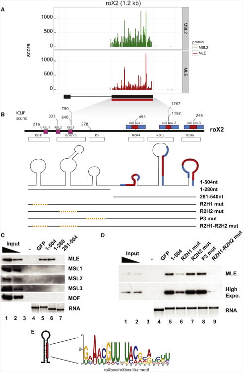
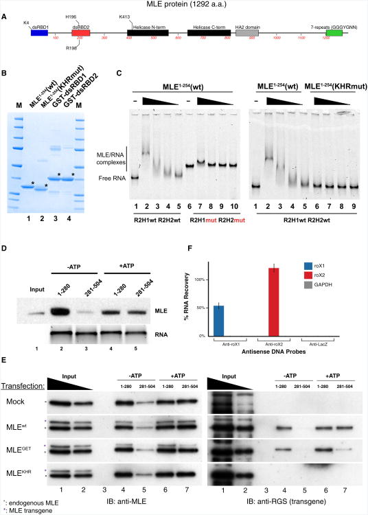
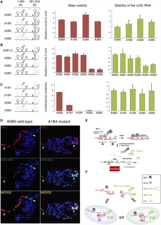
Dosage compensation in Drosophila is an epigenetic phenomenon utilizing proteins and long noncoding RNAs (lncRNAs) for transcriptional upregulation of the male X chromosome. Here, by using UV crosslinking followed by deep sequencing, we show that two enzymes in the Male-Specific Lethal complex, MLE RNA helicase and MSL2 ubiquitin ligase, bind evolutionarily conserved domains containing tandem stem-loops in roX1 and roX2 RNAs in vivo. These domains constitute the minimal RNA unit present in multiple copies in diverse arrangements for nucleation of the MSL complex. MLE binds to these domains with distinct ATP-independent and ATP-dependent behavior. Importantly, we show that different roX RNA domains have overlapping function, since only combinatorial mutations in the tandem stem-loops result in severe loss of dosage compensation and consequently male-specific lethality. We propose that repetitive structural motifs in lncRNAs could provide plasticity during multiprotein complex assemblies to ensure efficient targeting in cis or in trans along chromosomes.


Figures

Figure 1
Figure 1. MLE Is Enriched on High-Affinity Sites, and Its Chromatin Association Is RNase Sensitive
(A) A polytene squash staining of male third-instar larvae shows that MLE (green) coats the X chromosome. DNA is shown in blue (DAPI). (B) ChIP-seq analysis shows that MLE is enriched on the X chromosome. All genes are represented in their correct chromosomal location (gray). MLE binding sites are shown in green. Top (+) and bottom strand (−) are indicated. Scale at the bottom indicates the size chromosomal length in megabasepair (Mbp). (C) A browser snapshot for roX2 HAS. MLE binding (green) to HAS is more restricted than MOF (orange). The red box on top marks the HAS that overlaps the roX2 gene. (D) Almost all HASs are bound by MLE. The X chromosome is represented in three tracks: MLE-bound genes are shown in green (as in B), the HASs in red, and the MLE-bound HASs in orange (also see Figure S1E). Two specific HASs, the roX1 and roX2 genes, are specified by arrows. (E) Immunoprecipitation of MSL1 from an S2 nuclear extract, under mild conditions, results in coprecipitation of small amounts of MLE in comparison to MSL2, MSL3, and MOF (compare lanes 1 and 2 with lane 3). RNase A treatment (lane 4) leads to the loss of this weak MLE interaction. NXF1 served as a negative control. (F) RNAi-mediated depletion of MLE in S2 cells led to the destabilization of the MSL complex together with the loss of roX RNAs. (Left) Total RNA was isolated from double-stranded RNA (GFP [control] or MLE) treated S2 cells. Expression levels of roX1 and roX2 were determined by RT-qPCR analysis. PGK levels were used as a control. Error bars represent standard deviation (± SD) of three biological replicates. (Right) Western blot analysis of the corresponding experiment using whole-cell extracts. Tubulin and NXF1 served as loading controls. (G) The solubility of MSL1, MOF, MSL3, and MLE is tested in nuclei isolated from S2 cells, with or without RNase A incubation (ch, chromatin fraction; np, nucleoplasmic fraction). See Figure S1.
Figure 2
Figure 2. iCLIP Reveals that MLE and MSL2 Interact with RNA In Vivo, roX1 and roX2 Being the Major Interactors
(A) MLE or MSL2 immunoprecipitated from nuclear extracts prepared from clone8 cells under stringent conditions, with or without UV-C crosslinking, and treated with low or high concentrations of RNaseI. Bound RNA is radioactively labeled and visualized by autoradiography. RNA molecules above the molecular weight of the protein of interest (red box) are isolated from nitrocellulose membranes in triplicates, cloned into a library, and sequenced. (B) MLE and MSL2 libraries generated using the iCLIP approach. High-, medium-, and low-range libraries are prepared by gel fractionation of cDNA after reverse transcription and separate amplification with Illumina sequencing-compatible primers. (C) MLE iCLIP detects 2,447 crosslinked nucleotides. The genomic distribution of crosslinked nucleotides scored by number of crosslinking events shows no particular bias toward any chromosome. roX1 (blue) and roX2 (red) nucleotides score considerably higher than most other nucleotides. (D) MSL2 iCLIP detects 5,206 crosslinked nucleotides. Scores and distribution of roX1, roX2, and other nucleotides are similar to MLE. (E) Distribution of crosslinking events. The majority of crosslinking events fall on roX1 and roX2 (Figure S2E). See Figures S2 and S3.
Figure 3
Figure 3. SHAPE Structural Analysis Reveals the Architecture of roX1 and roX2
(A) SHAPE analysis of the R1H1 region of roX1. Full-length roX1 RNA was in vitro transcribed and treated with or without SHAPE reagent, (± NAI). cDNAs were reverse transcribed from the modified RNA to identify modified, flexible bases. Sequencing lanes identify the base position + 1, and the (−) NAI lane identifies spurious reverse transcription stops that are modification independent. The SHAPE reactivity score for each base is calculated by integrating gel band intensities, subtracting background (−NAI), and normalizing to top peaks. Paired bases have low reactivity (gray), and flexible unpaired bases have high reactivity (increasing from yellow to orange to red). (B) SHAPE-derived structural domains of roX1. From the SHAPE reactivity profile for the 3′-terminal domain of roX1, a structure model is built. The R1H1 structure is derived from the SHAPE gel in (A). For each structure, reactive bases are present in bulges and the terminal loop, whereas the helix of this stem -loop is characterized by stretches of low reactivity. (C) Structure model of the entire 3′ terminus of roX1. The 3′ terminus of roX1 is organized into three stable helices, two of which contain a core roX box motif (R1H1 and R1H2). The A-bulge likely participates in long-range base pairing with distal U-rich regions of roX1 that were not interrogated here. The very 3′ end lacks any well-defined secondary structure. (D) The SHAPE profile of the R2H1 region of roX2. The R2H1 domain of roX2 exhibits light-heavy-light modification, characteristic of a stable stem loop. (E) SHAPE-derived structural domains of roX2. SHAPE of roX2 exon-3 resolved many stable secondary structures. Three of the helices contain roX box-like motif (R2H1-3), and three contain the roX box motif (R2H4-6). (F) Structure model for roX2 exon-3. roX2 exon-3 is arranged into stable structural domains that are linked together by a flexible backbone. See Figures S4 and S5.
Figure 4
Figure 4. MLE and MSL2 Bind to Three Clustered Regions within roX1 RNA
(A) The iCLIP data show that MLE and MSL2 interact with three domains (D1–D3) of roX1. The red box marks roX1 D3, which shows the highest score of MLE binding. See also Figure S2. (B) A cartoon representation of roX1 RNA domain-3 (D3). roX1 region-3 is similar to roX2 exon-3 in its arrangement of stem oops and roX boxes (see Figure 5B). R1H1 contains an RBL element in its stem (pink box) and is followed by R2H2, which is formed by a long-range interaction between the inverted roX box (IRB, green box) element and roX1 box1 (RB1, red-in-blue box). Another stem-loop (P2) is predicted to form in the bulge separating IRB and roX1 box1. Some of MLE's top iCLIP scores are indicated on top. (C) The flow of a GRNA chromatography experiment. (D) (Top) A cartoon representation of roX1 RNA delineated by MLE/MSL2 binding shows the constructs used in GRNA experiments. Colored boxes represent the elements described in (B). All RNA fragments used were of similar length (428–585 nt). (Bottom) GRNA chromatography shows that MLE interacts with D1, D2, and D3 (lanes 4, 6, and 8, respectively) and not with regions U1 and U2, GFP RNA, or beads (lanes 5, 7, 3, and 2, respectively). Antibodies used for immunoblotting are indicated on the right. (E) (Top) The full-length roX1 region3 (D3) is split into four fragments for GRNA experiments. Colored boxes represent the elements described in Nucleotide is indicated as nt (B). (Bottom) Specific binding of MLE to D3 (1–450) (compare lane 2 [beads] and lane 3 [GFP] with lane 4) could be further reduced to first half of roX1 D3 (1–188) (lane 5). No significant binding was scored for the second half of D3 (230–450) (lanes 6). R1H1 (1–118) seems to be able to interact with MLE (lane 8); however, its deletion does not lead to a complete loss of MLE binding (119–450; lane 7), suggesting that P2 and/or R1H2 can also interact with MLE in this assay. RNA eluted from the beads was run on a 1.2% agarose gel and stained with SYBR Safe and is shown beneath the immunoblots.
Figure 5
Figure 5. MLE and MSL2 Bind Exclusively to roX2 Exon-3
(A) MSL2 and MLE interact with the evolutionarily conserved third exon of roX2 in vivo. See also Figure S2. (B) A cartoon representation of roX2 exon-3 shows that it has tandem helical regions at its 5′-end (R2H1, R2H2/3, and P3, white boxes), forming the first stem-loop cluster with RBL elements (pink boxes), and three roX-box elements at its 3′ end (RB1-3, red-in-blue boxes, indicated on top), which also resides in helical structures that form the second stem-loop cluster. MLE's top iCLIP scores are indicated on top. A schematic representation of fragments used in (C) and (D) is also shown. (C) GRNA chromatography shows MLE interaction with the full-length roX2 exon-3 (1–504 nt, lane 5) and not with the beads (lane 3) or GFP RNA (lane 4). Based on SHAPE, iCLIP, and HITS-CLIP data, RNA was split into two halves. MLE binding was retained on the first stem-loop cluster (1–280, lane 6), and not with the second stem-loop cluster (281-504, lane 7). The proteins are probed with the indicated antibodies on the right. (D) GRNA experiment, similar to (C), where the stem regions of R2H1, R2H2, or P3 are individually disrupted by point mutations in the context of the full-length exon-3 RNA in order to see if these are the regions that are responsible for MLE-roX2 interaction. High Expo. is higher exposure of the MLE blot. (C and D) RNA eluted from the beads was run on a 1.2% agarose gel and stained with SYBR Safe and is shown beneath the immunoblots. (E) The extended roXbox/roXbox-like motif found at MLE crosslinking sites with very high scores. See Figure S6.
Figure 6
Figure 6. MLE's Double-Stranded RNA Binding Domains Interact with roX2 RNA
(A) Schematic representation of MLE protein domains. Residues mutated in later experiments are also indicated. (B) MLE dsRBDs were expressed for EMSA. (Lane 1) His-tagged wild-type MLE1-254 (32 kDa) protein includes dsRBD 1 and dsRBD 2; (lane 2) a derivative expressing one point mutation in the dsRBD1 (K4E) and two in the dsRBD2 (H196E, R198E); (lanes 3 and 4) GST-tagged dsRBD1 and dsRBD2, respectively. M, molecular weight marker. Protein gel was stained with Coomassie blue. (C) MLE1-254 interacts with an RNA probe consisting of R2H1 and R2H2/3 regions ofroX2 exon-3 (lanes 2-5). Point mutations that disrupt the stability of the stems in both R2H1 and R2H2 (lanes 7-10, left), or point mutations predicted to render dsRBD1 (K4E) and dsRBD2 (H196E, R198E; lanes 7-10, right) incapable of binding RNA, severely reduce the interaction between MLE1-254(KHR mutant) and R2H1wt R2H2/3wt RNA. For each protein derivative, 125 nM, 250 nM, 500 nM, or 1 μM, respectively, was titrated (black triangle). Free RNA probe is shown in lanes 1 and 6 (left gel) and lane 1 (right gel). (D) The first (1-280) and the second (281-504) stem-loop clusters of roX2 RNA are used in a GRNA experiment as described in Figures 4 and 5. Here, ATP (3 mM) was added during the incubation of RNA with the nuclear extract (lanes 4 and 5). (E) GRNA chromatography using transgenic MLE constructs that are either wild-type (MLEwt), compromised in their ATP-binding pocket (MLEGET), or dsRBDs (MLEKHR). Immunoblots (IB) were probed either with an MLE antibody detecting endogenous (°) and ectopically expressed MLE derivatives (*) (left) or with an antibody that only detects the MLE derivatives (right). Lanes 1 and 2 show loading of 4% and 12% of input nuclear extracts. (F) roX1 and roX2 do not directly interact in vivo. Antisense oligos to roX1, roX2, and LacZ (negative control) were used to isolate RNA-protein-chromatin complexes with the targeted RNA. roX1 ChIRP recovers no roX2 RNA, and roX2 recovers no roX1 RNA. LacZ ChIRP enriches for none of the target RNAs. RNA was quantified by qRT-PCR. Error bars represent ± SD of three independent measurements. See Figure S6.
Figure 7
Figure 7. Cooperative Interaction of Tandem Stem Loops Is Important for roX Function In Vivo
(A–C) Schematic representation of wild-type and mutant roX2 exon-3 constructs are depicted (left). “A” and “B” refer to the first and second stem-loop clusters. Mutations introduced to the helical regions are indicated by the dashed orange line. (A) Helix-destabilizing mutations introduced to the first stem-loop cluster (A1B0, A2B0, and A3B0) rescue male lethality (middle, dark orange) and do not affect the stability of these RNAs (right, green). (B) Mutations in individual helices of the second stem-loop cluster also rescue male lethality (A0B1, A0B2, and A0B3). When the two terminal helices are disrupted at the same time (A0B4 and A0B5), male viability and RNA stability are reduced (middle and right). (C) However, combinatorial mutations affecting both the first and second half of roX2 exon-3 (A1B1, A1B4, A2B4, and A3B4) severely affect male viability (middle), without affecting RNA stability in vivo (right). (A-C) Error bars for male viability data represent ± SEM and for expression analysis ± SD of at least three biological replicates. (D) Polytene chromosomal immunostainings of male third-instar larvae show that the wild-type roX2 exon-3 construct (left) efficiently targets the MSL complex (MSL1, red) and H4K16 acetylation (green)to the X chromosome (X) in males. However, the A1B4 mutant shows a severe reduction in the number of MSL1-bound target sites on the X chromosome, and mistargeting to the chromocenter (arrow) is observed (right). DNA is stained with Hoechst 33342 (blue). (E) A summary of MLE-roX interactions described in this study. MLE engages with roX2 RNA stem loops in an ATP-independent and -dependent manner. Tandem stem loops in roX2 play an important role in dosage compensation as combinatorial mutations lead to severe male-specific lethality due to defective targeting of the MSL complex to the X chromosome. (F)A summary model using roX2 RNA as an example. We propose that roX RNAs play an important role in integration of MLE with the core MSL complex (MSL1-3, MOF). The dynamic interaction of MLE with different regions of roX RNAs in the absence or presence of ATP could provide a means to compose holo-MSL complexes where multiple molecules of either MLE or MSL core complex could be assembled. Such “heterogenous” assemblies could contribute to efficient targeting and also have the potential to mediate spreading of the MSL complex, enhancing the possibility of short- and long-range interactions on the X chromosome. See Figure S7.

Comment in

References

    1. Akhtar A, Zink D, Becker PB. Chromodomains are protein-RNA interaction modules. Nature. 2000;407:405–409. - PubMed
    1. Amrein H, Axel R. Genes expressed in neurons of adult male Drosophila. Cell. 1997;88:459–469. - PubMed
    1. Augui S, Nora EP, Heard E. Regulation of X-chromosome inactivation by the X-inactivation centre. Nat Rev Genet. 2011;12:429–442. - PubMed
    1. Chu C, Qu K, Zhong FL, Artandi SE, Chang HY. Genomic maps of long noncoding RNA occupancy reveal principles of RNA-chromatin interactions. Mol Cell. 2011;44:667–678. - PMC - PubMed
    1. Conrad T, Akhtar A. Dosage compensation in Drosophila melanogaster: epigenetic fine-tuning of chromosome-wide transcription. Nat Rev Genet. 2011;13:123–134. - PubMed

Publication types

MeSH terms