. 2013 Jul 16;105(2):320-7.
doi: 10.1016/j.bpj.2013.05.040.

Unraveling the nanoscale surface properties of chitin synthase mutants of Aspergillus fumigatus and their biological implications

Affiliations

Unraveling the nanoscale surface properties of chitin synthase mutants of Aspergillus fumigatus and their biological implications

David Alsteens et al. Biophys J. .

Abstract

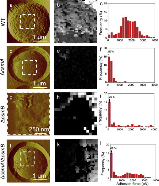
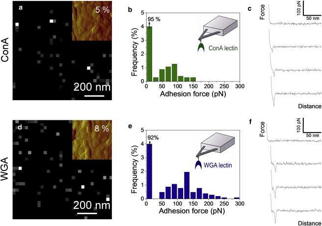
Understanding the surface properties of the human opportunistic pathogen Aspergillus fumigatus conidia is essential given the important role they play during the fungal interactions with the human host. Although chitin synthases with myosin motor-like domain (CSM) play a major role in cell wall biosynthesis, the extent to which deletion of the CSM genes alter the surface structural and biophysical-biological properties of conidia is not fully characterized. We used three complementary atomic force microscopy techniques-i.e., structural imaging, chemical force microscopy with hydrophobic tips, and single-molecule force spectroscopy with lectin tips-to gain detailed insights into the nanoscale surface properties (ultrastructure, hydrophobicity) and polysaccharide composition of the wild-type and the chitin synthase mutant (ΔcsmA, ΔcsmB, and ΔcsmA/csmB) conidia of A. fumigatus. Wild-type conidia were covered with a highly hydrophobic layer of rodlet nanostructures. By contrast, the surface of the ΔcsmA mutant was almost completely devoid of rodlets, leading to loss of hydrophobicity and exposure of mannan and chitin polysaccharides. The ΔcsmB and ΔcsmA/csmB mutants showed a different behavior, i.e., the surfaces featured poorly organized rodlet layers, yet with a low hydrophobicity and substantial amounts of exposed mannan and chitin at the surface. As the rodlet layer is important for masking recognition of immunogenic fungal cell wall components by innate immune cells, disappearance of rodlet layers in all three chitin synthase mutant conidia was associated with an activation of human dendritic cells. These nanoscale analyses emphasize the important and distinct roles that the CSMA and CSMB genes play in modulating the surface properties and immune interactions of A. fumigatus and demonstrate the power of atomic force microscopy in fungal genetic studies for assessing the phenotypic characteristics of mutants altered in cell surface organization.


Figures

Figure 1
Figure 1
AFM imaging reveals that deletion of the CSMA and CSMB genes leads to major remodeling of the cell wall architecture. AFM deflection images of the surface of WT (ac), ΔcsmA (df), ΔcsmB (gi), and ΔcsmA/csmB (jl) conidia recorded in acetate buffer at low (ad, g, j), medium (b, e, h, k), and high (c, f, il) resolution. Labels R and A” indicate regions made of rodlets and amorphous material. The arrow indicates artifacts resulting from the interaction between the tip and loosely bond material. For each strain, images are representative of those obtained for at least 15 cells from three independent cultures, and analyzed with three different tips (see also Fig. S1 for additional examples).
Figure 2
Figure 2
CFM demonstrates that rodlet layers on WT conidia are uniformly hydrophobic. (a) Deflection image of a WT conidial surface recorded in water with a silicon nitride tip. (bd). Representative force-distance curves (b), adhesion force map (z-range: 4 nN) (c), and adhesion force histogram (n = 512). (d) Recorded on the same cell with a hydrophobic tip (see also Fig. S1 for additional examples).
Figure 3
Figure 3
Structural changes in the mutants correlate with differences in cell surface hydrophobicity. Deflection images (a, d, g, j) recorded in water with silicon nitride tips, adhesion force maps (z -range: 4 nN) (b, e, h, k), and adhesion force histograms (n = 512) (c, f, i, l) recorded with hydrophobic tips on the surface of WT (ac), ΔcsmA (d–f), ΔcsmB (g–i), and ΔcsmA/csmB (j–l) conidia (see Fig. S1).
Figure 4
Figure 4
Mapping single polysaccharide residues on WT conidia using SMFS. Adhesion force maps (z-range: 300 pN; values in the top right corners correspond to the percentage of adhesive events) (a and d), adhesion force histograms (n = 512) (b and e) and representative force-distance curves (c and f) recorded on WT conidia in acetate buffer supplemented with Ca2+and Mn2+, using AFM tips bearing ConA (ac) and WGA (df) lectins. The insets in panels a and d show deflection images of the areas corresponding to the adhesion maps. See Fig. S2 for an independent set of data.
Figure 5
Figure 5
Chitin synthase mutations lead to the exposure of glycans. Adhesion force maps (z-range: 300 pN; the values correspond to the percentage of adhesive events) recorded on the surface of ΔcsmA (a and b), ΔcsmB (c and d), and ΔcsmA/csmB (e and f) conidia, using AFM tips bearing ConA (a, c, e) and WGA (b, d, f) lectins. The insets show deflection images of the areas corresponding to the adhesion maps. (g and h). Historgams of the mean (± SD) adhesion force measured on each mutant with ConA (g) and WGA (h) tips. See Figs. S3–S5 for independent sets of data.
Figure 6
Figure 6
Chitin synthase mutant conidia stimulate human dendritic cells. The expression of CD83, CD86 (both expressed as % positive cells) (a and b); CD80, CD86, HLA-DR, and CD40 (all expressed as mean fluorescence intensities) (cf, respectively) on DC that were cultured with cytokines alone or cytokines plus WT conidia, ΔcsmA, ΔcsmB, or ΔcsmA/csmB conidia for 48 h. Data (mean ± SE) from six independent donors. Statistical significance as determined by ANOVA test is indicated (P < 0.05; ∗∗P < 0.01; ∗∗∗P < 0.001).

References

    1. Chai L.Y.A., Hsu L.Y. Recent advances in invasive pulmonary aspergillosis. Curr. Opin. Pulm. Med. 2011;17:160–166. - PubMed
    1. Mahdavinia M., Grammer L.C. Management of allergic bronchopulmonary aspergillosis: a review and update. Ther. Adv. Respir. Dis. 2012;6:173–187. - PubMed
    1. Annaix V., Bouchara J.P., Tronchin G. Specific binding of human fibrinogen fragment D to Aspergillus fumigatus conidia. Infect. Immun. 1992;60:1747–1755. - PMC - PubMed
    1. Beever R.E., Dempsey G.P. Function of rodlets on the surface of fungal spores. Nature. 1978;272:608–610. - PubMed
    1. Cole G.T., Sekiya T., Kasai R. Surface ultrastructure and chemical composition of the cell walls of conidial fungi. Exp. Mycol. 1979;3:132–156.

Publication types

MeSH terms

LinkOut - more resources