Review
. 2013 Jul 20:10:239.
doi: 10.1186/1743-422X-10-239.

Medical virology of hepatitis B: how it began and where we are now

Affiliations
Review

Medical virology of hepatitis B: how it began and where we are now

Wolfram H Gerlich. Virol J. .

Abstract

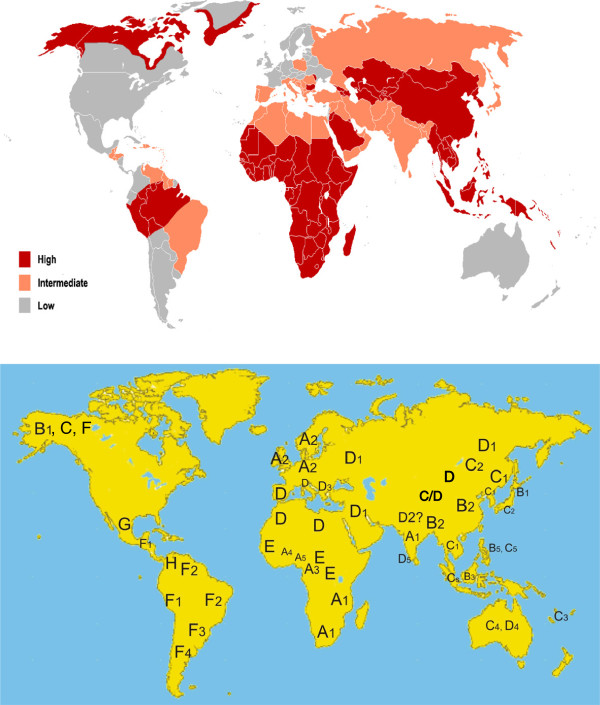
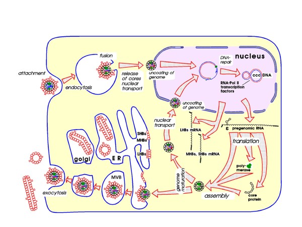
Infection with hepatitis B virus (HBV) may lead to acute or chronic hepatitis. HBV infections were previously much more frequent but there are still 240 million chronic HBV carriers today and ca. 620,000 die per year from the late sequelae liver cirrhosis or hepatocellular carcinoma. Hepatitis B was recognized as a disease in ancient times, but its etiologic agent was only recently identified. The first clue in unraveling this mystery was the discovery of an enigmatic serum protein named Australia antigen 50 years ago by Baruch Blumberg. Some years later this was recognized to be the HBV surface antigen (HBsAg). Detection of HBsAg allowed for the first time screening of inapparently infected blood donors for a dangerous pathogen. The need to diagnose clinically silent HBV infections was a strong driving force in the development of modern virus diagnostics. HBsAg was the first infection marker to be assayed with a highly sensitive radio immune assay. HBV itself was among the first viruses to be detected by assay of its DNA genome and IgM antibodies against the HBV core antigen were the first to be selectively detected by the anti-μ capture assay. The cloning and sequencing of the HBV genome in 1978 paved the way to understand the viral life cycle, and allowed development of efficient vaccines and drugs. Today's hepatitis B vaccine was the first vaccine produced by gene technology. Among the problems that still remain today are the inability to achieve a complete cure of chronic HBV infections, the recognition of occult HBV infections, their potential reactivation and the incomplete protection against escape mutants and heterologous HBV genotypes by HBV vaccines.


Figures

Figure 1
Figure 1
Electron microscopy images (negative staining) and approximate numbers of HBV associated particles in 1 ml of the serum from a highly viremic chronically infected HBV carrier.
Figure 2
Figure 2
Model of the hepatitis B virus (Dane particle) and the filamentous or spherical HBsAg particles. Dane described the virus as 42 nm particle but in the negative staining the outer preS1 and preS2 domains were not visible. 52 nm is the hydrodynamic diameter (Ch. Schüttler and W. Gerlich, unpublished) and also measured by cryo-EM as the outer diameter [16], suppl. Figure 2. HBsAg protein comes in three forms: large (L-) HBs protein with the preS1, preS2 and S domain, middle protein (M-) without preS1 and SHBs without preS1 and preS2.
Figure 3
Figure 3
Biochemical structure of HBV DNA. The molecule is open circular. The minus-strand has full length within the core particle and even a redundancy of 9 bases at the ends around the nick. The plus-strand is incomplete leaving a large single-stranded gap. The 5′ end of the minus-strand is covalently linked to the primase domain of the DNA polymerase which is present with its active center of the reverse transcriptase domain at the 3′ end of the plus-strand. The 5′ end of the plus-strand contains still its primer which is - in this case- derived from the 18 capped 5′ terminal bases of the degraded pregenomic HBV mRNA.
Figure 4
Figure 4
Structural components of HBV (left) and open reading frames (ORF) for encoding proteins in the covalently closed form of HBV DNA. The HBV core contains besides the HBV genome the HBV polymerase with the primase (pr) and the reverse transcriptase (RT) domain and the cellular protein kinase C alpha (PKC) [24]. Note the two start codons in the PreC/core ORF and the three start codons in the HBs ORF.
Figure 5
Figure 5
Schematic representation of the course of acute HBV infections with resolution. After the infecting event (time 0) follows a lag phase of several weeks without detectable markers. Thereafter HBV DNA (within the virus) and HBsAg increase exponentially in the serum. HBV DNA is detected earlier because its assay is much more sensitive. The peak of HBV DNA and HBsAg is reached before outbreak of the acute disease and both decrease after the onset of clinical symptoms. Initially, the HBV DNA decreases faster because it has a shorter half life time in serum than HBsAg. HBsAg finally disappears whereas HBV DNA may remain detectable in traces. Antibodies against the HBV core antigen (anti-HBc) appear with the onset of symptoms, the protective antibody against HBsAg (anti-HBs) appears very late, usually several weeks or months after disappearance of HBsAg. Disappearance of HBsAg is considered to be a sign of resolution but the virus often remains in occult form in the liver.
Figure 6
Figure 6
The three phases of chronic HBV infections.
Figure 7
Figure 7
Prevalence (top) and genotype distribution (bottom) of HBV infections. Please note that HBV subgenotype A2, present in the most popular hepatitis B vaccines, is only prevalent in the low endemic regions of the Americas and Europe. This means that >99% of all HBV carriers have other HBV subgenotypes.
Figure 8
Figure 8
Mutations in the HBsAg loop of a reactivated HBV variant. The complicated folded loop forms the surface of HBV and HBsAg particles. The exact topology and three-dimensional shape of the loop are unknown. One circle corresponds to one amino acid in the single letter code of the normal (wildtype) HBV, each square to a mutation. The boxed-in part is named a-determinant and is believed to be immunodominant, but immune escape induced mutations occurred in the entire HBsAg loop. Yellow (shaded) squares cause the loss of an immunodominant HBsAg subtype determinant. This variant replicated in a patient receiving lymphoma therapy. The patient was anti-HBc and anti-HBs positive before the immunosuppressive lymphoma therapy and developed severe acute hepatitis B after end of the therapy due to immunopathogenesis against the variant which had become abundant under immunosuppression. The serum from the acute reactivated hepatitis B phase had a high virus load, but was HBsAg negative in all assays.
Figure 9
Figure 9
Life cycle of HBV. Attachment to liver-specific receptors (heparansulfate proteoglycan and NTCP, see text) leads to endocytosis of HBV and release of HBV core particles. These are transported to the nucleus and arrested at the nuclear pore complex where the HBV genome is released to the nucleus. In the nucleus, the viral DNA is “repaired” to the covalently closed circular (ccc) DNA and complexed with nucleosomes (not shown). In interaction with transcription factors (not shown), the ccc DNA is transcribed to the pregenomic and subgenomic mRNAs. The mRNAs are transported, mainly without splicing, to the cytoplasm. The two subgenomic mRNAs for the three HBs proteins are translated at the endoplasmic reticulum, assemble to subviral HBsAg particles and are secreted via the Golgi apparatus. In parallel, the pregenomic mRNA is translated in the cytosol to the HBV core protein and the viral polymerase, whereby the three components assemble to the immature core particle. The HBV genomes mature within the core particles via reverse transcription of the pregenomic mRNA to DNA. The mature core particles can migrate again to the nuclear pore complex or are enveloped by the surface proteins and secreted via the multivesicular bodies (MVB).

References

    1. Blumberg BS. Hepatitis B - The hunt for a killer virus. Princeton: Princeton University Press; 2002.
    1. Lürmann A. Eine Icterusepidemie. Ber Klin Wochenschr. 1885. pp. 20–23.
    1. MacCallum FO. 1971 International symposium on viral hepatitis. Historical perspectives. Can Med Assoc J. 1972;10(Suppl):423–426. - PMC - PubMed
    1. Seeff LB, Beebe GW, Hoofnagle JH, Norman JE, Buskell-Bales Z, Waggoner JG, Kaplowitz N, Koff RS, Petrini JL Jr, Schiff ER. et al.A serologic follow-up of the 1942 epidemic of post-vaccination hepatitis in the United States Army. N Engl J Med. 1987;10:965–970. doi: 10.1056/NEJM198704163161601. - DOI - PubMed
    1. Barker LF, Murray R. Relationship of virus dose to incubation time of clinical hepatitis and time of appearance of hepatitis–associated antigen. Am J Med Sci. 1972;10:27–33. doi: 10.1097/00000441-197201000-00005. - DOI - PubMed

MeSH terms

Substances