Review
. 2013 Sep;12(9):894-905.
doi: 10.1016/S1474-4422(13)70136-4. Epub 2013 Jul 17.

Joubert syndrome: congenital cerebellar ataxia with the molar tooth

Affiliations
Review

Joubert syndrome: congenital cerebellar ataxia with the molar tooth

Marta Romani et al. Lancet Neurol. 2013 Sep.

Abstract

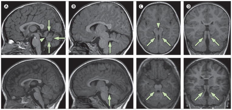
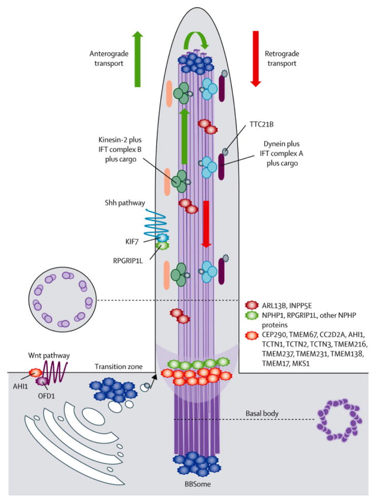
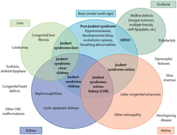
Joubert syndrome is a congenital cerebellar ataxia with autosomal recessive or X-linked inheritance, the diagnostic hallmark of which is a unique cerebellar and brainstem malformation recognisable on brain imaging-the so-called molar tooth sign. Neurological signs are present from the neonatal period and include hypotonia progressing to ataxia, global developmental delay, ocular motor apraxia, and breathing dysregulation. These signs are variably associated with multiorgan involvement, mainly of the retina, kidneys, skeleton, and liver. 21 causative genes have been identified so far, all of which encode for proteins of the primary cilium or its apparatus. The primary cilium is a subcellular organelle that has key roles in development and in many cellular functions, making Joubert syndrome part of the expanding family of ciliopathies. Notable clinical and genetic overlap exists between distinct ciliopathies, which can co-occur even within families. Such variability is probably explained by an oligogenic model of inheritance, in which the interplay of mutations, rare variants, and polymorphisms at distinct loci modulate the expressivity of the ciliary phenotype.


Conflict of interest statement

Conflicts of interest

The authors declare they have no conflict of interest.

Figures

Figure 1
Figure 1
Neuroimaging findings in a 2-year-old child with pure JS (upper panels) compared to a healthy control (lower panels). a) Parasagittal T1-weighted image shows the thickened, elongated and horizontally orientated superior cerebellar peduncles (white arrow). b) Midsagittal T1-weighted image demonstrates a moderate hypoplasia and dysplasia of the cerebellar vermis (white arrows) with secondary distortion and enlargement of the fourth ventricle with rostral shifting of the fastigium (white arrow head). A deepened interpeduncular fossa is also noted. c) Axial T1-weighted image at the level of the pontomesencephalic junction shows the molar tooth sign with a deepened interpeduncular fossa (white arrowhead) and elongated, thickened and horizontally orientated superior cerebellar peduncles (white arrows). Additionally, the cerebellar vermis appears to be hypoplastic and its remnants dysplastic. d) Coronal T1-weighted image reveals the thickened superior cerebellar peduncles (white arrows).
Figure 2
Figure 2
Schematic representation of the structure of the primary cilium and its protein complexes. Most proteins mutated in JS and MKS cluster in large complexes residing at the basal body or the transition zone. These complexes participate in the regulation of ciliogenesis, control the trafficking of specific pools of molecules targeted to the cilium and are implicated in signalling pathways mediated by the cilium. Other JS proteins are also found along the ciliary axoneme or interact with Shh or Wnt pathways.
Figure 3
Figure 3
Spectrum of organ involvement in JS and classification in clinical subgroups (in bold). Chorioretinal colobomas are more frequently found in the subgroup of JS with liver involvement, but can be present also in other subgroups. Similarly, polydactyly (expecially if pre- or meso-axial) is invariably present in the Oro-facio-digital type VI subgroup, but post-axial polydactyly is frequently observed also in association with other JS phenotypes. Other clinical features outside the circles occur more rarely, without a specific association to a clinical subgroup. Legend: CNS: central nervous system; COR: cerebello-oculo-renal; K: kidney involvement; L: liver involvement; MTS: Molar Tooth Sign; OFDVI: Oro-facio-digital type VI syndrome.

References

    1. Pazour GJ, Bloodgood RA. Targeting proteins to the ciliary membrane. Curr Top Dev Biol. 2008;85:115–49. - PubMed
    1. Williams CL, Li C, Kida K, et al. MKS and NPHP modules cooperate to establish basal body/transition zone membrane associations and ciliary gate function during ciliogenesis. J Cell Biol. 2011;192:1023–41. - PMC - PubMed
    1. Hu Q, Milenkovic L, Jin H, et al. A septin diffusion barrier at the base of the primary cilium maintains ciliary membrane protein distribution. Science. 2010;329:436–9. - PMC - PubMed
    1. Hildebrandt F, Benzing T, Katsanis N. Ciliopathies. N Engl J Med. 2011;364:1533–43. - PMC - PubMed
    1. Arts HH, Knoers NV. Current insights into renal ciliopathies: what can genetics teach us? Pediatr Nephrol. 2012;28:863–74. - PMC - PubMed

Publication types

MeSH terms

Supplementary concepts