. 2013 Jul 25;39(1):123-35.
doi: 10.1016/j.immuni.2013.07.001. Epub 2013 Jul 18.

The DHX33 RNA helicase senses cytosolic RNA and activates the NLRP3 inflammasome

Affiliations

The DHX33 RNA helicase senses cytosolic RNA and activates the NLRP3 inflammasome

Hiroki Mitoma et al. Immunity. .

Abstract

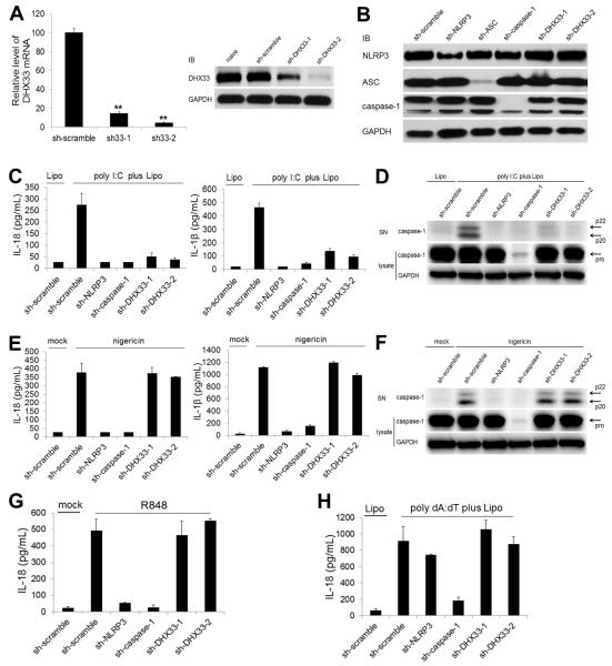
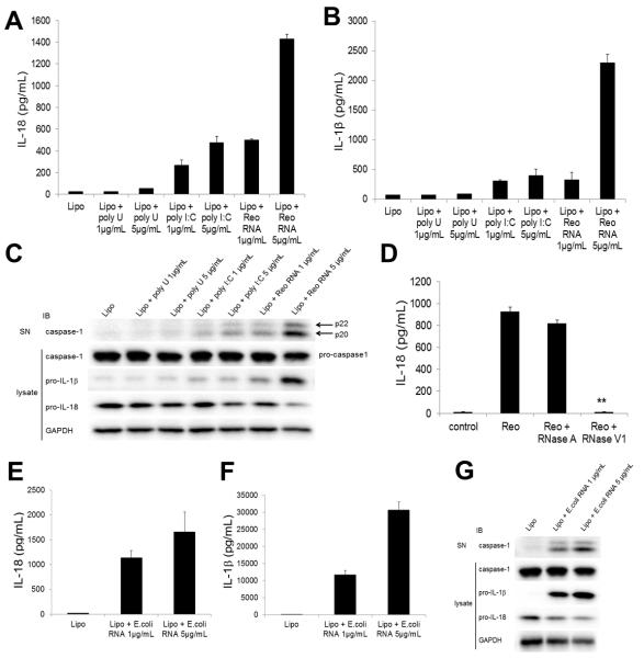
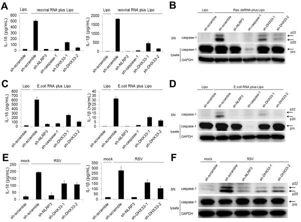
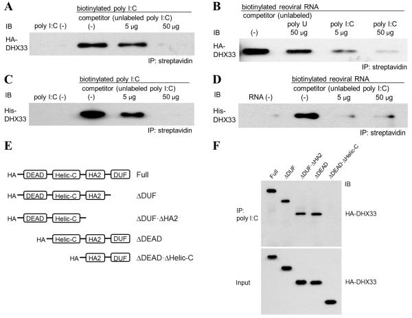
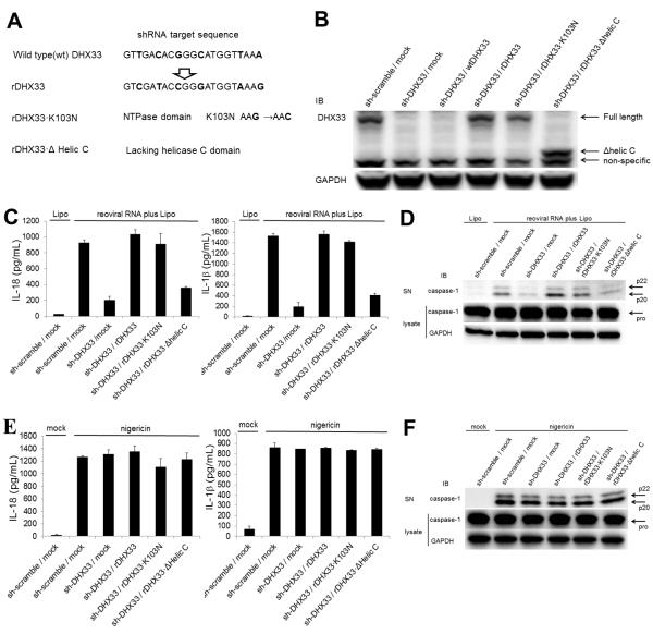
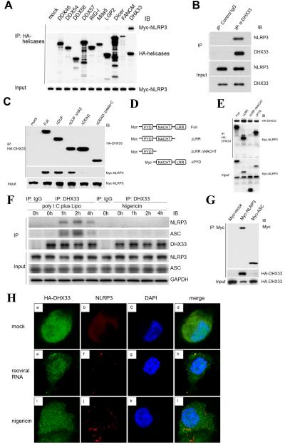
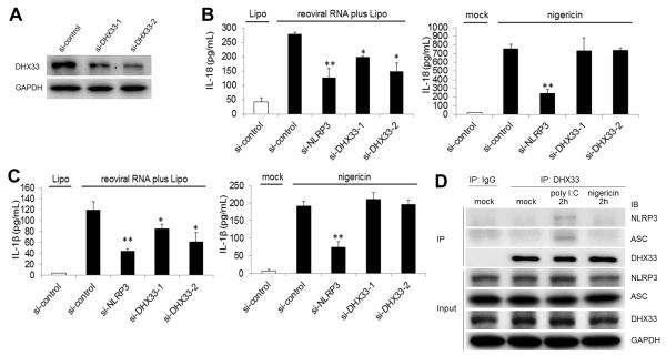
The NLRP3 inflammasome plays a major role in innate immune responses by activating caspase-1, resulting in secretion of interleukin-18 (IL-18) and IL-1β. Although cytosolic double-stranded RNA (dsRNA) and bacterial RNA are known to activate the NLRP3 inflammasome, the upstream sensor is unknown. We investigated the potential function of DExD/H-box RNA helicase family members (previously shown to sense cytosolic DNA and RNA to induce type 1 interferon responses) in RNA-induced NLRP3 inflammasome activation. Among the helicase family members tested, we found that targeting of DHX33 expression by short hairpin RNA efficiently blocked the activation of caspase-1 and secretion of IL-18 and IL-1β in human macrophages that were activated by cytosolic poly I:C, reoviral RNA, or bacterial RNA. DHX33 bound dsRNA via the helicase C domain. DHX33 interacted with NLRP3 and formed the inflammasome complex following stimulation with RNA. We therefore identified DHX33 as a cytosolic RNA sensor that activates the NLRP3 inflammasome.


Figures

Figure 1
Figure 1. Targeting of DHX33 expression abolishes activation of the NLPR3 inflammasome induced by cytosolic dsRNA
(A) DHX33 expression was analyzed by real-time PCR (left) and immunoblotting (IB) (right) in THP-1 cells that were naïve and expressing either scrambled shRNA (sh-scramble) or shRNA specific for DHX33 (sh-DHX33-1, sh-DHX33-2). GAPDH served as a loading control. (B) NLRP3, ASC and caspase-1 expression was analyzed by IB in THP-1 macrophages expressing sh-scramble or shRNA specific for NLRP3 (sh-NLRP3), ASC (sh-ASC), caspase-1 (sh-caspase-1) or DHX33. (C-F) THP-1 macrophages expressing shRNA were stimulated for 8 h with 5 μg/ml poly I:C delivered to the cytosol via Lipofectamine 2000 (Lipo) (C and D) or with 2 μM nigericin (E and F). Culture supernatants were analyzed for IL-18 and IL-β by ELISA (C and E) and for cleaved caspase-1 by IB (D and F). Cell lysates were analyzed by IB for pro-caspase-1 and GAPDH (D and F). (G and H) IL-18 secretion was measured by ELISA in culture supernatants of THP-1 macrophages stimulated for 8 h with 5 μg/ml R848 (G) or with 5 μg/ml poly dA:dT plus Lipo (H). Graph shows the mean ± SD of triplicate wells (**P<0.001 versus scramble). Data are representative of three independent experiments. See also Figure S1.
Figure 2
Figure 2. Reoviral dsRNA and bacterial RNA activate caspase-1 and induce secretion of IL-18 and IL-1β
(A-C) THP-1 macrophages were stimulated for 8 h with Lipo (control), poly U (1 or 5 μg/ml) plus Lipo, poly I:C (1 or 5 μg/ml) plus Lipo, or reoviral RNA (1 or 5 μg/ml) plus Lipo. Culture supernatants were analyzed for IL-18 (A) and IL-β (B) by ELISA and for cleaved caspase-1 by immunoblotting (IB) (C). Cell lysates were analyzed by IB for pro-caspase-1, pro-IL-1β, pro-IL-18 and GAPDH (C). (D) THP-1 macrophages were stimulated for 8 h with Lipo (control) or 5 μg/ml reoviral RNA plus Lipo pretreated without or with RNase A or RNase V1. Culture supernatants were analyzed for IL-18 by ELISA. (E-G) THP-1 macrophages were stimulated for 8 h with Lipo or bacterial RNA from E. coli (1 or 5 μg/ml) plus Lipo. Culture supernatants were analyzed for IL-18 (E) and IL-β (F) by ELISA and for cleaved caspase-1 by IB (G). Cell lysates were analyzed by IB for pro-caspase-1, pro-IL-1β, pro-IL-18 and GAPDH (G). Graph shows the mean ± SD of triplicate wells (**P<0.001 versus reoviral RNA without pre-treatment).
Figure 3
Figure 3. DHX33 is required for NLRP3 inflammasome activation induced by reoviral dsRNA, bacterial RNA and live virus
(A-F) THP-1 macrophages expressing shRNA were stimulated for 8 h with 5 μg/ml reoviral RNA with Lipo (A and B), or 5 μg/ml bacterial RNA from E. coli (C and D). THP-1 macrophages expressing shRNA were infected for 16 h with respiratory syncytial virus (RSV) (E and F). Culture supernatants were analyzed for IL-18 and IL-β (A, C, E) by ELISA and for cleaved caspase-1 by immunoblotting (IB) (B, D, F). Cell lysates were analyzed by IB for pro-caspase-1 and GAPDH (B, D, F). Graph shows the mean ± SD of triplicate wells. Data are representative of three independent experiments. See also Figure S2.
Figure 4
Figure 4. DHX33 requires the helicase C domain for binding to dsRNA
(A and B) HA-DHX33 was overexpressed in HEK293T cells and purified by anti-HA beads. Purified HA-DHX33 protein was incubated with biotinylated poly I:C (A) or biotinylated genomic reoviral RNA (B) in the absence (0 μg) or presence (A: 5 or 50 μg of poly I:C; B: 50 μg of poly U, 5 or 50 μg of poly I:C) of unlabeled competitor. Biotinylated poly I:C (A) or reoviral RNA (B) was precipitated by streptavidin beads, followed by immunoblotting (IB) for HA-DHX33. (C and D) Recombinant His-DHX33, generated in E. coli, was incubated with biotinylated poly I:C (C) or biotinylated reoviral genomic RNA (D) in the absence (0 μg) or presence (5 or 50 μg) of unlabeled poly I:C as a competitor. Biotinylated dsRNA was immunoprecipitated by streptavidin beads, followed by IB for DHX33. (E) Purified full-length and truncated HA-DHX33 proteins were incubated individually with biotinylated poly I:C. Biotinylated poly I:C was immunoprecipitated by streptavidin beads, followed by IB for HA-DHX33. DEAD, Asp-Glu-Ala-Asp domain; HELICc, helicase C-terminal domain; HA2, helicase-associated domain 2; DUF, domain of unknown function. See also Figure S3.
Figure 5
Figure 5. Reconstitution of DHX33 in DHX33-gene targeting cells rescues inflammasome activation induced by cytosolic dsRNA
(A and B) The shRNA-resistant DHX33 with 6-nucleotide substitution in the 21mer targeting sequence was constructed without changing amino acids (rDHX33). Using this rDHX33 construct, an NTPase dead mutant (rDHX33·K103N) and a helic C domain-deleted mutant (rDHX33·Δhelic C) were generated (A). The wild type DHX33 (wtDHX33) and shRNA-resistant DHX33 constructs were transfected into DHX33-gene targeting THP-1 cells, followed by immunoblotting (IB) for DHX33 (B). (C-F) Cells were stimulated for 8 h with 5 μg/ml reoviral RNA plus Lipo (C and D) or 2 μM nigericin (E and F). Culture supernatants were analyzed for IL-18 and IL-β by ELISA (C and E) and for cleaved caspase-1 by IB (D and F). Cell lysates were analyzed by IB for pro-caspase-1 and GAPDH (D and F). Graph shows the mean ± SD of triplicate wells.
Figure 6
Figure 6. DHX33 forms the inflammasome complex via direct binding to NLRP3
(A) HEK293T cells were co-transfected with a vector encoding Myc-NLRP3 plus a vector encoding an HA-DExD/H family member (DDX46, DDX54, DDX56, DDX57, RIG-I, Mda-5, LGP-2, Dicer, FANCM or DHX33) or a pCMV-HA empty vector (mock). HA-tagged helicases were immunoprecipitated, followed by immunoblotting (IB) for Myc-NLRP3 and HA-helicases. (B) A mixture of the purified GST-NLRP3 and His-DHX33 proteins generated in E. coli was immunoprecipitated with anti-DHX33 antibody or isotype control IgG, followed by IB for NLRP3 and DHX33. (C) HEK293T cells were co-transfected with a vector encoding Myc-NLRP3 along with a vector encoding a full-length or truncated HA-DHX33 (as in Figure 4E). HA-tagged helicases were immunoprecipitated, followed by IB for HA-DHX33 and Myc-NLRP3. (D) HEK293T cells were co-transfected with a vector encoding HA-DHX33 along with a vector encoding full-length or truncated Myc-NLRP3. HA-DHX33 was immunoprecipitated followed by IB for HA-DHX33 and Myc-NLRP3. (E) THP-1 macrophages were stimulated for 0, 1, 2 and 4 h with poly I:C plus Lipo or nigericin. Immunoprecipitation was performed by anti-DHX33 antibody or isotype control IgG, followed by IB for DHX33, NLRP3, ASC and GAPDH. (F) HEK293T cells were co-transfected with a vector encoding HA-DHX33 along with a pCMV-Myc empty vector (Myc-mock), or a vector encoding Myc-NLRP3 or Myc-ASC. Myc-tagged proteins were immunoprecipitated, followed by IB for Myc and HA-DHX33. (G) THP-1 macrophages transfected with HA-DHX33 were stimulated for 2 h with Lipo (mock) (a-d), reoviral RNA plus Lipo (reoviral RNA) (e-h) or nigericin (i-l) and analyzed for the localization of HA-DHX33 (red) (a, e, i), NLRP3 (green) (b, f, j), nucleus marker DAPI (blue) (c, g, k), and merge (d, h, l) using confocal microscopy. See also Figure S4.
Figure 7
Figure 7. DHX33 is involved in cytosolic RNA-induced NLRP3 inflammasome activation in human monocyte-derived macrophages
(A) DHX33 expression was analyzed by immunoblotting (IB) in human primary monocyte-derived macrophages (MDM) transfected with control siRNA (si-control) or siRNA specific for DHX33 (si-DHX33-1, si-DHX33-2). (B and C) MDM transfected with control siRNA or DHX33 siRNA were stimulated for 8 h with 5 μg/ml reoviral RNA plus Lipo (left) or 2 μM nigericin (right). Culture supernatants were analyzed for IL-18 (B) and IL-β (C) by ELISA. (D) MDM were stimulated without (mock) or with poly I:C plus Lipo or nigericin for 2 h. Immunoprecipitation was performed by anti-DHX33 antibody or isotype control IgG, followed by IB for DHX33, NLRP3, ASC and GAPDH. Graph shows the mean ± SD of triplicate wells (*P<0.05, **P<0.01 versus scramble siRNA). See also Figure S5.

Comment in

References

    1. Besch R, Poeck H, Hohenauer T, Senft D, Hacker G, Berking C, Hornung V, Endres S, Ruzicka T, Rothenfusser S, Hartmann G. Proapoptotic signaling induced by RIG-I and MDA-5 results in type I interferon-independent apoptosis in human melanoma cells. The Journal of clinical investigation. 2009;119:2399–2411. - PMC - PubMed
    1. Bryant C, Fitzgerald KA. Molecular mechanisms involved in inflammasome activation. Trends in cell biology. 2009;19:455–464. - PubMed
    1. Burckstummer T, Baumann C, Bluml S, Dixit E, Durnberger G, Jahn H, Planyavsky M, Bilban M, Colinge J, Bennett KL, Superti-Furga G. An orthogonal proteomic-genomic screen identifies AIM2 as a cytoplasmic DNA sensor for the inflammasome. Nature immunology. 2009;10:266–272. - PubMed
    1. Davis BK, Wen H, Ting JP. The inflammasome NLRs in immunity, inflammation, and associated diseases. Annual review of immunology. 2011;29:707–735. - PMC - PubMed
    1. Eigenbrod T, Franchi L, Munoz-Planillo R, Kirschning CJ, Freudenberg MA, Nunez G, Dalpke A. Bacterial RNA mediates activation of caspase-1 and IL-1beta release independently of TLRs 3, 7, 9 and TRIF but is dependent on UNC93B. Journal of immunology (Baltimore, Md.: 1950) 2012;189:328–336. - PMC - PubMed

Publication types

MeSH terms