. 2013 Sep;444(1-2):257-62.
doi: 10.1016/j.virol.2013.06.020. Epub 2013 Jul 18.

High-risk human papillomavirus E6 inhibits monocyte differentiation to Langerhans cells

Affiliations

High-risk human papillomavirus E6 inhibits monocyte differentiation to Langerhans cells

Norifumi Iijima et al. Virology. 2013 Sep.

Abstract

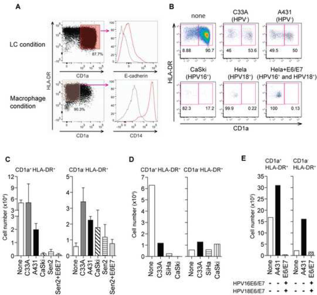
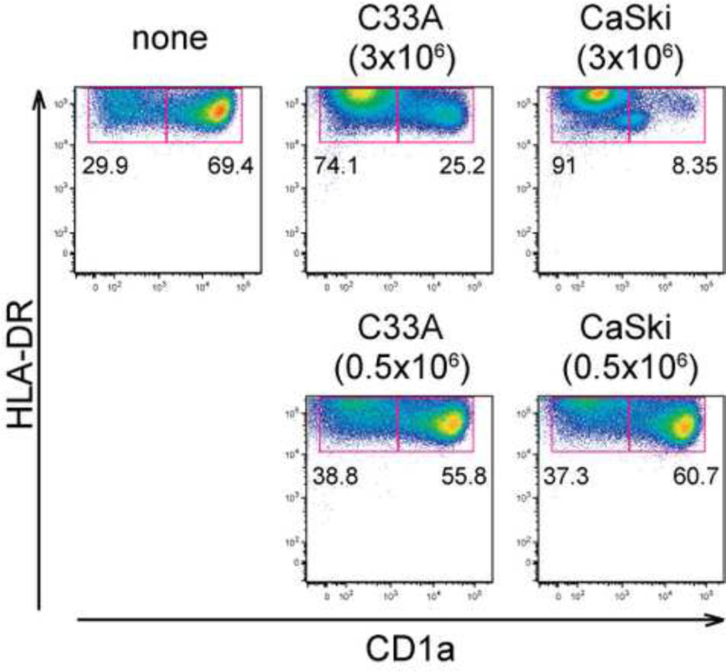
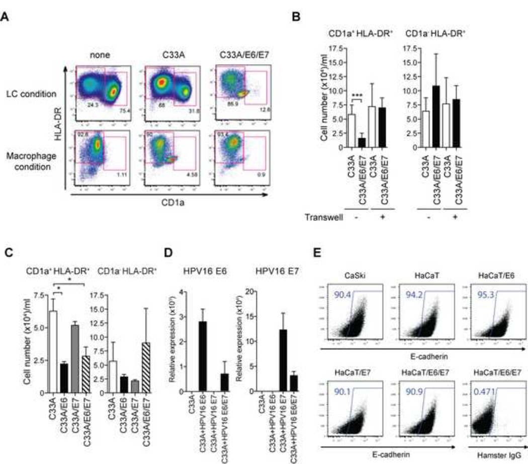
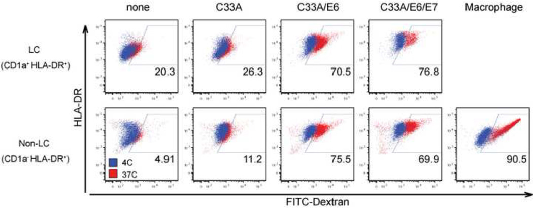
High-risk human papillomaviruses (HPVs) cause a variety of malignancies of the mucosal epithelium. However, the local immune evasion strategies used by HPV-transformed cells remain unclear. Here, we examined the effect of HPV-positive cancer cells on human peripheral blood monocytes, which are precursors of Langerhans cells, key antigen-presenting cells in the squamous epithelium. HPV-positive cervical cancer cells and HPV-E6 expressing cells inhibited monocyte differentiation to Langerhans cells in a contact-dependent manner. Unlike Langerhans cells, monocytes that differentiated in the presence of HPV16 E6-expressing cells exhibited high levels of endocytic activity. Our results suggest that cells infected by high-risk HPV evade immune surveillance by blocking the differentiation of monocytes into competent antigen presenting cells.

Keywords: Cervical cancer; Dendritic cell; Immune evasion; Monocytes.


Figures

Figure 1
Figure 1. High-risk HPV-positive cancer cell lines inhibit LC generation from human CD14+ monocytes
(A) CD14+ monocytes purified from human buffy coats were incubated in the presence of GM-CSF/IL-4/TGF-β (LC condition) or of GM-CSF (macrophage condition) for 7 days, and stained for LC markers (CD1a, E-cadherin, HLA-DR) and a monocyte marker (CD14). Red histograms indicate expression of E-cadherin or CD14 in the indicated condition, while gray histograms indicate expression in the other. (B) Purified monocytes were co-incubated with UV-treated HPV-negative (C33A and A431) or HPV-positive cancer cell lines (CaSki, HeLa, SiHa and HPV16 E6/E7-transduced HeLa), and analyzed for expression of CD1a and HLA-DR. (C) The average total numbers of differentiated LC (CD1a+ HLA-DR+) and non-LC (CD1a) obtained from at least four donors from co-culture with the indicated cancer cell lines are depicted. Error bars indicate standard error of the mean (SEM). (D, E) CD14+ monocytes were co-incubated with PFA-fixed (D) or live (E) HPV-negative (C33A, A431) or high-risk HPV-positive cell lines (SiHa and CaSki or HPV16 E6/E7-transduced HeLa) in the presence of GM-CSF/IL-4/TGF-β for 7 days. The average total numbers of differentiated LC (CD1a+ HLA-DR+) and non-LC (CD1a) obtained from at least four donors from co-culture with the indicated cancer cell lines are depicted.
Figure 2
Figure 2. Block of monocyte-to-LC differentiation by cancer cells occurs at high density
Differentiation of LC from CD14+ human monocytes (1 × 10⁶ cells/flask) in the presence of low (0.5 × 10⁶ cells/flask) or high (3 × 10⁶ cells) densities of cancer cell lines (C33A cells or CaSki cells) was compared. On day7, expression levels of CD1a and HLA-DR were analyzed by FACS. These data are representative of at least three separate experiments.
Figure 3
Figure 3. High-risk HPV16 E6 but not E7 impairs the differentiation of LCs from blood CD14+ monocytes in a contact-dependent manner
CD14+ monocytes were co-incubated with UV-treated HPV C33A cells containing the empty vector (C33A) or HPV16 E6 and E7-transduced C33A cells in the presence of GM-CSF/IL-4/TGF-β (LC condition) or GM-CSF (Macrophage condition) for 7 days. Cells were analyzed by flow cytometry for the expression of CD1a and HLA-DR. (B) CD14+ monocytes were co-incubated with UV-treated transduced C33A cells either together or separately in Transwell. The average numbers of differentiated CD1a+ HLA-DR+ LCs obtained from at least three donors are depicted. (C) CD14+ monocytes were co-incubated with UV-treated vector-transduced or HPV16 E6 and/or E7-transduced C33A cells in the presence of GM-CSF/IL-4/TGF-β for 7 days. The average numbers of LCs cultured in the presence of C33A transduced with the indicated viral genes obtained from at least three donors are depicted. (D) Expression levels of HPV16 E6 and/or E7 in C33A transduced with the indicated viral genes were analyzed by quantitative RT-PCR. (E) C33A, Caski, HaCaT or HaCaT expression HPV16 E6 and/or E7 were stained with antibody to E-cadherin or isotype control. Error bars indicate SEM. *, p < 0.05; **, p < 0.01; ***, p < 0.001.
Figure 4
Figure 4. Increased phagocytic capacity of monocytes differentiated in the presence of HPV16 E6-transduced cell line
CD14+ monocytes were co-incubated with UV-irradiated HPV-negative (C33A), HPV16 E6 C33A cells, or HPV16 E6 + E7 C33A cells in the presence of GM-CSF/ IL-4/TGF-β (LC condition) or GM-CSF (Macrophage condition) for 7 days. (A) Differentiated monocytes were incubated with FITC-Dextran for 1 hour at 37°C or 4°C. Phagocytic activity of CD1a+HLA-DR+ (upper row) and CD1a HLADR+ (bottom row) populations at 37°C (red dots) and 4°C (blue dots) was analyzed by flow cytometry. These data were representative of three separate experiments.

References

    1. zur Hausen H. Papillomaviruses in the causation of human cancers - a brief historical account. Virology. 2009 Feb 20;384(2):260–265. - PubMed
    1. Doorbar J. The papillomavirus life cycle. Journal of clinical virology : the official publication of the Pan American Society for Clinical Virology. 2005 Mar;32(Suppl 1):S7–S15. - PubMed
    1. Jeon S, Lambert PF. Integration of human papillomavirus type 16 DNA into the human genome leads to increased stability of E6 and E7 mRNAs: implications for cervical carcinogenesis. Proc Natl Acad Sci U S A. 1995 Feb 28;92(5):1654–1658. - PMC - PubMed
    1. Moody CA, Laimins LA. Human papillomavirus oncoproteins: pathways to transformation. Nat Rev Cancer. 2010 Aug;10(8):550–560. - PubMed
    1. Goodwin EC, DiMaio D. Repression of human papillomavirus oncogenes in HeLa cervical carcinoma cells causes the orderly reactivation of dormant tumor suppressor pathways. Proc Natl Acad Sci U S A. 2000 Nov 7;97(23):12513–12518. - PMC - PubMed

Publication types

Substances

LinkOut - more resources