. 2013 Aug 1;154(3):541-55.
doi: 10.1016/j.cell.2013.06.051. Epub 2013 Jul 18.

KDM4A lysine demethylase induces site-specific copy gain and rereplication of regions amplified in tumors

Affiliations

KDM4A lysine demethylase induces site-specific copy gain and rereplication of regions amplified in tumors

Joshua C Black et al. Cell. .

Abstract

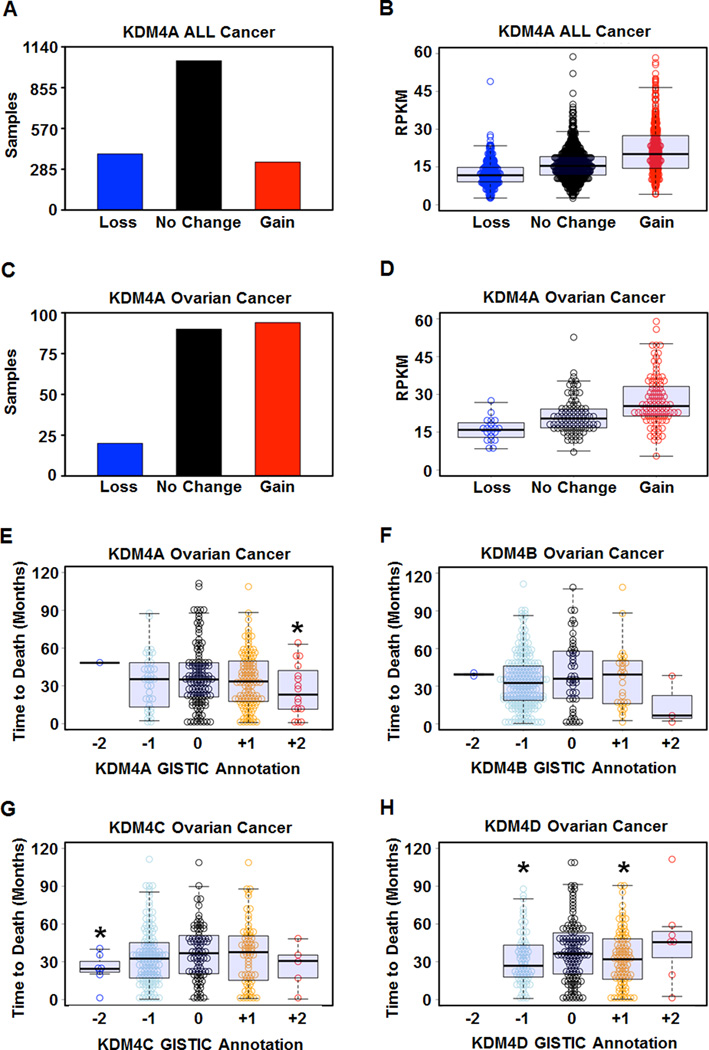
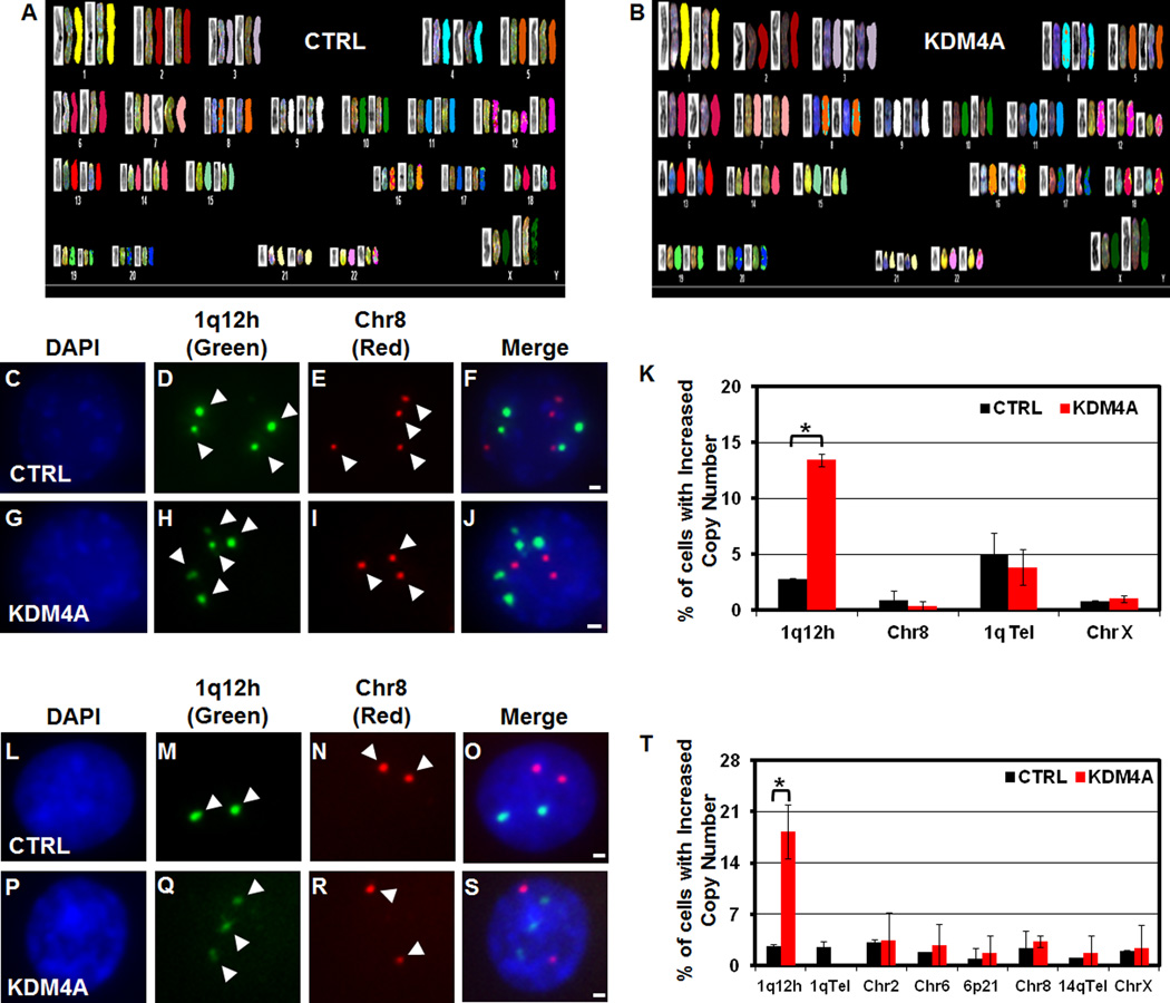
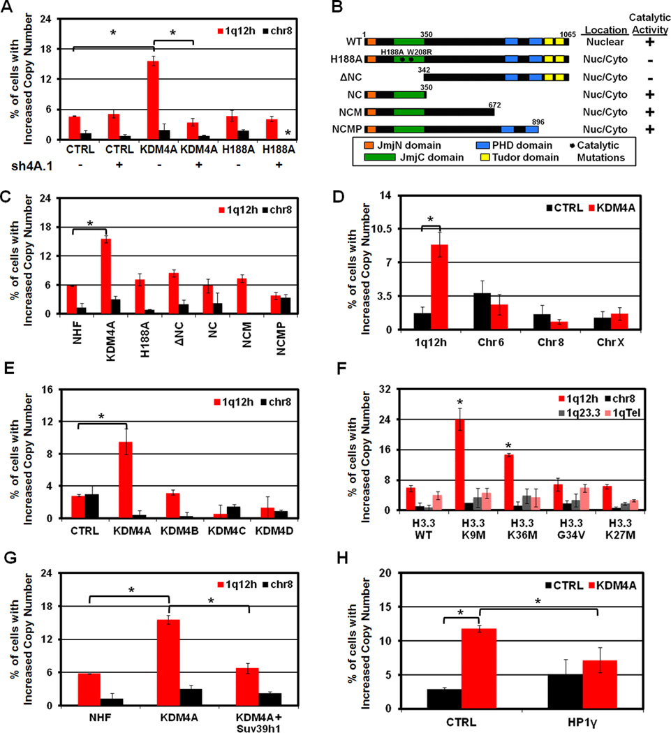
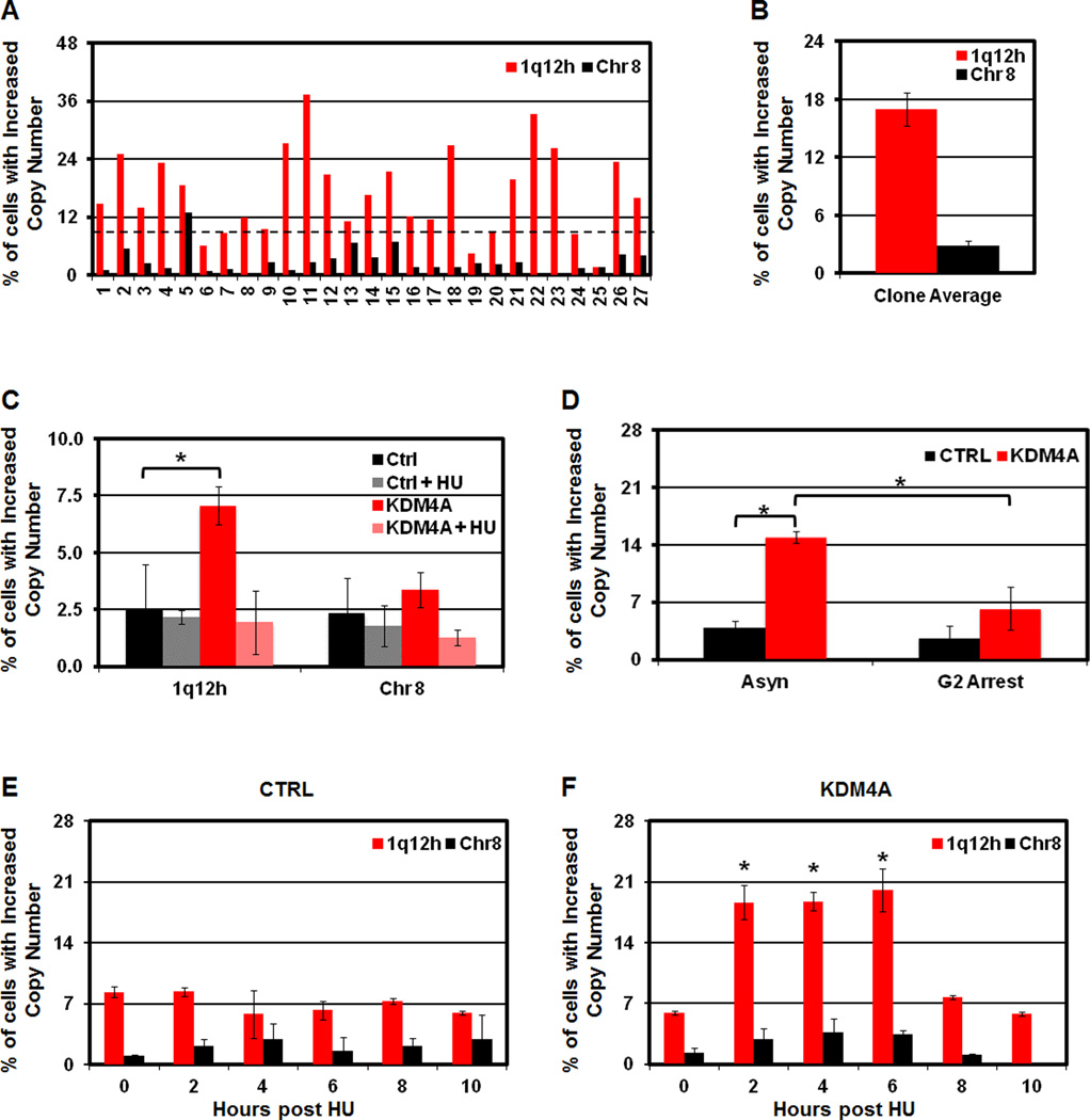
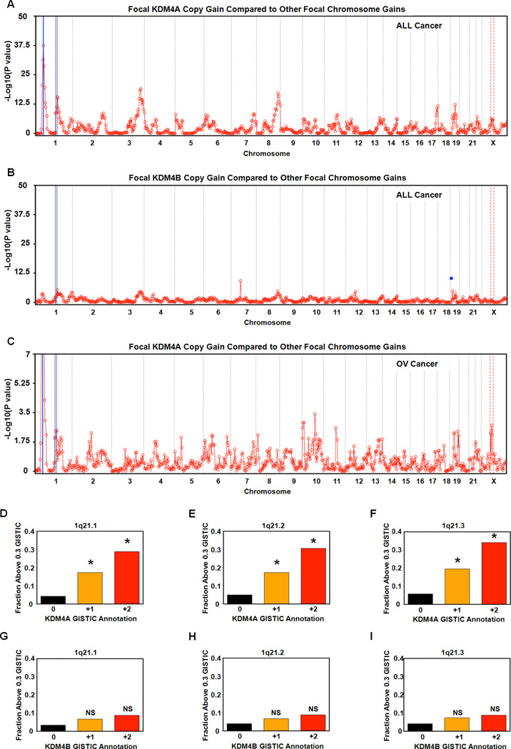
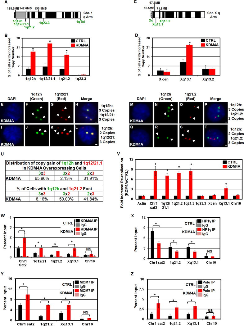
Acquired chromosomal instability and copy number alterations are hallmarks of cancer. Enzymes capable of promoting site-specific copy number changes have yet to be identified. Here, we demonstrate that H3K9/36me3 lysine demethylase KDM4A/JMJD2A overexpression leads to localized copy gain of 1q12, 1q21, and Xq13.1 without global chromosome instability. KDM4A-amplified tumors have increased copy gains for these same regions. 1q12h copy gain occurs within a single cell cycle, requires S phase, and is not stable but is regenerated each cell division. Sites with increased copy number are rereplicated and have increased KDM4A, MCM, and DNA polymerase occupancy. Suv39h1/KMT1A or HP1γ overexpression suppresses the copy gain, whereas H3K9/K36 methylation interference promotes gain. Our results demonstrate that overexpression of a chromatin modifier results in site-specific copy gains. This begins to establish how copy number changes could originate during tumorigenesis and demonstrates that transient overexpression of specific chromatin modulators could promote these events.


Figures

Figure 1
Figure 1. KDM4A is amplified and overexpressed in cancer and correlates with poor outcome in ovarian cancer
(A) Distribution of gain (GISTIC annotation +1 or +2) or loss (GISTIC annotation −1 or −2) of copy of KDM4A in 1770 cancer samples. (B) Amplification of KDM4A correlates with increased expression of KDM4A in TCGA. (C) KDM4A is frequently amplified in ovarian cancer (P=1.4×10⁻²¹ for Gain vs No change or Loss by Fisher’s exact test). (D) Amplification of KDM4A in ovarian cancer correlates with increased expression of KDM4A. (E) Focal amplification of KDM4A in ovarian cancer correlates with poor outcome in 285 deceased ovarian cancer samples (P = 0.02 by one-tailed Student’s t-test and 0.048 by one-tailed, Wilcoxon rank sum test for +2 vs 0). (F) Copy number of KDM4B does not correlate with outcome in ovarian cancer. (G) Deletion of KDM4C in ovarian cancer correlates with outcome (P=0.014 for Loss vs None). (H) Copy number loss and gain of KDM4D correlate with outcome in ovarian cancer (P=0.018 for Gain vs None and 0.013 for Loss vs None by Student’s t-test). * indicates significant difference from No Change samples (P< 0.05). RPKM denotes Reads per kilobase exon model per million reads of RNA seq data (see supplemental experimental procedures). See also Figure S1.
Figure 2
Figure 2. KDM4A overexpression results in 1q12h copy gain
(A–B) SKY analysis of RPE GFP-CTRL and GFP-KDM4A cells. (C–J) FISH of stable 293T cells overexpressing GFP-CTRL or GFP-KDM4A, respectively: (DAPI- C, G); 1q12h (Green- D, H); Chr 8 centromere (Red- E, I); merged images in F, J. (K) Quantification of FISH experiments in stable 293T GFP-CTRL (Black bars) and GFP-KDM4A cells (Red bars) with the indicated FISH probes. (L–S) FISH of stable RPE cells overexpressing GFP-CTRL or GFP-KDM4A, respectively: (DAPI- L, P); 1q12h (Green- M, Q); Chr 8 (Red- N, R); merged images in O, S. (T) Quantitation of RPE FISH experiments. Arrowheads indicate foci in FISH images. Error bars represent the S.E.M. * indicates significant difference from GFP-CTRL (P<0.05) by two-tailed students t-test. Scale bars represent 2µm. See also Figure S2.
Figure 3
Figure 3. 1q12h copy gain can be induced transiently, depends on KDM4A catalytic activity, and can be antagonized by Suv39h1 and HP1γ
(A) Quantification of FISH experiments in 293T cells overexpressing CTRL, KDM4A or catalytically inactive KDM4A (H188A) with (+) and without (-) depletion of endogenous KDM4A (sh4A.1) with the indicated FISH probes. (B) Schematic of NHF-tagged KDM4A constructs. The subcellular localization (“Location”- occurred in greater than 80% of assayed cells) and catalytic activity (“+” indicates strong reduction in total nuclear H3K36me3). (C,D) Quantification of FISH experiments with indicated probes in RPE cells transfected for 24 hours with the indicated constructs: (C) NHF-KDM4A constructs; (D) GFP-CTRL (Black bars) or GFP-KDM4A (Red bars). (E) 1q12h copy gain is specific to KDM4A overexpression and not other KDM4 family members. (F) Overexpression of H3.3 histone variants for H3K9 or H3K36 promotes 1q12h gain. (G,H) Co-expression of Suv39h1 (G) or HP1γ (H) abrogates KDM4A-dependent 1q12h gain. Error bars represent the S.E.M. * indicates significant difference from GFP-CTRL or NHF-CTRL (P<0.05) by two-tailed students t-test. See also Figure S3.
Figure 4
Figure 4. 1q12h copy gain is not stably inherited and requires S phase each cell cycle
(A) Increased 1q12h copy number in GFP-KDM4A RPE cells is not inherited. FISH of single cell clones derived from RPE KDM4A cells. (B) Average copy gain for 27 single cell clones from A is graphically depicted. (C) Increased copy number of 1q12h requires S phase. (D) 1q12h gain is lost by the end of G2. (E, F) 1q12h copy gain is generated in S phase. Stable GFP-CTRL (E) and GFP-KDM4A (F) RPE cells were arrested in hydroxyurea (HU) for 20 hours and released for the time indicated prior to FISH analysis. Error bars represent the S.E.M. * indicates significant difference from GFP-CTRL (P<0.05) by two-tailed students t-test. For the HU release, P values are based on the comparison of KDM4A to CTRL at each individual time point (F, E, respectively). See also Figure S4.
Figure 5
Figure 5. KDM4A interacts with replication machinery and KDM4A overexpression promotes re-replication
(A) Table depicting mass spectrometry analysis of KDM4A interacting proteins related to replication. (B) Western blots of co-immunoprecipitation of endogenous KDM4A and the indicated licensing and replication machinery in RPE Cells. (C) KDM4A overexpression in RPE cells leads to re-replication of Chr1 sat2. (D) KDM4A is enriched at Chr1 sat2 (1q12) in HU arrested KDM4A-overexpressing RPE cells. (E) H3K9me3, but not H3K36me3 decreases at Chr1 sat2 in HU arrested cells. (F) HP1γ enrichment decreases at Chr1 sat2 (1q12) in HU arrested KDM4A-overexpressing cells. (G, H) MCM7 and DNA polymerase α (Pol α) are enriched at Chr1 sat2 (1q12) in HU arrested KDM4A-overexpressing cells. Error bars represent the S.E.M. * indicates significant difference from GFP-CTRL (P<0.05) by two-tailed students t-test. See also Figure S5.
Figure 6
Figure 6. Identification of cytogenetic bands co-amplified with KDM4A in cancer
(A) Focal amplification of cytogenetic bands correlates with amplification of KDM4A in cancer. The blue line represents the locus of KDM4A and its gene-specific significance is P=1.5×10−37. (B) Focal amplification of cytogenetic bands with amplification of KDM4B. The blue dot represents the gene-specific significance of KDM4B. (C) Focal amplification of cytogenetic bands correlates with amplification of KDM4A in 547 ovarian cancer samples. The blue line represents the locus of KDM4A and its gene-specific significance is P=1.1×10⁻¹⁹. For each co-amplification plot, blue shaded regions indicates 1p11.2 through 1q21.3 and red dashed lines indicates Xp11.2 through Xq13.2. (D) Increased copy of KDM4A is associated with increased mean focal copy of 1q21.1 P=2×10⁻⁹ for +2 vs 0 and P= 2.04×10⁻²⁵ for +1 vs 0 by Fisher’s exact test. (E) Increased copy of KDM4A is associated with increased copy of 1q21.2 (P = 1.9×10⁻⁹ for +2 vs 0 and P=6.28×10⁻²² for +1 vs 0). (F) Increased copy of KDM4A is associated with increased copy of 1q21.3 (P=1.02×10⁻¹⁰ for +2 vs 0 and 3×10⁻²⁴ for +1 vs 0). (G) Increased copy number of KDM4B is not associated with increased copy of 1q21.1 (P=0.18 for +2 vs 0). (H) Increased copy number of KDM4B is not associated with increased copy of 1q21.2 (P=0.22 for +2 vs 0). (I) Increased copy number of KDM4B is not associated with increased copy number of 1q21.3 (P=0.24 for +2 vs 0). * indicates significant difference of +1 or +2 vs 0 by Fisher’s exact test. NS indicates not significantly different from 0. See also Figure S6.
Figure 7
Figure 7. KDM4A overexpression leads to copy gains and re-replication of regions co-amplified in tumors
(A) Chromosome arm schematic depicting location of FISH probes used on chromosome 1. (B) KDM4A overexpression increased copy number of 1q12h, 1q12/21.1 and 1q21.2 but not 1q23.3. (C) Chromosome arm schematic depicting location of FISH probes used on chromosome X. (D) KDM4A overexpression increases copy number of Xq13.1 but not X cen or Xq13.2 in RPE cells. (E–T) FISH of stable RPE cells overexpressing GFP-CTRL or GFP-KDM4A with indicated FISH probes. (U) Table summarizing co-amplification of 1q12h, 1q12/21.1 and 1q21.2 in panels E–T. Data are presented as % of amplified cells having 2 or 3+ copies of the indicated FISH probes. (V) KDM4A-dependent re-replication of chromosomal domains. (W) KDM4A ChIP in HU arrested cells. KDM4A is enriched in re-replicated regions in KDM4A-overexpressing RPE cells. (X) HP1γ enrichment decreases at re-replicated regions in KDM4A-overexpressing cells following 1 hour release from HU arrest. (Y,Z) MCM7 and DNA polymerase α (Pol α) are enriched at re-replicated regions in HU arrested KDM4A-overexpressing cells, respectively. Error bars represent the S.E.M. * indicates significant difference from GFP-CTRL (P<0.05) by two-tailed students t-test. For Re-replication (V) and ChIP experiments (W, Y, Z) Chr1 sat2, and Chr10 are the data presented in Figure 5 for reference. Scale bars represent 2µm. See also Figure S6,S7.

Comment in

References

    1. Alabert C, Groth A. Chromatin replication and epigenome maintenance. Nat Rev Mol Cell Biol. 2012;13:153–167. - PubMed
    1. Arias EE, Walter JC. Strength in numbers: preventing rereplication via multiple mechanisms in eukaryotic cells. Genes Dev. 2007;21:497–518. - PubMed
    1. Beck DB, Burton A, Oda H, Ziegler-Birling C, Torres-Padilla ME, Reinberg D. The role of PR-Set7 in replication licensing depends on Suv4-20h. Genes Dev. 2012;26:2580–2589. - PMC - PubMed
    1. Beroukhim R, Mermel CH, Porter D, Wei G, Raychaudhuri S, Donovan J, Barretina J, Boehm JS, Dobson J, Urashima M, et al. The landscape of somatic copy-number alteration across human cancers. Nature. 2010;463:899–905. - PMC - PubMed
    1. Berry WL, Shin S, Lightfoot SA, Janknecht R. Oncogenic features of the JMJD2A histone demethylase in breast cancer. Int J Oncol. 2012;41:1701–1706. - PubMed

Publication types

Substances